Electron Microscopy Public Image Archive
Electron Microscopy Public Image Archive
Due to a storage failure, some data files are currently inaccessible (Markted: Incomplete dataset).
We are currently working to restore, but we are accepting priority requests.(email, inquiry).
We are restoring lost files from backups in the following order:
We apologize for the inconvenience and appreciate your understanding.
The EMPIAR-PDBj team at Osaka University assists Asian EM researchers with the transfer of big EM image data to EMPIAR. Instead of sending the data directly to the EBI (UK) via the internet, hard drives can also be sent to Osaka University by postal mail or via a courier service. As an alternative, internet transfer to our server in Osaka is also available. If you would like to take advantage of our submission services, please contact us first by e-mail before sending the data to us.
| Release date | Imageset | Title | Authors and references | Size | Resolution |
|---|---|---|---|---|---|
| 2024-09-02 |  |
Cryo-ET of in vitro vaccinia core [38 tilt series in MRC format] | Liu J, Turoňová B [Pubmed: 38316878] [DOI: 10.1038/s41594-024-01218-5] |
42.9 GB | 7.7 - 13.4 Å |
| 2024-09-02 |  |
Cryo electron micrographs of 70S Escherichia coli ribosomes [4388 multi-frame micrographs composed of 35 frames each in TIFF format] | Gersteuer FG, Morici MM, Wilson DNW [Pubmed: 38503753] [DOI: 10.1038/s41467-024-46762-2] |
2.0 TB | 2.0 - 2.6 Å |
| 2024-08-29 |  |
Human calcium homeostasis modulator 1 (CALHM1) channel [3285 multi-frame micrographs composed of 30 frames each in TIFF format] | Syrjanen JL, Furukawa H [Pubmed: 37380652] [DOI: 10.1038/s41467-023-39388-3] |
553.5 GB | 3.76 Å |
| 2024-08-29 |  |
Cryo-EM micrographs of human DNA polymerase θ helicase domain bound to inhibitor AB25583 [4500 multi-frame micrographs composed of 2051 frames each in EER format] | Ito F, Li Z, Minakhin L, Chandramouly G, Tyagi M, Betsch R, Krais JJ, Tiberi B, Johnson N, Chen XS, Pomerantz RT [Pubmed: 39143110] [DOI: 10.1038/s41467-024-51351-4] |
4.9 TB | 3.21 Å |
| 2024-08-29 |  |
FAST-EM array tomography data of rat pancreas prepared with rOTO protocol and stained with neodymium acetate [688 micrographs in TIFF format] | Kievits AJ, Duinkerken BHP, Lane R, de Heus C, van Beijeren Bergen en Henegouwen D, Höppener T, Wolters AHG, Liv N, Giepmans BNG, Hoogenboom JP [Pubmed: 39119255] [DOI: 10.1515/mim-2024-0005] |
69.7 GB | — |
| 2024-08-29 |  |
SPOT-RASTR - a cryo-EM specimen preparation technique that overcomes problems with preferred orientation and the air/water interface [stack of 230000 particles in MRCS format] | Ghazi Esfahani BGE, Randolph PR, Peng RP, Stroupe MES, Grant TG, Stagg SMS [Pubmed: 39108302] [DOI: 10.1093/pnasnexus/pgae284] |
304.6 GB | 3.58 Å |
| 2024-08-29 |  |
The Prothrombin-Prothrombinase Interaction [2390 micrographs in MRC format] | Stojanovski BM, Mohammed BM, Ruben EA, Summers B, Rau MJ, Fitzpatrick JAJ, Di Cera E [Pubmed: 35427420] [DOI: 10.1182/blood.2022015807] |
149.4 GB | 5.3 - 6.47 Å |
| 2024-08-29 |  |
Low-dose cryo-electron ptychography of Apoferritin at 4.0 mrad [90 diffraction images in BIG DATA VIEWER HDF5 format] | Küçükoğlu BK, Mohammed IM, Guerrero-Ferreira RCG, Kube MK, Radecke JR, Ribet SR, Varnavides GV, Leidl MLL, Lau KL, Nazarov SN, Myasnikov AM, Sachse CS, Müller-Caspary KMC, Ophus CO, Stahlberg HS [DOI: 10.1101/2024.02.12.579607] |
83.8 GB | 5.8 - 12.3 Å |
| 2024-08-28 |  |
CryoEM micrographs of HK97 small terminase in complex with DNA (2 datasets:550 and 1874 multi-frame micrographs composed of 50 frames in tif format) [multiple data sets in TIFF format] | Antson AA, Chechik M, Greive SJ, Jenkins HT [Pubmed: 39116131] [DOI: 10.1073/pnas.2406138121] |
549.9 GB | 2.92 - 3.0 Å |
| 2024-08-28 |  |
Human calcium homeostasis modulator 1 (CALHM1) channel, I109W point mutation [8664 multi-frame micrographs composed of 30 frames each in TIFF format] | Syrjanen JL, Furukawa H [Pubmed: 37380652] [DOI: 10.1038/s41467-023-39388-3] |
1.8 TB | 2.8 Å |
| 2024-08-28 |  |
Human calcium homeostasis modulator 1 (CALHM1) channel, I109W point mutation in complex with ruthenium red [multiple data sets in TIFF format] | Syrjanen JL, Furukawa H [Pubmed: 37380652] [DOI: 10.1038/s41467-023-39388-3] |
4.6 TB | 3.91 - 4.73 Å |
| 2024-08-28 |  |
Cryoelectron microscopy of IL-21/IL-21R/common gamma signaling complex [3684 multi-frame micrographs composed of 51 frames each in TIFF format] | Abhiraman G.C., Jude K.M., Caveney N.A., Garcia K.C. [Pubmed: 37339051] [DOI: 10.1016/j.celrep.2023.112657] |
4.2 TB | 3.7 Å |
| 2024-08-28 |  |
streptavidin on GSAMs [2144 multi-frame micrographs composed of 32 frames each in MRC format] | Liu N, Xu J, Wang HW | 525.5 GB | 2.56 Å |
| 2024-08-28 |  |
Cryo electron micrographs of CCP5 decorated microtubules [4643 multi-frame micrographs composed of 22 frames each in TIFF format] | Chen J, Zehr EA, Gruschus JM, Szyk A, Liu Y, Tanner ME, Tjandra N, Roll-Mecak A [Pubmed: 39020174] [DOI: 10.1038/s41586-024-07699-0] |
1.3 TB | 2.9 - 3.6 Å |
| 2024-08-28 |  |
Full-length mouse 5-HT3A receptor in complex with SMP100, pre-activated and open-like [multiple data sets in TIFF format] | Felt KCF, Chakrapani SC [Pubmed: 38177669] [DOI: 10.1038/s41594-023-01140-2] |
6.3 TB | 2.7 - 3.79 Å |
| 2024-08-27 |  |
Cryo-EM micrographs of anti-CRISPR associated protein Aca2 bound to its RNA [11232 multi-frame micrographs composed of 40 frames each in TIFF format] | Wilkinson ME, Birkholz N, Kimanius D, Fineran PC [Pubmed: 38987591] [DOI: 10.1038/s41586-024-07644-1] |
1.9 TB | 2.61 Å |
| 2024-08-27 |  |
CryoEM of near-native eisosome/Membrane Compartment containing Can1 (MCC) membrane microdomain scaffolded by Pil1/Lsp1 [2827 multi-frame micrographs composed of 40 frames each in TIFF format] | Kefauver JM, Zou L, Loewith R [Pubmed: 39048819] [DOI: 10.1038/s41586-024-07720-6] |
559.9 GB | 3.2 - 3.67 Å |
| 2024-08-26 |  |
Cryo-EM structure of human pseudouridine synthase 3 [8211 micrographs in TIFF format] | Lin T.-Y., Glatt S. [Pubmed: 38996458] [DOI: 10.1016/j.molcel.2024.06.013] |
3.4 TB | 6.5 Å |
| 2024-08-26 |  |
Molecular model of a bacterial flagellar motor in situ reveals a “parts-list” of protein adaptations to increase torque [multiple data sets in MRC format] | Beeby M, Cohen EJ, Drobnič T, Nans A, Rosenthal PB [Pubmed: 40595286] [DOI: 10.1038/s41564-025-02012-9] |
10.1 TB | 7.9 - 9.36 Å |
| 2024-08-26 |  |
Single particle cryo-EM analysis of cystic fibrosis transmembrane conductance regulator E1371Q mutant [10159 multi-frame micrographs composed of 40 frames each in TIFF format] | Gao X, Hwang T [Pubmed: 39107303] [DOI: 10.1038/s41467-024-50641-1] |
5.0 TB | 3.4 Å |
| 2024-08-26 |  |
Single particle cryo-EM analysis of inhibitor 172 bound cystic fibrosis transmembrane conductance regulator E1371Q mutant [10700 multi-frame micrographs composed of 50 frames each in TIFF format] | Gao X, Hwang T [Pubmed: 39107303] [DOI: 10.1038/s41467-024-50641-1] |
6.4 TB | 3.6 Å |
| 2024-08-24 |  |
E. coli clamp loader with a closed clamp [multiple data sets in TIFF format] | Xu ZQ, Jergic S, Lo ATY, Pradhan AC, Brown HJ, Bouwer JC, Ghodke H, Lewis PJ, Tolun G, Oakley AJ, Dixon NE [Pubmed: 39333521] [DOI: 10.1038/s41467-024-52623-9] |
1.3 TB | 3.7 Å |
| 2024-08-23 |  |
Cryo-EM structure of the histamine-bound histamine H4 receptor and Gq complex [multiple data sets in TIFF format] | Im D, Asada H, Iwata S [Pubmed: 37863901] [DOI: 10.1038/s41467-023-42260-z] |
3.1 TB | 3.0 - 3.1 Å |
| 2024-08-23 | 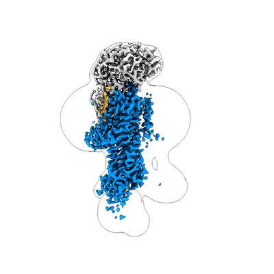 |
Cyo-EM structure of wildtype non-gastric proton pump in the presence of Na+, AlF and ADP [multiple data sets in TIFF format] | Abe K, Artigas P [Pubmed: 37482134] [DOI: 10.1016/j.bbamcr.2023.119543] |
1.7 TB | 2.62 - 3.65 Å |
| 2024-08-23 | 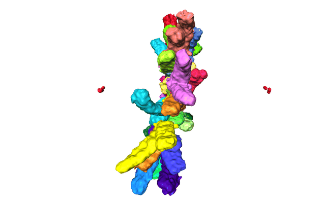 |
Exploring the 3D structure of chromosomes in mitotic cells [multiple data sets in TIFF and DM4 formats] | Cisneros- Soberanis F, Beckett AJ, Simpson E, Pucekova N, Prior IA, Booth DG, Earnshaw WC [Pubmed: 39186086] [DOI: 10.1083/jcb.202403165] |
31.6 GB | — |
