Electron Microscopy Public Image Archive
Electron Microscopy Public Image Archive
Due to a storage failure, some data files are currently inaccessible (Markted: Incomplete dataset).
We are currently working to restore, but we are accepting priority requests.(email, inquiry).
We are restoring lost files from backups in the following order:
We apologize for the inconvenience and appreciate your understanding.
The EMPIAR-PDBj team at Osaka University assists Asian EM researchers with the transfer of big EM image data to EMPIAR. Instead of sending the data directly to the EBI (UK) via the internet, hard drives can also be sent to Osaka University by postal mail or via a courier service. As an alternative, internet transfer to our server in Osaka is also available. If you would like to take advantage of our submission services, please contact us first by e-mail before sending the data to us.
| Release date | Imageset | Title | Authors and references | Size | Resolution |
|---|---|---|---|---|---|
| 2026-04-07 |  |
Cryo-EM of connexin-46/50 in DMPC nanodiscs at low pH (Collection #4) [6057 multi-frame micrographs composed of 45 frames each in TIFF format] | Jarodsky JM, Myers JB, Reichow SL [Pubmed: 41526355] [DOI: 10.1038/s41467-026-68311-9] |
2.3 TB | 2.0 - 2.7 Å |
| 2026-04-07 |  |
Cryo-EM of connexin-46/50 in DMPC nanodiscs at neutral pH (Collection #3) [3261 multi-frame micrographs composed of 45 frames each in TIFF format] | Jarodsky JM, Myers JB, Reichow SL [Pubmed: 41526355] [DOI: 10.1038/s41467-026-68311-9] |
1.3 TB | 2.0 - 2.7 Å |
| 2026-03-30 |  |
Movies of PARP1-DNA Complexes [multiple data sets in TIFF and EER formats] | Sverzhinsky A, Pascal JM [Pubmed: 41698892] [DOI: 10.1038/s41467-026-69375-3] |
27.2 TB | 3.6 - 4.3 Å |
| 2026-03-30 |  |
Borna disease virus 1 nucleoprotein complexes [multiple data sets in TIFF format] | Sugita Y, Hirai Y, Goto SH, Horie M | 8.0 TB | 2.78 - 9.12 Å |
| 2026-03-30 |  |
Solubilized octameric pore of actinoporin Fav prepared on DOPC:sphingomyelin membranes [2073 multi-frame micrographs composed of 40 frames each in TIFF format] | Šolinc G, Srnko M, Švigelj T, Podobnik M, Anderluh G [Pubmed: 40140423] [DOI: 10.1038/s41467-025-58334-z] |
2.1 TB | 2.6 Å |
| 2026-03-30 |  |
Aligned, dose-weighted electron micrographs of His-tagged Exo84 and Sro7 on NI-NTA lipid monolayer grids [4970 micrographs in MRC format] | Brennwald P, Strauss JD, Baker RW [Pubmed: 41083086] [DOI: 10.1016/j.jsb.2025.108253] |
436.4 GB | 3.1 Å |
| 2026-03-30 |  |
C. thermocellum UvrA-UvrB in complex with DNA with a fluorescein modification and AMPPNP [multiple data sets in TIFF and MRCS formats] | Nirwal S, Czarnocki-Cieciura M, Zajko W, Skowronek K, Szczepanowski RH, Nowotny M [Pubmed: 41381534] [DOI: 10.1038/s41467-025-67075-y] |
3.6 TB | 2.97 - 3.18 Å |
| 2026-03-27 |  |
Cryo-electron tomograms of an oxygen-induced tubular nanocompartment in Pyrococcus furiosus [5 tilt series in TIFF format] | Song W, Skalidis I, Howes SC, Foerster F | 13.8 GB | — |
| 2026-03-27 |  |
C. thermocellum UvrA [multiple data sets in TIFF and MRCS formats] | Nirwal S, Czarnocki-Cieciura M, Zajko W, Skowronek K, Szczepanowski RH, Nowotny M [Pubmed: 41381534] [DOI: 10.1038/s41467-025-67075-y] |
666.2 GB | 2.97 Å |
| 2026-03-27 |  |
C. thermocellum UvrA in complex with AMPPNP [multiple data sets in TIFF and MRCS formats] | Nirwal S, Czarnocki-Cieciura M, Zajko W, Skowronek K, Szczepanowski RH, Nowotny M [Pubmed: 41381534] [DOI: 10.1038/s41467-025-67075-y] |
764.0 GB | 2.96 Å |
| 2026-03-27 |  |
C. thermocellum UvrA in complex with unmodified DNA and AMPPNP [multiple data sets in TIFF and MRCS formats] | Nirwal S, Czarnocki-Cieciura M, Zajko W, Skowronek K, Szczepanowski RH, Nowotny M [Pubmed: 41381534] [DOI: 10.1038/s41467-025-67075-y] |
4.0 TB | 3.1 - 3.23 Å |
| 2026-03-27 |  |
C. thermocellum UvrA in complex with DNA with an abasic site [multiple data sets in TIFF and MRCS formats] | Nirwal S, Czarnocki-Cieciura M, Zajko W, Skowronek K, Szczepanowski RH, Nowotny M [Pubmed: 41381534] [DOI: 10.1038/s41467-025-67075-y] |
3.1 TB | 3.84 Å |
| 2026-03-27 |  |
C. thermocellum UvrA in complex with DNA with an abasic site and AMPPNP [multiple data sets in TIFF and MRCS formats] | Nirwal S, Czarnocki-Cieciura M, Zajko W, Skowronek K, Szczepanowski RH, Nowotny M [Pubmed: 41381534] [DOI: 10.1038/s41467-025-67075-y] |
2.7 TB | 2.74 - 3.04 Å |
| 2026-03-27 |  |
C. thermocellum UvrA in complex with DNA with a fluorescein modification [multiple data sets in TIFF and MRCS formats] | Nirwal S, Czarnocki-Cieciura M, Zajko W, Skowronek K, Szczepanowski RH, Nowotny M [Pubmed: 41381534] [DOI: 10.1038/s41467-025-67075-y] |
1.2 TB | 3.14 Å |
| 2026-03-27 |  |
C. thermocellum UvrA in complex with DNA with a fluorescein modification and AMPPNP [multiple data sets in TIFF and MRCS formats] | Nirwal S, Czarnocki-Cieciura M, Zajko W, Skowronek K, Szczepanowski RH, Nowotny M [Pubmed: 41381534] [DOI: 10.1038/s41467-025-67075-y] |
2.2 TB | 3.1 Å |
| 2026-03-27 |  |
C. thermocellum UvrA (K647A) in complex with DNA with a fluorescein modification [multiple data sets in TIFF and MRCS formats] | Nirwal S, Czarnocki-Cieciura M, Zajko W, Skowronek K, Szczepanowski RH, Nowotny M [Pubmed: 41381534] [DOI: 10.1038/s41467-025-67075-y] |
2.1 TB | 4.81 Å |
| 2026-03-27 |  |
C. thermocellum UvrA in complex with DNA with a fluorescein modification and ADP [multiple data sets in TIFF and MRCS formats] | Nirwal S, Czarnocki-Cieciura M, Zajko W, Skowronek K, Szczepanowski RH, Nowotny M [Pubmed: 41381534] [DOI: 10.1038/s41467-025-67075-y] |
1.4 TB | 3.87 Å |
| 2026-03-27 |  |
C. thermocellum UvrA (K39A) in complex with DNA with a fluorescein modification and AMPPNP [multiple data sets in TIFF and MRCS formats] | Nirwal S, Czarnocki-Cieciura M, Zajko W, Skowronek K, Szczepanowski RH, Nowotny M [Pubmed: 41381534] [DOI: 10.1038/s41467-025-67075-y] |
1.1 TB | 3.63 Å |
| 2026-03-26 |  |
Clostridium perfringens iota toxin membrane-binding component Ib-prepore and Ib-pore [11549 multi-frame micrographs composed of 58 frames each in TIFF format] | Yamada TY, Sugita YS, Yoshida TY, Noda TN, Tsuge HT | 2.7 TB | 2.47 - 3.73 Å |
| 2026-03-24 |  |
Influenza A virus Hemagglutinin H3/Darwin/6/2021 in complex with Fab ADI-85647 [7772 micrographs in MRC format] | Ferreira Ramos AS, Bajic G [Pubmed: 40267182] [DOI: 10.1126/sciadv.adu9140] |
682.4 GB | 3.01 Å |
| 2026-03-24 |  |
Influenza A virus Hemagglutinin H3/Darwin/6/2021 in complex with Fab ADI-85666 [4435 micrographs in MRC format] | Ferreira Ramos AS, Bajic G [Pubmed: 40267182] [DOI: 10.1126/sciadv.adu9140] |
389.4 GB | 3.01 Å |
| 2026-03-24 | 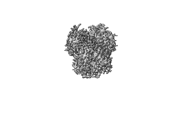 |
Single particle cryo-EM dataset of influenza hemagglutinin (A/Hong Kong/1/1968, H3N2) jetted control sample [4882 multi-frame micrographs composed of 50 frames each in TIFF format] | Williams HM, Curtis WA, Haubner M, Hruby J, Drabbels M, Lorenz UJ [Pubmed: 41000704] [DOI: 10.1101/2025.09.14.676144] |
2.8 TB | 2.7 Å |
| 2026-03-24 |  |
Single particle cryo-EM dataset of the light-driven sodium pump ErNaR prepared by jet vitrification [13007 multi-frame micrographs composed of 50 frames each in EER format] | Haubner M, Williams HM, Hruby J, Straub MS, Guskov A, Kovalev K, Drabbels M, Lorenz UJ [Pubmed: 41332690] [DOI: 10.1101/2025.11.21.689681] |
3.7 TB | 2.1 Å |
| 2026-03-24 |  |
Single particle cryo-EM dataset of the light-driven sodium pump ErNaR in the K2 state prepared by jet vitrification [30180 multi-frame micrographs composed of 50 frames each in EER format] | Haubner M, Williams HM, Hruby J, Straub MS, Guskov A, Kovalev K, Drabbels M, Lorenz UJ [Pubmed: 41332690] [DOI: 10.1101/2025.11.21.689681] |
8.6 TB | 2.8 Å |
| 2026-03-23 |  |
Single particle cryo-EM dataset of the 50S ribosomal subunit following ultrasonic excitation [4637 multi-frame micrographs composed of 50 frames each in TIFF format] | Williams HM, Curtis WA, Haubner M, Hruby J, Drabbels M, Lorenz UJ [Pubmed: 41000704] [DOI: 10.1101/2025.09.14.676144] |
2.6 TB | 2.8 Å |
