Electron Microscopy Public Image Archive
Electron Microscopy Public Image Archive
Due to a storage failure, some data files are currently inaccessible (Markted: Incomplete dataset).
We are currently working to restore, but we are accepting priority requests.(email, inquiry).
We are restoring lost files from backups in the following order:
We apologize for the inconvenience and appreciate your understanding.
The EMPIAR-PDBj team at Osaka University assists Asian EM researchers with the transfer of big EM image data to EMPIAR. Instead of sending the data directly to the EBI (UK) via the internet, hard drives can also be sent to Osaka University by postal mail or via a courier service. As an alternative, internet transfer to our server in Osaka is also available. If you would like to take advantage of our submission services, please contact us first by e-mail before sending the data to us.
| Release date | Imageset | Title | Authors and references | Size | Resolution |
|---|---|---|---|---|---|
| 2025-01-28 | 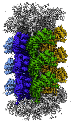 |
Scytonema hofmannii TnsC bound to AMPPNP and DNA [multiple data sets in TIFF and DM4 formats] | Querques I, Schmitz M, Oberli S, Chanez C, Jinek M [Pubmed: 34759315] [DOI: 10.1038/s41586-021-04030-z] |
1.3 TB | 3.57 Å |
| 2025-01-28 | 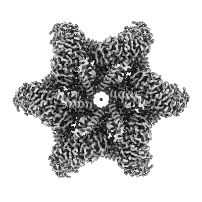 |
Cryo electron micrographs of disease resistance protein NRC2 from Nicotiana benthamiana [14049 multi-frame micrographs composed of 50 frames each in TIFF format] | Webster MW, Madhuprakash J, Kamoun S [DOI: 10.1101/2024.06.18.599586] |
2.7 TB | 2.9 Å |
| 2025-01-28 | 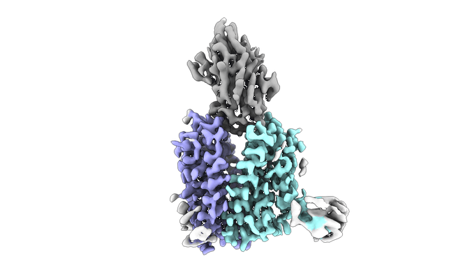 |
Cryo-EM structure of apo human SLC19A3 in outward-open state [5859 multi-frame micrographs composed of 45 frames each in TIFF format] | Gabriel FG [Pubmed: 39358356] [DOI: 10.1038/s41467-024-52872-8] |
2.5 TB | 3.09 Å |
| 2025-01-27 |  |
Movies of cagT∆ mutant Cag T4SS from Helicobacter pylori [3443 multi-frame micrographs composed of 34 frames each in TIFF format] | Roberts JR [Pubmed: 38631913] [DOI: 10.26508/lsa.202302560] |
741.3 GB | 8.0 Å |
| 2025-01-24 | 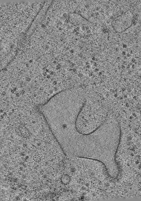 |
Cryo-ET of Dictyostelium discoideum in distilled H2O (hypoosmotic stress) [59 tilt series in MRC format] | Hoffmann PC, Beck M [Pubmed: 39729993] [DOI: 10.1016/j.molcel.2024.11.038] |
241.1 GB | 37.0 Å |
| 2025-01-24 | 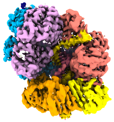 |
E. coli clamp loader with primer-template DNA and a closed clamp [7201 multi-frame micrographs composed of 60 frames each in MRC format] | Xu ZQ, Jergic S, Dixon NE, Lo ATY, Pradhan AC, Brown HJ, Bouwer JC, Ghodke H, Lewis PJ, Tolun G, Oakley AJ [Pubmed: 39333521] [DOI: 10.1038/s41467-024-52623-9] |
1.0 TB | 2.6 Å |
| 2025-01-23 | 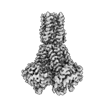 |
Cryo-EM of hMRS2-BRIL with EDTA [3477 multi-frame micrographs in MRC format] [3477 multi-frame micrographs composed of 42 frames each in MRC format] | He Z, Mount JW, Zhang J, Yuan P [Pubmed: 39609651] [DOI: 10.1038/s41594-024-01420-5] |
2.7 TB | 2.95 Å |
| 2025-01-23 | 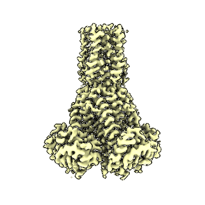 |
Cryo-EM of hMRS2-BRIL with Mg2+ [2631 multi-frame micrographs in MRC format] [2631 multi-frame micrographs composed of 42 frames each in MRC format] | He Z, Mount JW, Zhang J, Yuan P [Pubmed: 39609651] [DOI: 10.1038/s41594-024-01420-5] |
2.0 TB | 2.92 Å |
| 2025-01-23 |  |
Cryogenic electron microscopy of blot-plunged acetyl-CoA decarbonylase/synthase [14078 multi-frame micrographs composed of 21 frames each in TIFF format] | Biester A, Drennan CL [Pubmed: 39361653] [DOI: 10.1073/pnas.2410995121] |
1.8 TB | 3.3 Å |
| 2025-01-23 | 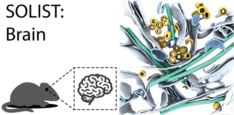 |
In situ cryo-electron tomograms #1-#2 of native mouse corpus callosum from SOLIST procedure. [2 tilt series in MRC format] | Dung NHT, Perone G, Klena N, Vazzana R, Kaluthantrige Don F, Silva M, Sorrentino S, Swuec P, Leroux F, Kelebic N, Coscia F, Erdmann PS [Pubmed: 39271806] [DOI: 10.1038/s41592-024-02384-6] |
5.1 GB | — |
| 2025-01-23 | 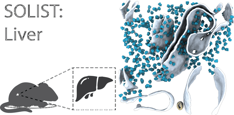 |
Motion-corrected tilt series of native mouse liver using SOLIST procedure. (Minimal dataset for 8.3 Å ribosome) [43 tilt series in MRC format] | Dung NHT, Perone G, Klena N, Vazzana R, Kaluthantrige Don F, Silva M, Sorrentino S, Swuec P, Leroux F, Kelebic N, Coscia F, Erdmann PS [Pubmed: 39271806] [DOI: 10.1038/s41592-024-02384-6] |
112.2 GB | — |
| 2025-01-23 | 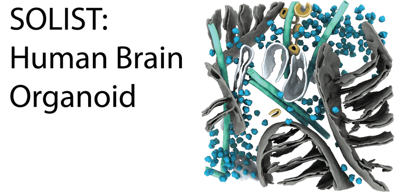 |
In situ cryo-electron tomograms #1-#2 of human brain organoid from SOLIST procedure. [multiple data sets in MRC format] | Dung NHT, Perone G, Klena N, Vazzana R, Kaluthantrige Don F, Silva M, Sorrentino S, Swuec P, Leroux F, Kelebic N, Coscia F, Erdmann PS [Pubmed: 39271806] [DOI: 10.1038/s41592-024-02384-6] |
5.1 GB | — |
| 2025-01-23 | 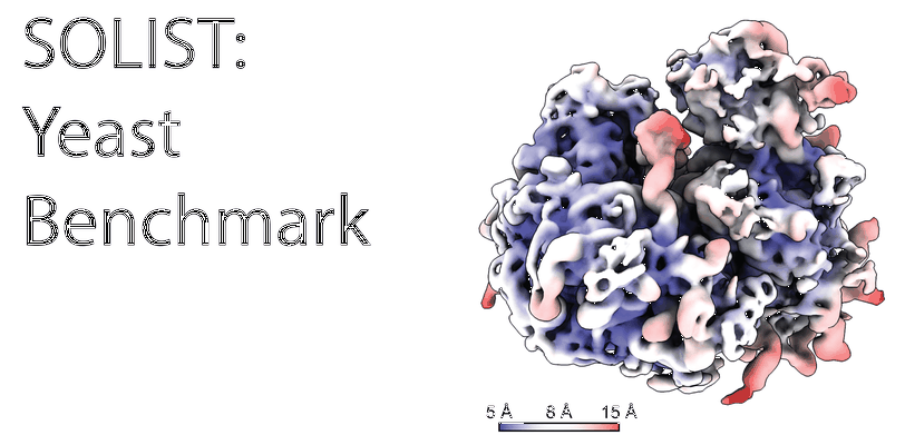 |
In situ cryo-electron tomograms of S. cerevisiae from SOLIST procedure. [65 tilt series in MRC format] | Dung NHT, Perone G, Klena N, Vazzana R, Kaluthantrige Don F, Silva M, Sorrentino S, Swuec P, Leroux F, Kelebic N, Coscia F, Erdmann PS [Pubmed: 39271806] [DOI: 10.1038/s41592-024-02384-6] |
160.4 GB | 7.3 Å |
| 2025-01-23 |  |
Single particle cryo-EM data of spinach cytochrome b6f with decylplastoquinone bound at plastoquinol reduction site [4613 multi-frame micrographs composed of 40 frames each in TIFF format] | Pintscher S, Pietras R, Mielecki B, Indyka P, Rawski M, Koziej L, Jaciuk M, Wazny G, Glatt S, Osyczka A [Pubmed: 39362993] [DOI: 10.1038/s41477-024-01804-x] |
1.6 TB | 2.24 Å |
| 2025-01-23 |  |
Movies of cagM∆ mutant Cag T4SS from Helicobacter pylori [2877 multi-frame micrographs composed of 60 frames each in TIFF format] | Roberts JR [Pubmed: 38631913] [DOI: 10.26508/lsa.202302560] |
885.9 GB | 8.5 Å |
| 2025-01-21 |  |
Type six secretion system exported effector 5 (Tse5) [10244 multi-frame micrographs composed of 50 frames each in EER format] | Gonzalez-Magana A, Tascon I, Altuna-Alvarez J, Velazquez C, Zabala M, Ubarretxena-Belandia I, Albesa-Jove D [Pubmed: 38016939] [DOI: 10.1038/s41467-023-43585-5] |
11.3 TB | 2.45 Å |
| 2025-01-21 |  |
Cryo-EM images of NINJ1 disks [multiple data sets in MRC format] | Liron LD, Wu HW [Pubmed: 38614101] [DOI: 10.1016/j.cell.2024.03.008] |
2.9 TB | 4.3 Å |
| 2025-01-21 | 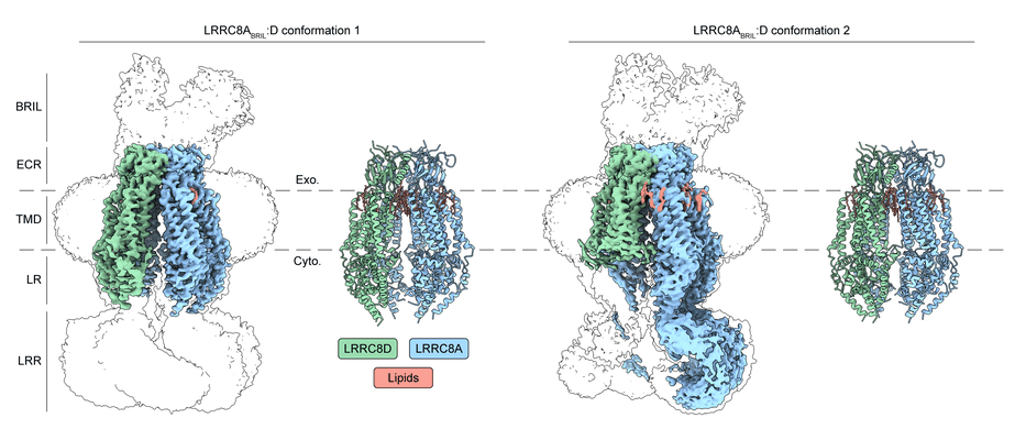 |
Mouse LRRC8A(BRIL):D in GDN [multiple data sets in TIFF, MRCS and MRC formats] | Lurie A, Brohawn SG [DOI: 10.1101/2024.11.24.625074] |
3.0 TB | 3.3 - 3.4 Å |
| 2025-01-20 |  |
Single particle dataset of respiratory complex I from Paracoccus dentrificans in MSP2N2 nanodiscs plus Fluorinated Octyl Maltoside (FOM) [4561 multi-frame micrographs composed of 40 frames each in TIFF format] | Ivanov BS, Bridges HR [Pubmed: 39472559] [DOI: 10.1038/s41467-024-53679-3] |
2.1 TB | 2.3 - 2.5 Å |
| 2025-01-20 |  |
Cryo-EM structure of human TUT1 complexed with U6 snRNA [multiple data sets in TIFF format] | Yamashita S, Tomita K [DOI: 10.1093/nar/gkae1314] |
3.4 TB | 3.21 - 3.83 Å |
| 2025-01-20 |  |
CryoEM of E. coli RNA polymerase and lambda PR promoter DNA 500 ms after mixing (1.5 mM FC8F) [20770 multi-frame micrographs composed of 38 frames each in TIFF format] | Saecker RM, Mueller AU, Malone B, Chen J, Budell WC, Dandey VP, Maruthi K, Mendez JH, Molina N, Eng ET, Yen LY, Potter CS, Carragher B, Darst SA [Pubmed: 38951624] [DOI: 10.1038/s41594-024-01349-9] |
3.6 TB | 2.8 - 2.9 Å |
| 2025-01-20 |  |
Cryo-ET dataset of FIB-milled mock infected (control) human monocyte-derived macrophages [multiple data sets in MRC format] | Kreysing JP, Welsch S, Turonova B, Beck M [Pubmed: 39826544] [DOI: 10.1016/j.cell.2024.12.008] |
324.5 GB | 27.9 - 36.7 Å |
| 2025-01-17 |  |
CryoEM of E. coli RNA polymerase and lambda PR promoter DNA 500 ms after mixing (8 mM CHAPSO) [27923 multi-frame micrographs composed of 50 frames each in TIFF format] | Saecker RM, Mueller AU, Malone B, Chen J, Budell WC, Dandey VP, Maruthi K, Mendez JH, Molina N, Eng ET, Yen LY, Potter CS, Carragher B, Darst SA [Pubmed: 38951624] [DOI: 10.1038/s41594-024-01349-9] |
5.9 TB | 2.8 - 2.9 Å |
| 2025-01-17 |  |
CryoEM of E. coli RNA polymerase and lambda PR promoter DNA 120 ms after mixing [multiple data sets in TIFF format] | Saecker RM, Mueller AU, Malone B, Chen J, Budell WC, Dandey VP, Maruthi K, Mendez JH, Molina N, Eng ET, Yen LY, Potter CS, Carragher B, Darst SA [Pubmed: 38951624] [DOI: 10.1038/s41594-024-01349-9] |
6.1 TB | 2.8 - 2.9 Å |
| 2025-01-17 |  |
Cryo-EM structure of the decameric TraT surface exclusion lipoprotein from Escherichia coli (F plasmid) [8061 multi-frame micrographs composed of 40 frames each in TIFF format] | Beis K, Seddon C [DOI: 10.1101/2024.08.09.607165] |
1023.7 GB | 2.66 Å |
