Electron Microscopy Public Image Archive
Electron Microscopy Public Image Archive
The EMPIAR-PDBj team at Osaka University assists Asian EM researchers with the transfer of big EM image data to EMPIAR. Instead of sending the data directly to the EBI (UK) via the internet, hard drives can also be sent to Osaka University by postal mail or via a courier service. As an alternative, internet transfer to our server in Osaka is also available. If you would like to take advantage of our submission services, please contact us first by e-mail before sending the data to us.
| Release date | Imageset | Title | Authors and references | Size | Resolution |
|---|---|---|---|---|---|
| 2020-02-18 | 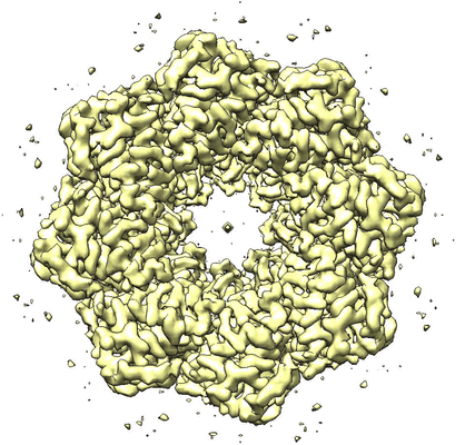 |
Structure of an undocked hemichannel of the N-terminal-deleted INX-6 in a nanodisc [300 micrographs in MRC format] | Burendei B, Shinozaki R, Watanabe M, Terada T, Tani K, Fujiyoshi Y, Oshima A [Pubmed: 32095518] [DOI: 10.1126/sciadv.aax3157] |
15.9 GB | 3.6 Å |
| 2020-02-24 | 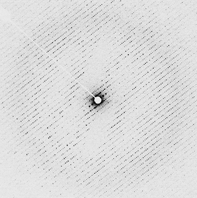 |
Electron energy-filtered diffraction (eEFD) of catalase 3D crystal with CRYO ARM 300 [84 micrographs in MRC format] | Yonekura K, Ishikawa T, Maki-Yonekura S [Pubmed: 30928615] [DOI: 10.1016/j.jsb.2019.03.009] |
5.3 GB | — |
| 2020-02-28 | 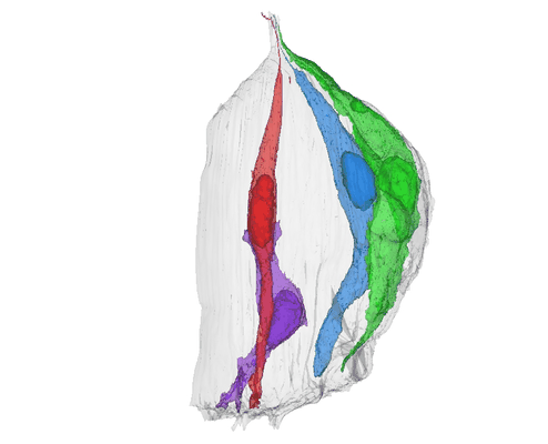 |
Three-Dimensional Reconstructions of Mouse Circumvallate Taste Buds Using Serial Blockface Scanning Electron Microscopy: I. Cell Types and the Apical Region of the Taste Bud [1194 multi-frame micrographs composed of 1 frames each in TIFF format] | Yang R, Dzowo YK, Wilson CE, Russell RL, Kidd GJ, Salcedo E, Lasher RS, Kinnamon JC, Finger TE [Pubmed: 31587284] [DOI: 10.1002/cne.24779] |
184.9 GB | — |
| 2020-03-02 | 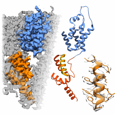 |
Tilt-schemes benchmarking for cryo electron tomography of HIV-1 CA-SP1 [multiple data sets in MRC format] | Turoňová B, Hagen WJH, Obr M, Mosalaganti S, Beugelink JW, Zimmerli CE, Kräusslich HG, Beck M [Pubmed: 32054835] [DOI: 10.1038/s41467-020-14535-2] |
45.6 GB | 4.2 Å |
| 2020-03-02 | 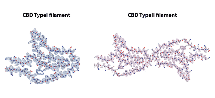 |
Cryo-EM reconstruction of tau filaments extracted from the brains of three individuals with Corticobasal degeneration [multiple data sets in TIFF format] | Zhang W, Tarutani A, Newell KL, Murzin AG, Matsubara T, Falcon B, Vidal R, Garringer HJ, Shi Y, Ikeuchi T, Murayama S, Ghetti B, Hasegawa M, Goedert M, Scheres SHW [Pubmed: 32050258] [DOI: 10.1038/s41586-020-2043-0] |
2.8 TB | 3.0 - 3.2 Å |
| 2020-03-04 |  |
Two new polymorphic structures of human full-length alpha-synuclein fibrils solved by cryo-electron microscopy [1713 micrographs in MRC format] | Guerrero-Ferreira R, Taylor NM, Arteni AA, Kumari P, Mona D, Ringler P, Britschgi M, Lauer ME, Makky A, Verasdonck J, Riek R, Melki R, Meier BH, Böckmann A, Bousset L, Stahlberg H [Pubmed: 31815671] [DOI: 10.7554/eLife.48907] |
90.9 GB | 2.98 - 3.4 Å |
| 2020-03-10 |  |
Cryo-EM of GLP-1 receptor bound to TT-OAD2 non-peptidic agonist [multiple data sets in TIFF format] | Zhao P, Liang YL, Belousoff MJ, Deganutti G, Fletcher MM, Willard FS, Bell MG, Christe ME, Sloop KW, Inoue A, Truong TT, Clydesdale L, Furness SGB, Christopoulos A, Wang MW, Miller LJ, Reynolds CA, Danev R, Sexton PM, Wootten D [Pubmed: 31915381] [DOI: 10.1038/s41586-019-1902-z] |
6.2 TB | 3.0 Å |
| 2020-03-20 |  |
Cryo Electron Tomograms of Membrane Fractions of Rabbit Skeletal Muscle for Structural Determination of RyR1 in SR Vesicles [82 tilt series in MRC format] | Chen W, Kudryashev M [Pubmed: 32147968] [DOI: 10.15252/embr.201949891] |
254.2 GB | 12.6 - 38.0 Å |
| 2020-03-20 | 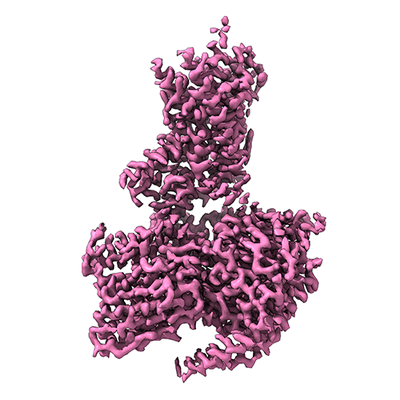 |
Cryo-EM of CRF1 receptor bound to CRF and Gs protein [4300 multi-frame micrographs composed of 62 frames each in TIFF format] | Liang YL, Belousoff MJ, Zhao P, Koole C, Fletcher MM, Truong TT, Julita V, Christopoulos G, Xu HE, Zhang Y, Khoshouei M, Christopoulos A, Danev R, Sexton PM, Wootten D [Pubmed: 32004469] [DOI: 10.1016/j.molcel.2020.01.012] |
2.6 TB | 2.7 - 2.91 Å |
| 2020-03-23 | 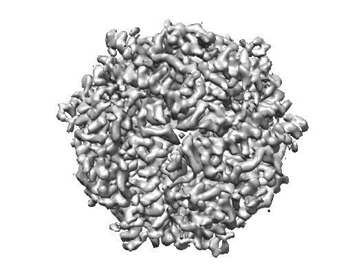 |
Micrographs of DPS collected at 100 keV using a hybrid pixel direct electron detector [739 multi-frame micrographs composed of 32 frames each in MRCS format] | Naydenova K, McMullan G, Peet MJ, Lee Y, Edwards PC, Chen S, Leahy E, Scotcher S, Henderson R, Russo CJ [Pubmed: 31709064] [DOI: 10.1107/S2052252519012612] |
23.3 GB | 3.4 Å |
| 2020-03-23 | 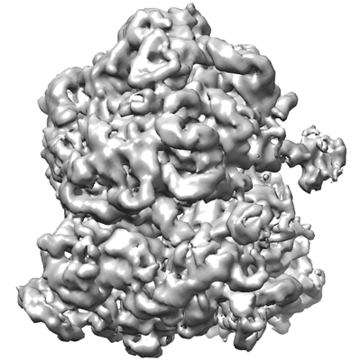 |
Micrographs of E. coli 70S ribosomes collected at 100 keV using a hybrid pixel direct electron detector [127 multi-frame micrographs composed of 32 frames each in MRCS format] | Naydenova K, McMullan G, Peet MJ, Lee Y, Edwards PC, Chen S, Leahy E, Scotcher S, Henderson R, Russo CJ [Pubmed: 31709064] [DOI: 10.1107/S2052252519012612] |
4.0 GB | 7.0 Å |
| 2020-04-03 | 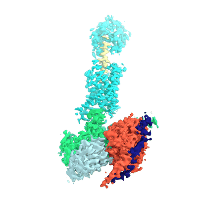 |
Cryo-EM structure of the human PAC1 receptor coupled to an engineered heterotrimeric G protein [2895 multi-frame micrographs composed of 64 frames each in TIFF format] | Kobayashi K, Shihoya W, Nishizawa T, Kadji FMN, Aoki J, Inoue A, Nureki O [Pubmed: 32157248] [DOI: 10.1038/s41594-020-0386-8] |
3.5 TB | 3.9 Å |
| 2020-04-03 | 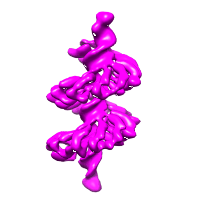 |
BurrH bound to DNA Origami Goniometer [multiple data sets in MRC and MRCS formats] | Aksel T, Yu Z, Cheng Y, Douglas SM [Pubmed: 33077960] [DOI: 10.1038/s41587-020-0716-8] |
1.3 TB | 6.5 Å |
| 2020-04-09 | 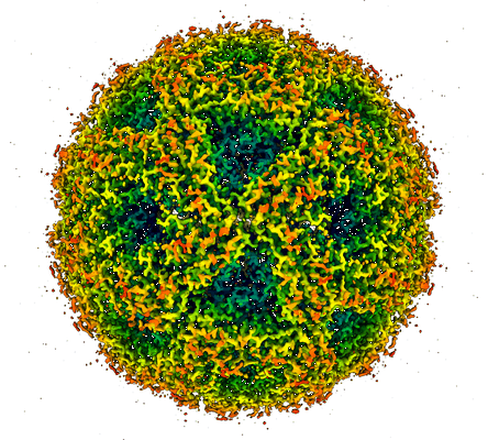 |
Cryo-EM structure of Lumazine Synthase [1198 multi-frame micrographs composed of 41 frames each in MRC format] | Bhella D, Streetley J, Clarke M, Cowton V, Patel A [Pubmed: 31359340] [DOI: 10.1007/s12551-019-00571-w] |
3.0 TB | 2.0 Å |
| 2020-04-09 | 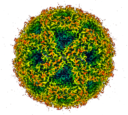 |
Cryo-EM structure of Lumazine Synthase [2874 multi-frame micrographs composed of 39 frames each in MRC format] | Bhella D, Streetley J, Clarke M, Cowton V, Patel A [Pubmed: 31359340] [DOI: 10.1007/s12551-019-00571-w] |
6.8 TB | 2.0 Å |
| 2020-04-14 | 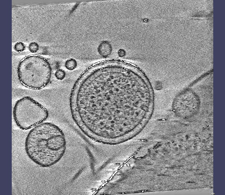 |
Cryo-electron tomography of E. coli minicells [17 multi-frame micrographs composed of 5 frames each in MRC format] | Burt A., Desfosses A., Gutsche I., Clare D. K [Pubmed: 32029744] [DOI: 10.1038/s41467-020-14350-9] |
141.7 GB | 16.0 Å |
| 2020-04-17 | 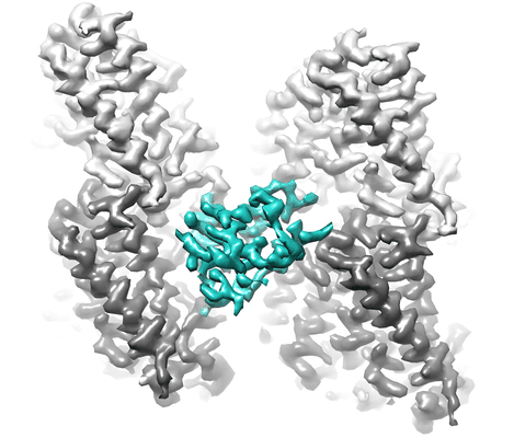 |
Cryo micrographs of microtubules (GDP state) decorated with NDC-NDC chimera of human doublecortin [950 multi-frame micrographs composed of 32 frames each in MRC format] | Cook AD, Manka SW, Wang S, Moores CA, Atherton J [Pubmed: 31610239] [DOI: 10.1016/j.jsb.2019.10.004] |
50.4 GB | 4.5 Å |
| 2020-04-21 | 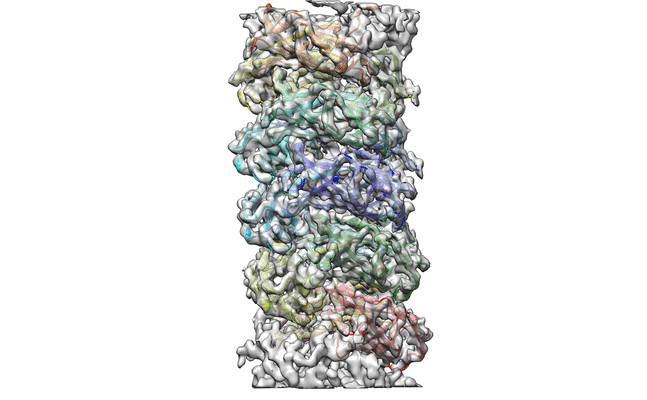 |
Cryo-EM reconstruction of CFA/I pili [2884 micrographs in SPIDER format] | Zheng W, Andersson M, Mortezaei N, Bullitt E, Egelman E [Pubmed: 31576215] [DOI: 10.1107/S2052252519007966] |
183.2 GB | 4.0 Å |
| 2020-04-21 | 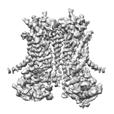 |
Cryo-EM structure of TMEM16F in digitonin without calcium bound [stack of 2249 particles in MRCS format] | Feng S, Dang S, Han T, Ye W, Jin P, Cheng T, Li J, Jan YN, Jan LY, Cheng Y [Pubmed: 31291589] [DOI: 10.1016/j.celrep.2019.06.023] |
289.5 GB | 3.9 Å |
| 2020-04-21 | 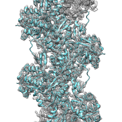 |
Cardiac thin filament in low calcium state [8820 multi-frame micrographs composed of 50 frames each in TIFF format] | Oda T, Yanagisawa HA, Wakabayashi T [Pubmed: 31954841] [DOI: 10.1016/j.jsb.2020.107450] |
2.5 TB | 3.0 - 12.0 Å |
| 2020-04-24 | 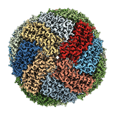 |
Mouse heavy-chain apoferritin movies obtained using a Talos Arctica (200 kV) equipped with a K2 [1679 multi-frame micrographs composed of 90 frames each in TIFF format] | Wu M, Lander GC, Herzik MA [Pubmed: 32647824] [DOI: 10.1016/j.yjsbx.2020.100020] |
549.5 GB | 1.75 Å |
| 2020-04-24 | 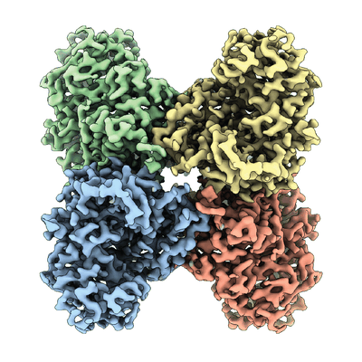 |
Rabbit muscle aldolase movies obtained using a Talos Arctica (200 kV) equipped with a K2 [3316 multi-frame micrographs composed of 44 frames each in TIFF format] | Wu M, Lander GC, Herzik MA [Pubmed: 32647824] [DOI: 10.1016/j.yjsbx.2020.100020] |
543.3 GB | 2.13 Å |
| 2020-05-01 | 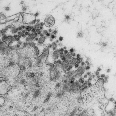 |
SARS-CoV-2 productively infects human gut enterocytes [multiple data sets in TIFF format] | Lamers MM, Beumer J, van der Vaart J, Knoops K, Puschhof J, Breugem T, Ravelli RBG, van Schayck JP, Mykytyn AZ, Duimel HQ, van Donselaar E, Riesebosch S, Kuijpers HJH, Schipper D, van de Wetering WJ, de Graaf M, Koopmans M, Cuppen E, Peters PJ, Haagmans B, Clevers H [Pubmed: 32358202] [DOI: 10.1126/science.abc1669] |
156.2 GB | — |
| 2020-05-13 | 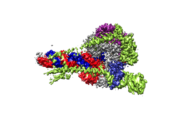 |
Structure of replicating SARS-CoV-2 polymerase [multiple data sets in TIFF and MRCS formats] | Hillen HS, Kokic G, Farnung L, Dienemann C, Tegunov D, Cramer P [Pubmed: 32438371] [DOI: 10.1038/s41586-020-2368-8] |
3.0 TB | 2.9 Å |
| 2020-05-14 | 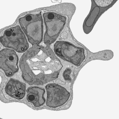 |
Three-dimensional ultrastructure of Plasmodium falciparum throughout cytokinesis [multiple data sets in MRC and TIFF formats] | Rudlaff RM, Kraemer S, Marshman J, Dvorin JD [Pubmed: 32511279] [DOI: 10.1371/journal.ppat.1008587] |
3.9 GB | — |
