Electron Microscopy Public Image Archive
Electron Microscopy Public Image Archive
The EMPIAR-PDBj team at Osaka University assists Asian EM researchers with the transfer of big EM image data to EMPIAR. Instead of sending the data directly to the EBI (UK) via the internet, hard drives can also be sent to Osaka University by postal mail or via a courier service. As an alternative, internet transfer to our server in Osaka is also available. If you would like to take advantage of our submission services, please contact us first by e-mail before sending the data to us.
| Release date | Imageset | Title | Authors and references | Size | Resolution |
|---|---|---|---|---|---|
| 2023-06-30 | 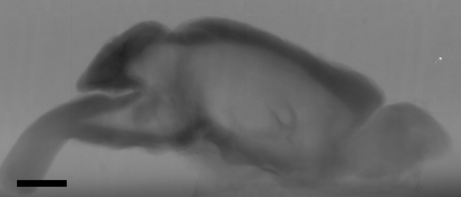 |
In situ X-ray assisted electron microscopy staining for large biological samples [33 multi-frame micrographs composed of 1 frames each in TIFF format] | Ströh S, Hammerschmith EW, Tank DW, Seung HS, Wanner AA [Pubmed: 36263931] [DOI: 10.7554/elife.72147] |
981.6 GB | — |
| 2022-04-19 | 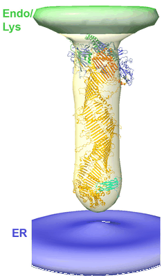 |
In situ architecture of the lipid transport protein VPS13C at ER-lysosomes membrane contacts [5 tilt series in MRC format] | Cai S [Pubmed: 35858323] [DOI: 10.1073/pnas.2203769119] |
29.5 GB | 47.0 Å |
| 2024-04-09 |  |
In situ cryo-ET dataset of Chlamydomonas reinhardtii prepared using cryo-plasmaFIB milling [multiple data sets in EER and MRC formats] | Kelley R, Khavnekar S, Zhang X, Obr M, Chakraborty S, Koh AF, Heebner J, Righetto R, Waltz F, McCafferty C, Van den Hoek H, Wietrzynski W, Van Der Stappen P, Michael A, Van Dorst S, Tagiltsev G, Beck F, Zhong E, Wan W, Briggs J, Plitzko J, Engel B, Kotecha A [Pubmed: 37613825] [DOI: 10.1093/micmic/ozad067.480] |
27.7 TB | — |
| 2023-08-24 |  |
In situ cryo-ET dataset of S. cerevisiae prepared using cryo-plasmaFIB milling [260 tilt series in MRC format] | Khavnekar S, Kelley R, Kotecha A [DOI: 10.1101/2023.08.18.553799] |
3.0 TB | 4.3 - 4.7 Å |
| 2024-01-11 |  |
In situ cryo-ET structure of phycobilisome–photosystem II supercomplex from red alga [60 reconstructed volumes in MRC format] | Meijing Li ML, Jianfei Ma JM [Pubmed: 34515634] [DOI: 10.7554/eLife.69635] |
113.9 GB | — |
| 2021-04-14 |  |
In situ cryo-electron tomogram of a pyrenoid inside a Chlamydomonas reinhardtii cell (tilt series) [1 tilt series in MRC format] | Cuellar LK, Schaffer M, Martinez-Sanchez A, Plitzko JM, Foerster F, Engel BD [Pubmed: 28938114] [DOI: 10.1016/j.cell.2017.08.008] |
5.8 GB | — |
| 2023-10-03 | 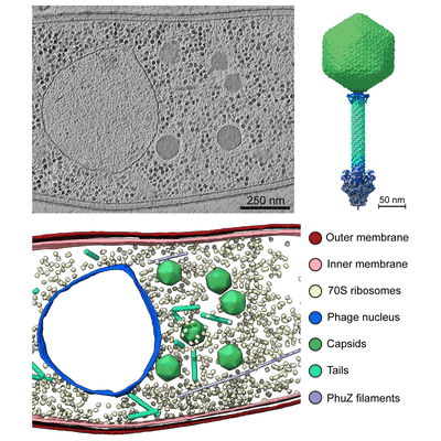 |
In situ cryo-electron tomography of E. amylovora cells infected by the jumbo bacteriophage RAY [multiple data sets in TIFF format] | Prichard A, Lee J, Laughlin TG, Lee A, Thomas KP, Sy A, Spencer T, Asavavimol A, Cafferata A, Cameron M, Chiu N, Davydov D, Desai I, Diaz G, Guereca M, Hearst K, Huang L, Jacobs E, Johnson A, Kahn S, Koch R, Martinez A, Norquist M, Pau T, Prasad G, Saam K, Sandhu M, Sarabria AJ, Schumaker S, Sonin S, Sonin A, Uyeno A, Zhao A, Corbett K, Pogliano K, Meyer J, Grose JH, Villa E, Dutton R, Pogliano J [Pubmed: 36865095] [DOI: 10.1101/2023.02.24.529968] |
244.3 GB | 8.9 - 38.0 Å |
| 2022-07-26 | 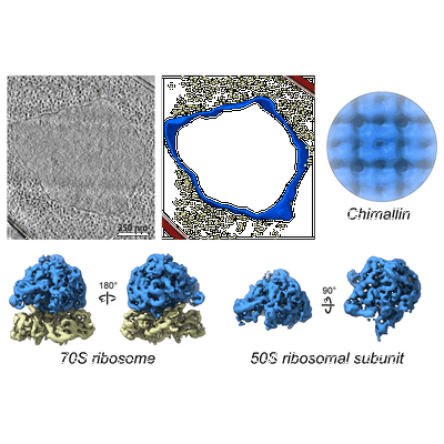 |
In situ cryo-electron tomography of E. coli APEC 2248 infected by Goslar [1073 tilt series in TIFF format] | Laughlin TG, Deep A, Prichard AM, Seitz C, Gu Y, Enustun E, Suslov S, Khanna K, Birkholz EA, Armbruster E, McCammon JA, Amaro RE, Pogliano J, Corbett KD, Villa E [Pubmed: 35922510] [DOI: 10.1038/s41586-022-05013-4] |
52.2 GB | 8.53 - 27.0 Å |
| 2022-07-26 |  |
In situ cryo-electron tomography of P. chlororaphis infected by 201phi2-1 [269 multi-frame micrographs composed of 12 frames each in TIFF format] | Laughlin TG, Deep A, Prichard AM, Seitz C, Gu Y, Enustun E, Suslov S, Khanna K, Birkholz EA, Armbruster E, McCammon JA, Amaro RE, Pogliano J, Corbett KD, Villa E [Pubmed: 35922510] [DOI: 10.1038/s41586-022-05013-4] |
14.5 GB | 10.2 - 24.0 Å |
| 2024-05-16 |  |
In situ cryo-electron tomography of Saccharomyces cerevisiae [37 multi-frame micrographs composed of 252 frames each in EER format] | Comet M, Dijkman PM, Boer Iwema R, Franke T, Masiulis S, Schampers R, Raschdorf O, Grollios F, Pryor Jr. EE, Drulyte I [Pubmed: 38512070] [DOI: 10.1107/S2059798324001840] |
3.1 GB | — |
| 2022-07-12 |  |
In situ cryo-electron tomography of T. kivui cells [multiple data sets in MRC and TIFF formats] | Dietrich HM, Righetto RD, Kumar A, Wietrzynski W, Trischler R, Schuller SK, Wagner J, Schwarz FM, Engel BD, Müller V, Schuller JM [Pubmed: 35859174] [DOI: 10.1038/s41586-022-04971-z] |
373.4 GB | 17.0 Å |
| 2022-09-26 |  |
In situ cryo-electron tomography of autophagic structures in S. cerevisiae [84 tilt series in MRC format] | Bieber A, Capitanio C, Erdmann PS, Schulman BA, Baumeister W, Wilfling F [Pubmed: 36122245] [DOI: 10.1073/pnas.2209823119] |
249.0 GB | — |
| 2022-08-12 | 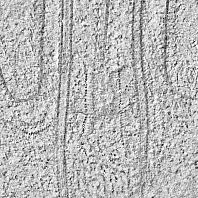 |
In situ cryo-electron tomography of the C. reinhardtii ciliary transition zone [multiple data sets in MRC and EM formats] | van den Hoek H, Klena N, Jordan MA, Alvarez Viar G, Righetto RD, Schaffer M, Erdmann PS, Wan W, Geimer S, Plitzko JM, Baumeister W, Pigino G, Hamel V, Guichard P, Engel BD [Pubmed: 35901159] [DOI: 10.1126/science.abm6704] |
302.0 GB | — |
| 2022-05-03 | 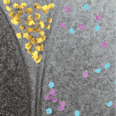 |
In situ single particle classification reveals distinct 60S maturation intermediates in cells [multiple data sets in MRC format] | Lucas BA, Zhang K, Loerch S, Grigorieff N [DOI: 10.1101/2022.04.10.487797] |
10.5 GB | — |
| 2015-11-04 | 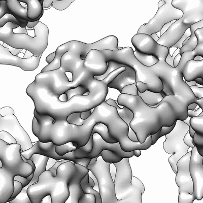 |
In vitro assembled bacteriophage phi6 polymerase complex [stack of 798 particles in MRC format] | Ilca SL, Kotecha A, Sun X, Poranen MP, Stuart DI, Huiskonen JT [Pubmed: 26534841] [DOI: 10.1038/ncomms9843] |
8.1 GB | 7.9 Å |
| 2022-01-12 | 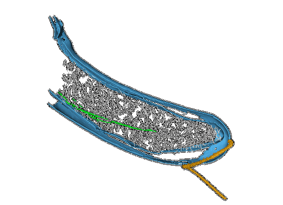 |
In vivo architecture of the polar organizing protein Z (PopZ) meshwork in the Alphaproteobacteria Magnetospirillum gryphiswaldense and Caulobacter crescentus [multiple data sets in MRC format] | Toro-Nahuelpan M, Plitzko JM, Schüler D, Pfeiffer D [Pubmed: 34971672] [DOI: 10.1016/j.jmb.2021.167423] |
6.4 GB | — |
| 2023-09-22 |  |
In-tissue cryo electron tomography of App^NL-G-F amyloid plaques [23 tilt series in MRC format] | Leistner C, Wilkinson M, Burgess A, Lovatt M, Goodbody S, Xu Y, Deuchars S, Radford SE, Ranson NA, Frank RAW [Pubmed: 37198197] [DOI: 10.1002/pro.3943] |
38.0 GB | 3.0 Å |
| 2022-07-27 |  |
Infectious mouse-adapted RML scrapie prion fibrils purified from terminally-infected mouse brains [6039 micrographs in MRC format] | Manka SW, Zhang W, Wenborn A, Betts J, Joiner S, Saibil HR, Collinge J, Wadsworth JDF [Pubmed: 35831275] [DOI: 10.1038/s41467-022-30457-7] |
1.0 TB | 2.7 Å |
| 2023-08-18 | 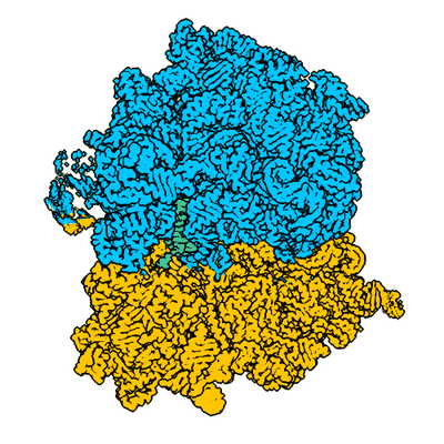 |
Initiation complex consisting of E. coli 70S ribosome with AAA mRNA codon in the A-site [10115 multi-frame micrographs composed of 40 frames each in TIFF format] | Koziej L, Glatt S [Pubmed: 37553384] [DOI: 10.1038/s41467-023-40422-7] |
3.8 TB | 2.04 Å |
| 2023-08-18 | 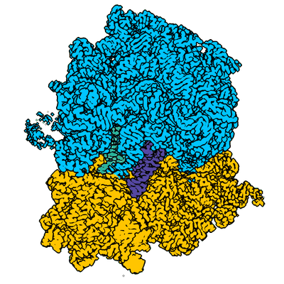 |
Initiation complex consisting of E. coli 70S ribosome with AAA mRNA codon in the A-site incubated with a ternary complex containing cognate acylated tRNA(Lys) [8435 multi-frame micrographs composed of 40 frames each in TIFF format] | Koziej L, Glatt S [Pubmed: 37553384] [DOI: 10.1038/s41467-023-40422-7] |
3.2 TB | 2.33 - 2.88 Å |
| 2023-08-18 |  |
Initiation complex consisting of E. coli 70S ribosome with AAm6A mRNA codon in the A-site [10276 multi-frame micrographs composed of 40 frames each in TIFF format] | Koziej L, Glatt S [Pubmed: 37553384] [DOI: 10.1038/s41467-023-40422-7] |
4.0 TB | 2.04 Å |
| 2023-08-18 | 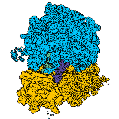 |
Initiation complex consisting of E. coli 70S ribosome with AAm6A mRNA codon in the A-site incubated with a ternary complex containing cognate acylated tRNA(Lys) [7207 multi-frame micrographs composed of 40 frames each in TIFF format] | Koziej L, Glatt S [Pubmed: 37553384] [DOI: 10.1038/s41467-023-40422-7] |
2.7 TB | 2.11 - 2.62 Å |
| 2023-08-18 | 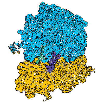 |
Initiation complex consisting of E. coli 70S ribosome with Am6AA mRNA codon in the A-site incubated with a ternary complex containing cognate acylated tRNA(Lys) [7172 multi-frame micrographs composed of 40 frames each in TIFF format] | Koziej L, Glatt S [Pubmed: 37553384] [DOI: 10.1038/s41467-023-40422-7] |
2.7 TB | 2.21 - 2.81 Å |
| 2023-08-18 | 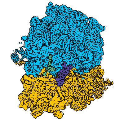 |
Initiation complex consisting of E. coli 70S ribosome with m6AAA mRNA codon in the A-site incubated with a ternary complex containing cognate acylated tRNA(Lys) [9366 multi-frame micrographs composed of 40 frames each in TIFF format] | Koziej L, Glatt S [Pubmed: 37553384] [DOI: 10.1038/s41467-023-40422-7] |
3.3 TB | 2.37 - 2.85 Å |
| 2024-05-03 |  |
Initiation factor 3 bound to the 30S ribosomal subunit in an initial step of translation [multiple data sets in MRC and MRCS formats] | Uday AB, Mishra RK, Hussain T [Pubmed: 38148682] [DOI: 10.1002/prot.26655] |
4.8 TB | 4.4 - 4.6 Å |
