Electron Microscopy Public Image Archive
Electron Microscopy Public Image Archive
Due to a storage failure, some data files are currently inaccessible (Markted: Incomplete dataset).
We are currently working to restore, but we are accepting priority requests.(email, inquiry).
We are restoring lost files from backups in the following order:
We apologize for the inconvenience and appreciate your understanding.
The EMPIAR-PDBj team at Osaka University assists Asian EM researchers with the transfer of big EM image data to EMPIAR. Instead of sending the data directly to the EBI (UK) via the internet, hard drives can also be sent to Osaka University by postal mail or via a courier service. As an alternative, internet transfer to our server in Osaka is also available. If you would like to take advantage of our submission services, please contact us first by e-mail before sending the data to us.
| Release date | Imageset | Title | Authors and references | Size | Resolution |
|---|---|---|---|---|---|
| 2024-02-13 | 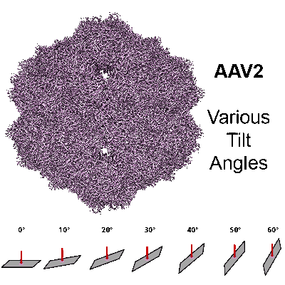 |
Single-Particle Cryo-EM of AAV2 at Various Tilts [multiple data sets in MRCS and MRC formats] | Aiyer S, Baldwin PR, Tan SM, Shan Z, Oh J, Mehrani A, Bowman ME, Louie G, Passos DO, Đorđević-Marquardt S, Mietzsch M, Hull JA, Hoshika S, Barad BA, Grotjahn DA, McKenna R, Agbandje-McKenna M, Benner SA, Noel JAP, Wang D, Tan YZ, Lyumkis D [Pubmed: 38195598] [DOI: 10.1038/s41467-023-44555-7] |
730.4 GB | 2.1 - 2.2 Å |
| 2024-02-13 | 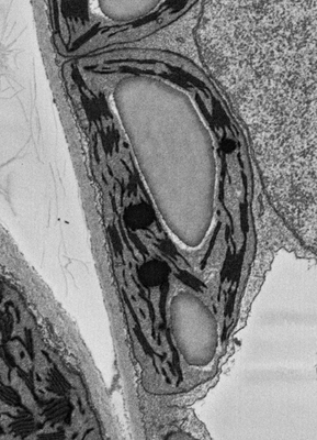 |
Plant SBF-SEM - Tobacco Leaf Chloroplast [130 micrographs in TIFF format] | Wickramanayake JS, Czymmek KJ [Pubmed: 37451777] [DOI: 10.1016/bs.mcb.2023.04.008] |
544.4 MB | — |
| 2024-02-09 | 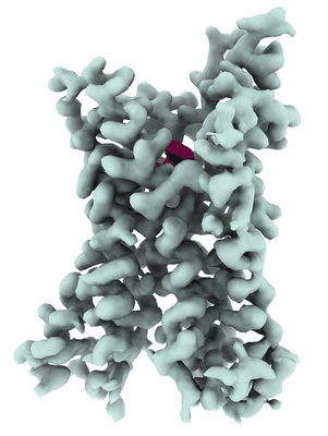 |
5-HT2B receptor bound to LSD obtained by cryo-electron microscopy (cryoEM) [12751 multi-frame micrographs composed of 50 frames each in TIFF format] | Barros-Alvarez X, Cao C, Panova O, Roth BL, Skiniotis G [Pubmed: 36087581] [DOI: 10.1016/j.neuron.2022.08.006] |
6.3 TB | 2.7 Å |
| 2024-02-09 | 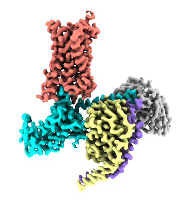 |
5-HT2B receptor bound to LSD in complex with heterotrimeric mini-Gq protein obtained by cryo-electron microscopy (cryoEM) [3916 multi-frame micrographs composed of 57 frames each in TIFF format] | Barros-Alvarez X [Pubmed: 36087581] [DOI: 10.1016/j.neuron.2022.08.006] |
2.0 TB | 2.9 Å |
| 2024-02-09 |  |
5-HT2B receptor bound to LSD in complex with beta-arrestin1 obtained by cryo-electron microscopy (cryoEM) [9617 multi-frame micrographs composed of 50 frames each in TIFF format] | Barros-Alvarez X [Pubmed: 36087581] [DOI: 10.1016/j.neuron.2022.08.006] |
5.0 TB | 3.3 Å |
| 2024-02-09 | 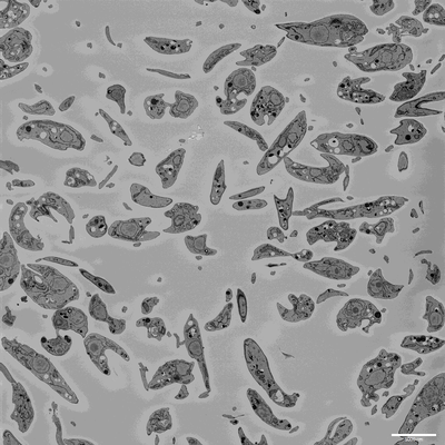 |
SBF-SEM imaging of Leishmania mexicana culture derived promastigotes [708 multi-frame micrographs composed of 1 frames each in MRC format] | Hair M [DOI: 10.1101/2023.11.28.568992] |
131.9 GB | — |
| 2024-02-09 | 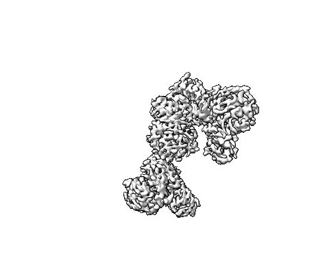 |
Single particle Cryo EM of the C-terminal half LRRK2 G2019S mutant bound to GZD-824 [7988 multi-frame micrographs composed of 40 frames each in EER format] | Villagran Suarez A, Leschziner A [Pubmed: 38039358] [DOI: 10.1126/sciadv.adk6191] |
7.0 TB | 2.99 Å |
| 2024-02-08 | 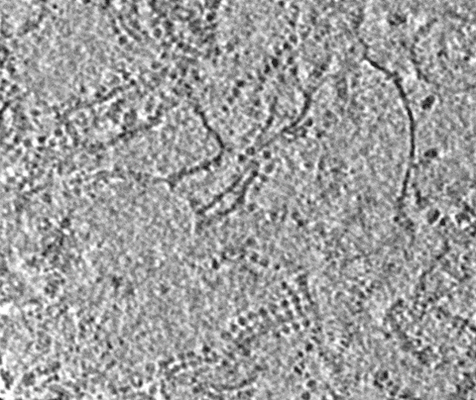 |
Cryo electron tomography of whole mitochondria and released crista membranes isolated from Caenorhabditis elegans [multiple data sets in MRC format] | Buzzard E, McLaren M, Gold VAM [Pubmed: 38164968] [DOI: 10.1042/BCJ20230450] |
159.0 GB | 38.6 Å |
| 2024-02-08 |  |
Single particle Cryo EM of the LRRK2 I2020T mutant bound to GZD-824 [4102 multi-frame micrographs composed of 40 frames each in EER format] | Villagran Suarez A, Leschziner A [Pubmed: 38039358] [DOI: 10.1126/sciadv.adk6191] |
4.6 TB | 3.4 Å |
| 2024-02-08 | 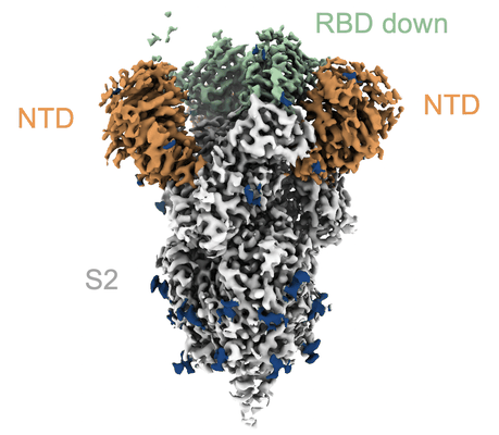 |
Single-particle cryo-EM unaligned micrographs of prefusion SARS-CoV-2 spike omicron B.1.1.529 variant [13697 multi-frame micrographs composed of 50 frames each in TIFF format] | Cerutti G, Guo Y, Liu L, Liu L, Zhang Z, Luo Y, Huang Y, Wang HH, Ho DD, Sheng Z, Shapiro L [Pubmed: 35172173] [DOI: 10.1016/j.celrep.2022.110428] |
3.4 TB | 3.11 Å |
| 2024-02-06 | 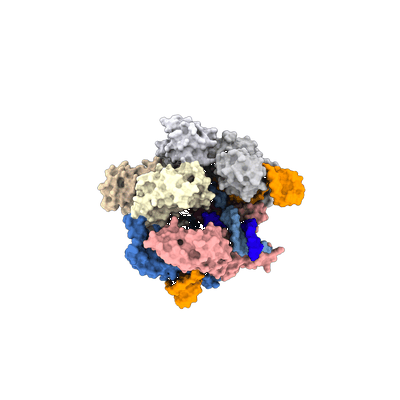 |
SpCas9 bound to 18 nucleotide complementary DNA substrate in the checkpoint state [multiple data sets in EER format] | Pacesa M, Loeff L, Querques I, Muckenfuss LM, Sawicka M, Jinek M [Pubmed: 36002571] [DOI: 10.1038/s41586-022-05114-0] |
7.7 TB | 2.54 Å |
| 2024-02-06 | 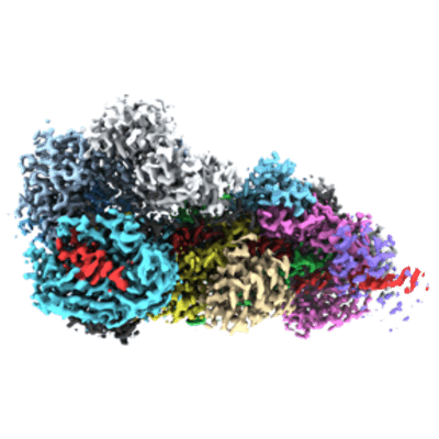 |
RNA-triggered protein cleavage and cell growth arrest by the type III-E CRISPR nuclease-protease [multiple data sets in TIFF format] | Kato K, Okazaki S, Ishikawa J, Isayama Y, Nishizawa T, Nishimasu H [Pubmed: 36423304] [DOI: 10.1126/science.add7347] |
2.6 TB | 2.49 - 2.84 Å |
| 2024-02-06 | 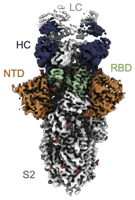 |
Cryo-EM structure of neutralizing antibody 1-57 in complex with prefusion SARS-CoV-2 spike glycoprotein [2735 multi-frame micrographs composed of 50 frames each in TIFF format] | Cerutti G, Rapp M, Guo Y, Bahna F, Bimela J, Reddem ER, Yu J, Wang P, Liu L, Huang Y, Ho DD, Kwong PD, Sheng Z, Shapiro L [Pubmed: 34111408] [DOI: 10.1016/j.str.2021.05.014] |
746.5 GB | 3.42 Å |
| 2024-02-06 |  |
Raw micrographs of Form1-N2 peptide nanotube [7917 multi-frame micrographs composed of 40 frames each in TIFF format] | Wang F, Gnewou O, Conticello VP, Egelman EH [Pubmed: 35133794] [DOI: 10.1021/acs.chemrev.1c00753] |
1.8 TB | 3.4 Å |
| 2024-02-06 |  |
CryoEM structures of the human CLC-2-AK42 voltage gated chloride channel reveal a ball and chain gating mechanism [stack of 11498 particles in MRC format] | Xu M, Pintilie G, Liu Y, Chiu W, Maduke M [Pubmed: 38345841] [DOI: 10.7554/eLife.90648] |
1.2 TB | 2.46 Å |
| 2024-02-06 |  |
CryoEM structures of the human CLC-2 voltage gated chloride channel reveal a ball and chain gating mechanism [stack of 11404 particles in MRC format] | Xu M, Pintilie G, Liu Y, Chiu W, Maduke M [Pubmed: 38345841] [DOI: 10.7554/eLife.90648] |
1.2 TB | 2.46 Å |
| 2024-02-06 |  |
Single-particle cryo-EM unaligned micrographs of NTD-directed neutralizing antibody 4-18 in complex with prefusion SARS-CoV-2 spike glycoprotein [7711 multi-frame micrographs composed of 50 frames each in TIFF format] | Cerutti G, Guo Y, Zhou T, Gorman J, Lee M, Rapp M, Reddem ER, Yu J, Bahna F, Bimela J, Huang Y, Katsamba PS, Liu L, Nair MS, Rawi R, Olia AS, Wang P, Zhang B, Chuang GY, Ho DD, Sheng Z, Kwong PD, Shapiro L [Pubmed: 33789084] [DOI: 10.1016/j.chom.2021.03.005] |
2.0 TB | 2.97 Å |
| 2024-02-06 |  |
Human CPSF160-WDR33-CPSF30 complex bound to the PAS AAUAAA motif [multiple data sets in TIFF and DM4 formats] | Muckenfuss LM, Jinek M [Pubmed: 29358758] [DOI: 10.1038/s41594-017-0020-6] |
1.2 TB | 3.07 Å |
| 2024-02-01 |  |
Purified tails from bacteriophage T5 after interaction with E. coli receptor FhuA inserted into nanodiscs - dataset 2 [5733 multi-frame micrographs composed of 40 frames each in MRC format] | Linares R, Arnaud C, Effantin G, Darnault C, Epalle NH, Erba EB, Schoehn G, Breyton C [Pubmed: 36961893] [DOI: 10.1126/sciadv.ade9674] |
917.9 GB | 3.45 - 4.32 Å |
| 2024-02-01 |  |
Cryo-EM micrographs of Sweet Potato Mild Mottle Virus [2260 micrographs in MRC format] | Javed A, Byrne MJ, Ranson NA [Pubmed: 37076658] [DOI: 10.1038/s42003-023-04799-x] |
141.3 GB | 2.9 Å |
| 2024-02-01 |  |
Cryo-EM micrographs of Sweet Potato Feathery Mosaic Virus [3276 micrographs in MRC format] | Javed A, Byrne MJ, Ranson NA [Pubmed: 37076658] [DOI: 10.1038/s42003-023-04799-x] |
204.8 GB | 2.6 Å |
| 2024-02-01 |  |
Cryo-EM structure NDUFS4 knockout complex I from Mus musculus kidney [3373 multi-frame micrographs composed of 59 frames each in MRC format] | Yin Z, Agip ANA, Bridges HR, Hirst J [Pubmed: 38177503] [DOI: 10.1038/s44318-023-00001-4] |
3.8 TB | 6.2 Å |
| 2024-02-01 |  |
Structure and engineering of the type III-E CRISPR-Cas7-11 effector complex [2781 multi-frame micrographs composed of 64 frames each in TIFF format] | Kato K, Okazaki S, Isayama Y, Nishizawa T, Nishimasu H [Pubmed: 35643083] [DOI: 10.1016/j.cell.2022.05.003] |
853.6 GB | 2.45 Å |
| 2024-02-01 |  |
Structure of BARD1 ARD-BRCTs in complex with H2AKc15ub nucleosomes [multiple data sets in EER and MRC formats] | Foglizzo M, Burdett H, Wilson MD, Zeqiraj E [Pubmed: 37823591] [DOI: 10.1093/nar/gkad793] |
10.4 TB | 3.4 - 3.75 Å |
| 2024-01-23 |  |
Cryo electron microscopy micrographs of high molecular weight fractions from yeast native cell extracts [multiple data sets in MRC format] | Schmidt L, Tueting C, Kyrilis F, Hamdi F, Semchonok DA, Kastritis PL [Pubmed: 38730276] [DOI: 10.1038/s42003-024-06204-7] |
4.7 TB | 3.78 - 8.1 Å |
