3D density maps used as templates to detect 50S ribosomal subunits the 2D images and 3D tomograms of Mycoplasma pneumoniae cells by 2D and 3D template matching, respectively. The pixel size of the map used for 2D template matching is 1.27 Å. The map used for 3D template matching has a pixel size of 6.80 Å.
Files:
Available download options:
Archives and downloads the selected files into an uncompressed zip file.
Depending on the number and size of files to be downloaded, this can take an enormous amount of time.
Also, this feature is unstable and may not work properly, and checksums of downloaded files are not verified.
We recommend using rsync, aspera, globus, etc.
Download a list of selected files.
It is possible to download files by specifying the file list with rsync command, etc.
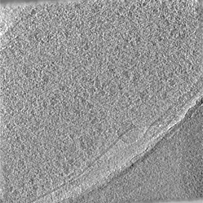
19 micrographs of Mycoplasma pneumoniae cells, using movies of 20 frames and a total exposures of 32 e-/Å^2. The movies were aligned using cisTEM, and exposure weighting was applied to calculate the frame average.
Files:
Available download options:
Archives and downloads the selected files into an uncompressed zip file.
Depending on the number and size of files to be downloaded, this can take an enormous amount of time.
Also, this feature is unstable and may not work properly, and checksums of downloaded files are not verified.
We recommend using rsync, aspera, globus, etc.
Download a list of selected files.
It is possible to download files by specifying the file list with rsync command, etc.
19 aligned tomographic tilt series of Mycoplasma pneumoniae cells collected using SerialEM. Tilts ranged from -60 to 60 deg using 3 deg angular steps. The total exposure was 129 e-/Å^2. The tilt series were recorded after a pre-exposure of 32 e-/Å^2 to collect a 2D image of each cell at higher resolution.
Files:
Available download options:
Archives and downloads the selected files into an uncompressed zip file.
Depending on the number and size of files to be downloaded, this can take an enormous amount of time.
Also, this feature is unstable and may not work properly, and checksums of downloaded files are not verified.
We recommend using rsync, aspera, globus, etc.
Download a list of selected files.
It is possible to download files by specifying the file list with rsync command, etc.
19 tomographic reconstructions of Mycoplasma pneumoniae cells
Files:
Available download options:
Archives and downloads the selected files into an uncompressed zip file.
Depending on the number and size of files to be downloaded, this can take an enormous amount of time.
Also, this feature is unstable and may not work properly, and checksums of downloaded files are not verified.
We recommend using rsync, aspera, globus, etc.
Download a list of selected files.
It is possible to download files by specifying the file list with rsync command, etc.