Electron Microscopy Public Image Archive
Electron Microscopy Public Image Archive
The EMPIAR-PDBj team at Osaka University assists Asian EM researchers with the transfer of big EM image data to EMPIAR. Instead of sending the data directly to the EBI (UK) via the internet, hard drives can also be sent to Osaka University by postal mail or via a courier service. As an alternative, internet transfer to our server in Osaka is also available. If you would like to take advantage of our submission services, please contact us first by e-mail before sending the data to us.
| Release date | Imageset | Title | Authors and references | Size | Resolution |
|---|---|---|---|---|---|
| 2018-02-08 |  |
Tilt-series of salmonella enterica wild-type bacterial flagellar motor [1 tilt series in MRC format] | Beeby M, Ribardo DA, Brennan CA, Ruby EG, Jensen GJ, Hendrixson DR [Pubmed: 26976588] [DOI: 10.1073/pnas.1518952113] |
328.0 MB | 69.4 Å |
| 2021-11-15 | 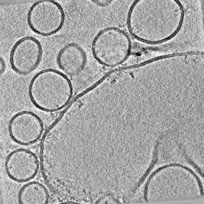 |
Arrangements of proteins at reconstituted synaptic vesicle fusion sites depend on membrane separation. [multiple data sets in MRC format] | Ginger L, Malsam J, Sonnen A.F.P., Morado D, Scheutzow A, Söllner T.H., Briggs J.A.G. [Pubmed: 32860428] [DOI: 10.1002/1873-3468.13916] |
70.0 GB | — |
| 2021-11-30 |  |
Entropy Regularized Deconvolution of Cellular Cryo-Transmission Electron Tomograms [3 tilt series in MRC format] | Croxford MW, Elbaum ME, Arigovindan M, Kam Z, Agard DA, Villa E, Sedat J [Pubmed: 34876518] [DOI: 10.1073/pnas.2108738118] |
6.4 GB | — |
| 2022-11-04 | 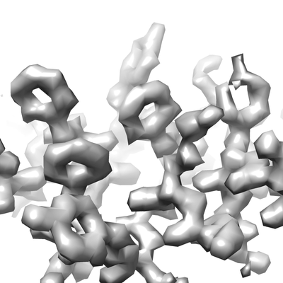 |
Tilt series of mouse heavy chain apoferritin acquired on Krios G4 equipped with SelectrisX and Falcon4i [3300 multi-frame micrographs composed of 153 frames each in EER format] | Obr M, Yang W, Karia D, Koh FA, Kotecha A | 131.0 GB | — |
| 2021-11-30 |  |
Entropy Regularized Deconvolution of Cellular Cryo-Transmission Electron Tomograms [2 tilt series in MRC format] | Croxford MW, Elbaum ME, Arigovindan M, Kam Z, Agard DA, Villa E, Sedat J [Pubmed: 34876518] [DOI: 10.1073/pnas.2108738118] |
5.2 GB | — |
| 2022-10-28 | 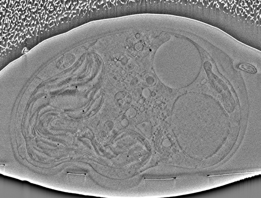 |
Chlamydomonas Cryo-Slice and View on Thermo Scientific Helios 5 Hydra PFIB [477 micrographs in TIFF format] | Kelley R, Khavnekar S, Wietrzynski W, Plitzko J, Kotecha A | 4.4 GB | — |
| 2023-05-22 | 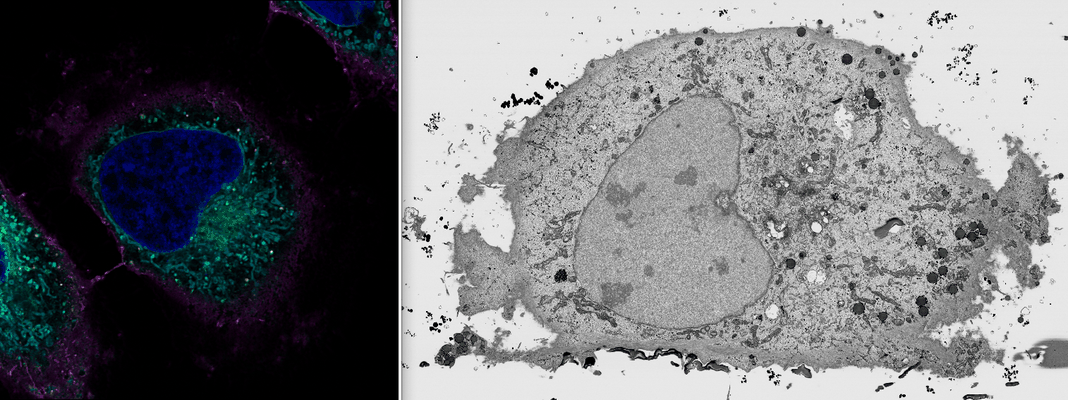 |
Benchmark FIB SEM data (#2) of HeLa cells previously imaged by Zeiss LSM900 Airyscan microscopy [multiple data sets in TIFF format] | Peddie CJ, Domart MC, Collinson LM [DOI: 10.1101/2023.05.11.540445] |
511.9 GB | — |
| 2020-11-06 | 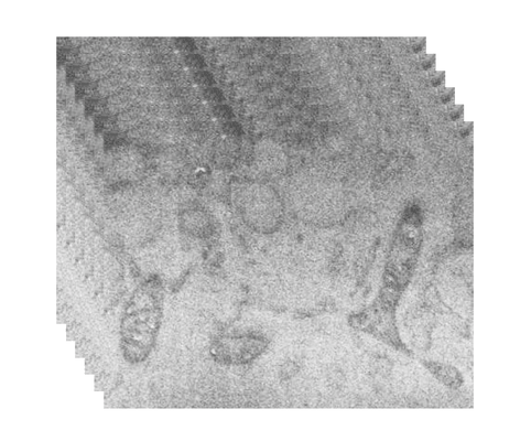 |
Serial cryoFIB/SEM reveals cytoarchitectural disruptions in Leigh syndrome patient cells [multiple data sets in TIFF format] | Zhu Y, Sun D, Schertel A, Fu X, Gwo P, Watson A, Zanetti-Domingues LC, Marisa L. Martin-Fernandez ML, Freyberg Z, Zhang P [Pubmed: 33096015] [DOI: 10.1016/j.str.2020.10.003] |
17.3 GB | — |
| 2023-05-12 | 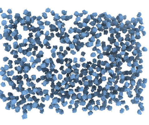 |
Quantification of gallium cryo-FIB milling damage in biological lamella [multiple data sets in MRC format] | Lucas BA, Grigorieff N [Pubmed: 37216561] [DOI: 10.1073/pnas.2301852120] |
8.1 GB | — |
| 2022-05-20 |  |
CEM1.5M : a cellular EM dataset containing ~1.5 x 10⁶ unlabeled 2D image patches curated for deep learning [1592753 micrographs in TIFF format] | Narayan K | 57.6 GB | — |
| 2022-05-10 |  |
CEM-MitoLab: a dataset of ~22K cellular EM 2D images with label maps of ~135K mitochondrial instances, for deep learning [43720 micrographs in TIFF format] | Narayan K, Conrad RW | 2.8 GB | — |
| 2023-06-30 | 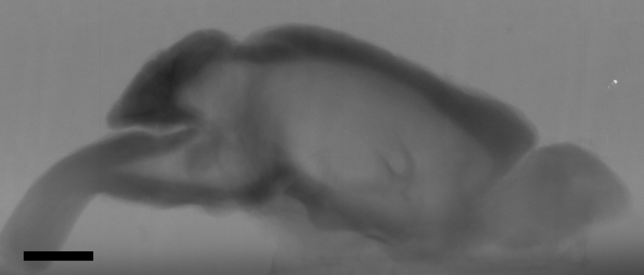 |
In situ X-ray assisted electron microscopy staining for large biological samples [33 multi-frame micrographs composed of 1 frames each in TIFF format] | Ströh S, Hammerschmith EW, Tank DW, Seung HS, Wanner AA [Pubmed: 36263931] [DOI: 10.7554/elife.72147] |
981.6 GB | — |
| 2021-10-19 | 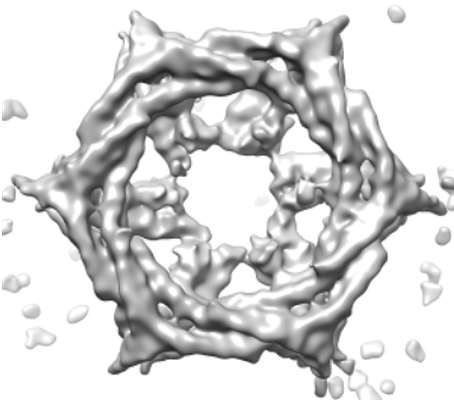 |
Multi-modal adaptor-clathrin contacts drive coated vesicle assembly [stack of 5720 particles in MRCS format] | Smith SM, Larocque G, Wood KM, Morris KL, Roseman AM, Sessions RB, Royle SJ, Smith CJ [Pubmed: 34487371] [DOI: 10.15252/embj.2021108795] |
1.9 GB | — |
| 2023-05-22 | 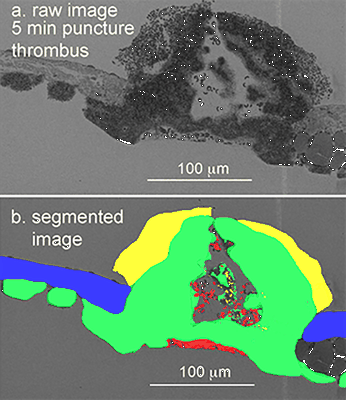 |
Venous puncture wound thrombi, 1, 5, 20 min post-puncture, 100 nm XY raw images, 20 nm XY pixels every 20 micons, 3 nm wide area TEM montages at selected depth [15 multi-frame micrographs composed of 1 frames each in DM4 format] | Storrie B, Leapman RD [Pubmed: 34531522] [DOI: 10.1038/s42003-021-02615-y] |
381.2 GB | — |
| 2022-10-07 | 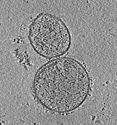 |
Tilt series of SARS-CoV-2 spike-bearing virus-like particles (VLPs) interacting with hACE2-bearing extracellular vesicles (tEVs), showing various intermediate states of the SARS-CoV-2 spike protein [6 tilt series in MRC format] | Marcink TC, Porotto M, des Georges A, Moscona A [Pubmed: 35984891] [DOI: 10.1126/sciadv.abo3153] |
9.6 GB | — |
| 2020-11-18 |  |
SARS-CoV-2 infection in human adult lung alveolar stem cells [multiple data sets in TIFF format] | Youk J, Kim T, Evans KV, Jeong YI, Hur Y, Hong SP, Kim JH, Yi K, Kim SY, Na KJ, Bleazard T, Kim HM, Fellows M, Mahbubani KT, Saeb-Parsy K, Kim SY, Kim YT, Koh GY, Choi BS, Ju YS, Lee JH [Pubmed: 33142113] [DOI: 10.1016/j.stem.2020.10.004] |
20.8 GB | — |
| 2021-11-08 |  |
Monitoring reversion of hepatitis C virus-induced cellular alterations by Direct-Acting Antivirals using cryo Soft X-ray Tomography and Infrared Microscopy [7 reconstructed volumes in TIFF format] | Perez-Berna AJ, Benseny-Cases N, Rodríguez MJ, Valcarcel R, Carrascosa JL, Gastaminzab P, Pereiroa E [Pubmed: 34726165] [DOI: 10.1107/S2059798321009955] |
260.2 MB | — |
| 2022-01-14 |  |
High resolution 3D imaging of liver subcellular architecture and its link to metabolic function [multiple data sets in TIFF format] | Parlakgul G, Hotamisligil GS [Pubmed: 35264794] [DOI: 10.1038/s41586-022-04488-5] |
2.1 TB | — |
| 2015-06-18 |  |
A simulated cryoEM data set of GroEL particles [stack of 10000 particles in MRC format] | Deng Y, Sun F | 1.5 GB | — |
| 2022-06-01 | 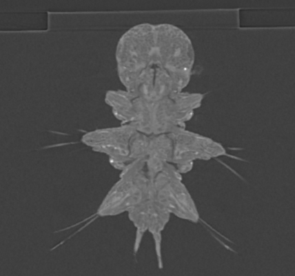 |
Crosshair, semi-automated targeting for electron microscopy with a motorised ultramicrotome [multiple data sets in TIFF and PNG formats] | Meechan K, Guan W, Riedinger A, Stankova V, Yoshimura A, Pipitone R, Milberger A, Schaar H, Romero-Brey I, Templin R, Peddie C J, Schieber N L, Jones M L, Collinson L, Schwab Y | 151.0 GB | — |
| 2022-12-19 | 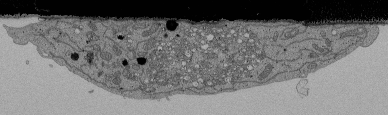 |
CLEMSite, a software for automated phenotypic screens using light microscopy and FIB-SEM. [multiple data sets in TIFF format] | Lleti JMSL, Steyer AMS, Schwab YS | 19.7 GB | — |
| 2024-02-09 | 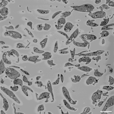 |
SBF-SEM imaging of Leishmania mexicana culture derived promastigotes [708 multi-frame micrographs composed of 1 frames each in MRC format] | Hair M [DOI: 10.1101/2023.11.28.568992] |
131.9 GB | — |
| 2021-11-01 | 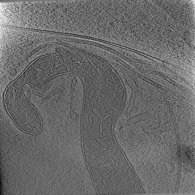 |
electron cryo-tomograms of axons from human cerebral organoids, expressing GFP-ESYT1 [multiple data sets in MRC and TIFF formats] | Hoffmann P, Giandomenico S, Ganeva I, Wozny M, Sutcliffe M, Lancaster M, Kukulski W [Pubmed: 34698018] [DOI: 10.7554/eLife.70269] |
40.9 GB | — |
| 2015-10-09 | 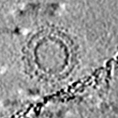 |
Cryo-electron tomography and subtomogram averaging of Rous-Sarcoma-Virus deltaMBD virus-like particles [1 class averages in MRC format] | Schur FKM, Dick RA, Hagen WJH, Vogt VM, Briggs JAG [Pubmed: 26223638] [DOI: 10.1128/JVI.01502-15] |
248.1 MB | — |
| 2020-02-24 | 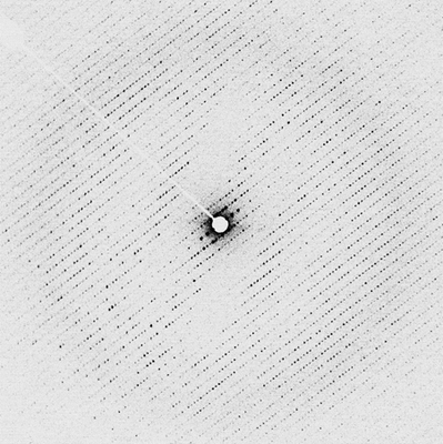 |
Electron energy-filtered diffraction (eEFD) of catalase 3D crystal with CRYO ARM 300 [84 micrographs in MRC format] | Yonekura K, Ishikawa T, Maki-Yonekura S [Pubmed: 30928615] [DOI: 10.1016/j.jsb.2019.03.009] |
5.3 GB | — |
