EMPIAR-10785
Venous puncture wound thrombi, 1, 5, 20 min post-puncture, 100 nm XY raw images, 20 nm XY pixels every 20 micons, 3 nm wide area TEM montages at selected depth [15 multi-frame micrographs composed of 1 frames each in DM4 format]
Publication:

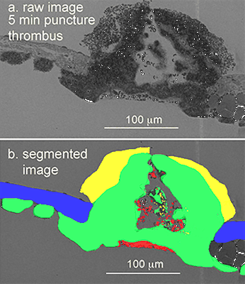
Venous puncture wound hemostasis results in a vaulted thrombus structured by locally nucleated platelet aggregates

Rhee SW, Pokrovskaya ID, Ball KK, Ling K, Vedanaparti Y, Cohen J, Cruz DRD, Zhao OS, Aronova MA, Zhang G, Kamykowski JA, Leapman RD, Storrie B

Communications biology 4 (2021)

PMID: 34531522

Related EMDB entry:
Deposited:
2021-07-22
Released:
2023-05-22
Last modified:
2023-05-23
Imageset size:
381.22 GB
Imageset DOI:
Experimental metadata:
Download xml json
Contains:
  • micrographs - multiframe
1. SBF-SEM imaging of jugular vein thrombi, 30 gauge needle puncture wounds, at 20 nm and 100 pixel, 20 micron and 200 nm step size, and 3 nm pixel wide TEM at ~mid-thrombus across full width montage
Category:
micrographs - multiframe
Image format:
DM4
No. of images or tilt series:
15
Image size:
(12500, 20000)
Pixel type:
UNSIGNED 16 BIT INTEGER
Pixel spacing:
(20.0, 20.0)
Details:
SBF-SEM image sets of individual jugular vein puncture wound thrombi (clots) at 1, 5, and 20 min post puncture. 20 nm image sets are at 20,20 nm XY and 20 micron Z step. 100 nm image sets are at 100,100 nm XY and 200 nm spacing. 20 nm sets are 10-30 images each and 100 nm sets are often about 2500 images each.
Files:
Loading...
Files:
Loading...

Ito F, Alvarez-Cabrera AL, Liu S, Yang H, Shiriaeva A, Zhou ZH, Chen XS. (2023)
Rigden DJ, Fernández XM. (2023)
Iudin A, Korir PK, Somasundharam S, Weyand S, Cattavitello C, Fonseca N, Salih O, Kleywegt GJ, Patwardhan A. (2023)
Serra Lleti JM, Steyer AM, Schieber NL, Neumann B, Tischer C, Hilsenstein V, Holtstrom M, Unrau D, Kirmse R, Lucocq JM, Pepperkok R, Schwab Y. (2023)
Caldwell BJ, Norris AS, Karbowski CF, Wiegand AM, Wysocki VH, Bell CE. (2022)