Electron Microscopy Public Image Archive
Electron Microscopy Public Image Archive
The EMPIAR-PDBj team at Osaka University assists Asian EM researchers with the transfer of big EM image data to EMPIAR. Instead of sending the data directly to the EBI (UK) via the internet, hard drives can also be sent to Osaka University by postal mail or via a courier service. As an alternative, internet transfer to our server in Osaka is also available. If you would like to take advantage of our submission services, please contact us first by e-mail before sending the data to us.
| Release date | Imageset | Title | Authors and references | Size | Resolution |
|---|---|---|---|---|---|
| 2023-07-25 |  |
Human dimeric alpha-catenin binding to F-actin [multiple data sets in MRC and TIFF formats] | Rangarajan ES, Smith EW, Izard T [Pubmed: 36928388] [DOI: 10.1038/s42003-023-04610-x] |
10.6 TB | 2.7 - 2.77 Å |
| 2023-03-13 | 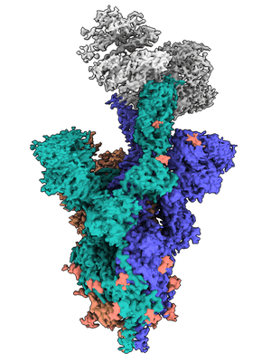 |
mouse ACE2 receptor bound to SARS-CoV-2 variant Omicron BA.1 Spike [20892 multi-frame micrographs composed of 1568 frames each in EER format] | Ni D, Myasnikov A, Stahlberg S, Lau K [DOI: 10.1371/journal.ppat.1011206] |
10.6 TB | 3.03 - 3.22 Å |
| 2024-02-01 | 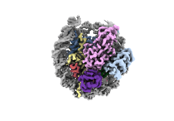 |
Structure of BARD1 ARD-BRCTs in complex with H2AKc15ub nucleosomes [multiple data sets in EER and MRC formats] | Foglizzo M, Burdett H, Wilson MD, Zeqiraj E [Pubmed: 37823591] [DOI: 10.1093/nar/gkad793] |
10.4 TB | 3.4 - 3.75 Å |
| 2024-07-19 | 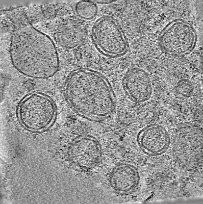 |
Cryo-ET dataset of purified SARS-CoV-2 double membrane vesicles formed by nsp3-4 [4635 tilt series in MRC format] | Huang YX, Ni T [DOI: 10.1016/s41586-024-07817-y] |
10.3 TB | — |
| 2025-01-16 |  |
Cryo-EM of the CCT-PhLP1-Gb5 complex treated with ATP-AlFx [25457 multi-frame micrographs composed of 40 frames each in TIFF format] | Sass MI, Wang S, Mack D, Cottam SL, Shen PS, Willardson BM [Pubmed: 37852256] [DOI: 10.1016/j.molcel.2023.09.032] |
10.3 TB | 2.7 - 3.36 Å |
| 2022-01-28 |  |
Anaplastic lymphoma kinase (ALK) extracellular fragment of ligand binding region 648-1025 in complex with AUG-alpha [multiple data sets in TIFF and MRC formats] | Reshetnyak AV, Myasnikov AG, Kalodimos CG [Pubmed: 34819673] [DOI: 10.1038/s41586-021-04140-8] |
10.2 TB | 2.27 Å |
| 2024-12-12 |  |
Cryo-EM of human CAK: Microscope comparisons [multiple data sets in EER format] | Cushing VI, Koh AF, Feng J, Ali S, Kotecha A, Greber BJ [Pubmed: 38480681] [DOI: 10.1038/s41467-024-46375-9] |
10.2 TB | 2.0 Å |
| 2024-08-26 |  |
Molecular model of a bacterial flagellar motor in situ reveals a “parts-list” of protein adaptations to increase torque [multiple data sets in MRC format] | Beeby M, Cohen EJ, Drobnič T, Nans A, Rosenthal PB | 10.1 TB | — |
| 2018-09-10 |  |
Cryo electron microscopy micrographs of yeast Exocyst complex [6472 multi-frame micrographs composed of 32 frames each in MRCS format] | Wang HW, Guo W, Li Y, Mei KR [Pubmed: 29335562] [DOI: 10.1038/s41594-017-0016-2] |
10.0 TB | 4.4 Å |
| 2025-01-08 |  |
Micrographs that led to the structure of Dia1 on F-actin [multiple data sets in TIFF format] | Palmer NJ, Barrie KR, Dominguez R [Pubmed: 38843827] [DOI: 10.1038/s41586-024-07637-0] |
10.0 TB | 3.41 - 3.51 Å |
| 2021-11-26 |  |
Cryo electron microscopy of hIAPP fibrils seeded by patient-extracted fibrils [20390 multi-frame micrographs composed of 30 frames each in TIFF format] | Cao Q, Boyer DR, Sawaya MR, Abskharon R, Saelices L, Nguyen BA, Lu J, Murray KA, Kandeel F, Eisenberg DS [Pubmed: 34518699] [DOI: 10.1038/s41594-021-00646-x] |
9.9 TB | 3.8 - 4.1 Å |
| 2023-06-02 |  |
f1-derived nanorods [25065 multi-frame micrographs composed of 40 frames each in TIFF format] | Conners R, McLaren MJ, Gold V [Pubmed: 37169795] [DOI: 10.1038/s41467-023-37915-w] |
9.9 TB | 2.58 - 2.97 Å |
| 2022-06-17 |  |
Cryo electron micrographs of Tetrahymena thermophila solubilized mitochondrial membrane complexes - Glacios data [multiple data sets in TIFF and MRC formats] | Letts JA, Zhou L, Guo F [Pubmed: 35357889] [DOI: 10.1126/science.abn7747] |
9.9 TB | 3.02 Å |
| 2025-07-01 |  |
CryoEM dataset of Retron-Eco1 WT filaments in the presence of NAD+ [multiple data sets in MRC format] | Carabias A, Camara-Wilpert S, Mestre MR, Lopéz-Méndez B, Hendriks IA, Zhao R, Pape T, Fuglsang A, Luk SH, Nielsen ML, Pinilla-Redondo R, Montoya G [Pubmed: 38788717] [DOI: 10.1016/j.molcel.2024.05.001] |
9.9 TB | 2.99 - 3.13 Å |
| 2023-09-19 |  |
Spraguea lophii 100S ribosome [multiple data sets in TIFF format] | McLaren MJ, Gil Diez P, Isupov M, Conners R, Gambelli L, Gold V, Connell S, Walter A, Williams B, Daum B [Pubmed: 37709902] [DOI: 10.1038/s41564-023-01469-w] |
9.8 TB | 2.79 Å |
| 2025-07-01 |  |
CryoEM dataset of Retron-Eco1 E106A filaments [7748 multi-frame micrographs composed of 40 frames each in MRC format] | Carabias A, Camara-Wilpert S, Mestre MR, Lopéz-Méndez B, Hendriks IA, Zhao R, Pape T, Fuglsang A, Luk SH, Nielsen ML, Pinilla-Redondo R, Montoya G [Pubmed: 38788717] [DOI: 10.1016/j.molcel.2024.05.001] |
9.7 TB | — |
| 2025-06-27 | 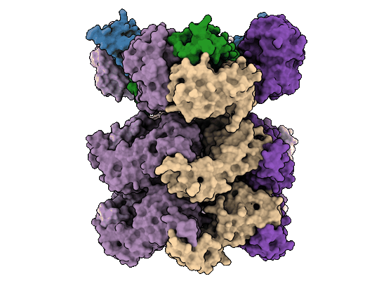 |
Structure of the 55LCC ATPase complex [multiple data sets in EER and MRC formats] | Foglizzo M, Degtjarik O, Zeqiraj E [Pubmed: 38554706] [DOI: 10.1016/j.cell.2024.03.002] |
9.7 TB | 4.5 Å |
| 2025-05-13 | 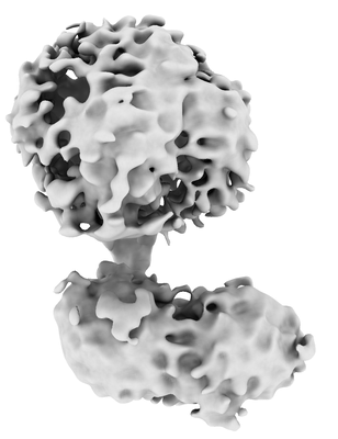 |
Micrographs of carvedilol-B1AR-arrestin2-V2Rpp complex [29650 multi-frame micrographs composed of 729 frames each in EER format] | Tatli M, Abiko LA, Petrovic I, Stahlberg H, Grzesiek S, Desai S, Spang A [Pubmed: 40372433] [DOI: 10.1073/pnas.2501487122] |
9.5 TB | 6.5 Å |
| 2024-02-16 | 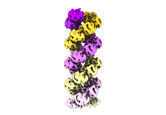 |
Cryo-EM reconstruction of the influenza A virus helical ribonucleoprotein-like [26515 multi-frame micrographs composed of 40 frames each in TIFF format] | Chenavier F, Ruigrok RWH, Schoehn G, Ballandras-Colas A, Crépin T [Pubmed: 38100595] [DOI: 10.1126/sciadv.adj9974] |
9.5 TB | 5.3 - 8.7 Å |
| 2024-04-12 | 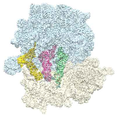 |
Escherichia coli 70S ribosome in complex with A-site tRNAIle(LAU) bound to the cognate AUA codon (Structure III) [13536 multi-frame micrographs composed of 39 frames each in MRC format] | Rybak MY, Gagnon MG [Pubmed: 38538914] [DOI: 10.1038/s41594-024-01236-3] |
9.4 TB | 2.8 Å |
| 2025-03-21 | 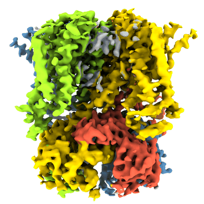 |
Unaligned cryo-EM micrographs of HCN4 channel bound to Ivabradine [31670 multi-frame micrographs composed of 40 frames each in TIFF format] | Saponaro A, Chaves-Sanjuan A, Sharifzadeh AS, Clarke OB, Marabelli C, Bolognesi M, Thiel G, Moroni A [Pubmed: 38917012] [DOI: 10.1073/pnas.2402259121] |
9.3 TB | 3.6 Å |
| 2022-08-16 | 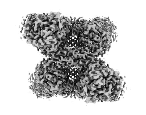 |
Cryo-EM structure of the human GS-GN complex in the inhibited state [multiple data sets in EER and MRC formats] | Marr L, Biswas D, Daly LA, Browning C, Vial SCM, Maskell DP, Hudson C, Bertrand JA, Pollard J, Ranson NA, Khatter H, Eyers CE, Sakamoto K, Zeqiraj E [Pubmed: 35690592] [DOI: 10.1038/s41467-022-31109-6] |
9.2 TB | 2.62 Å |
| 2020-07-06 | 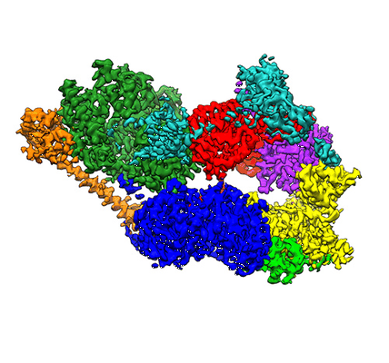 |
Phase-plate cryo-EM of human TFIIH [multiple data sets in TIFF format] | Greber BJ, Toso DB, Fang J, Nogales E [Pubmed: 30860024] [DOI: 10.7554/eLife.44771] |
9.2 TB | 3.7 Å |
| 2024-04-12 | 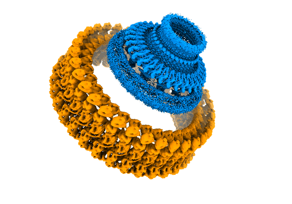 |
Structural basis for directional rotation of the Salmonella flagellum [34381 multi-frame micrographs composed of 50 frames each in TIFF format] | Singh PK, Iverson TM | 9.1 TB | 3.4 - 6.7 Å |
| 2024-10-10 | 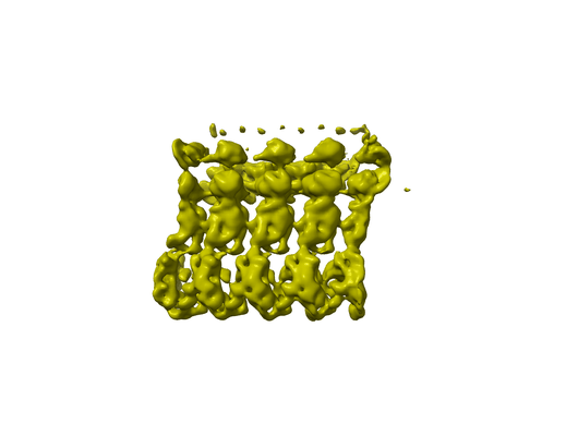 |
Structural basis for directional rotation of the Salmonella flagellum [39322 multi-frame micrographs composed of 50 frames each in TIFF format] | Singh PK, Iverson TM [Pubmed: 38632342] [DOI: 10.1038/s41564-024-01674-1] |
9.1 TB | 3.4 - 6.7 Å |
