Electron Microscopy Public Image Archive
Electron Microscopy Public Image Archive
Due to a storage failure, some data files are currently inaccessible (Markted: Incomplete dataset).
We are currently working to restore, but we are accepting priority requests.(email, inquiry).
We are restoring lost files from backups in the following order:
We apologize for the inconvenience and appreciate your understanding.
The EMPIAR-PDBj team at Osaka University assists Asian EM researchers with the transfer of big EM image data to EMPIAR. Instead of sending the data directly to the EBI (UK) via the internet, hard drives can also be sent to Osaka University by postal mail or via a courier service. As an alternative, internet transfer to our server in Osaka is also available. If you would like to take advantage of our submission services, please contact us first by e-mail before sending the data to us.
| Release date | Imageset | Title | Authors and references | Size | Resolution |
|---|---|---|---|---|---|
| 2025-06-05 | 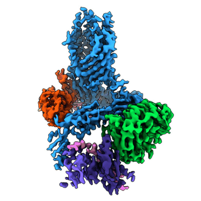 |
E. coli BAM complex bound to SurA [15993 multi-frame micrographs composed of 47 frames each in TIFF format] | Fenn KF, Ranson NA [Pubmed: 39218969] [DOI: 10.1038/s41467-024-51358-x] |
8.2 TB | 4.1 - 4.2 Å |
| 2025-06-03 |  |
Cryo-EM structure of human MPC in complex with TZD [7457 multi-frame micrographs composed of 40 frames each in TIFF format] | Zhang J, Feng L [Pubmed: 40044865] [DOI: 10.1038/s41586-025-08667-y] |
3.1 TB | 3.35 Å |
| 2025-06-02 | 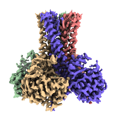 |
Single-particle cryo-EM dataset of the glycosyltransferase ArnC from Salmonella enterica in the apo state from Krios microscope [multiple data sets in TIFF and MRC formats] | Ashraf KU, Punetha A, Kaelber JT, Petrou VI [Pubmed: 39974898] [DOI: 10.1101/2025.01.29.634835] |
5.9 TB | 2.74 Å |
| 2025-06-02 |  |
full length AP5:SPG11-SPG15 [10869 micrographs in MRC format] | Mai X., Su M.-Y., Stjepanovic G. [Pubmed: 40175557] [DOI: 10.1038/s41594-025-01500-0] |
954.4 GB | 3.3 Å |
| 2025-06-02 |  |
SPG11-SPG15 [6660 micrographs in MRC format] | Mai X., Su M.-Y., Stjepanovic G. [Pubmed: 40175557] [DOI: 10.1038/s41594-025-01500-0] |
584.8 GB | 4.02 Å |
| 2025-06-02 | 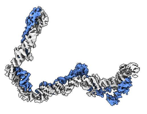 |
SPG11-SPG15 [6849 micrographs in MRC format] | Mai X., Su M.-Y., Stjepanovic G. [Pubmed: 40175557] [DOI: 10.1038/s41594-025-01500-0] |
601.4 GB | 4.02 Å |
| 2025-06-02 | 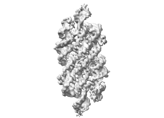 |
Rous sarcoma virus frameshifting pseudoknot [multiple data sets in MRC and TIFF formats] | Jones CP, Ferre-D'Amare AR [Pubmed: 40172966] [DOI: 10.1073/pnas.2418418122] |
4.4 TB | — |
| 2025-06-02 | 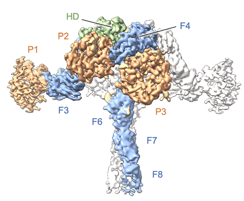 |
Single-particle cryo-EM unaligned micrographs of Sevenless extracellular domain (pH 4.6) [6120 multi-frame micrographs composed of 50 frames each in TIFF format] | Cerutti G [Pubmed: 39510067] [DOI: 10.1016/j.molcel.2024.10.017] |
1.5 TB | 3.67 - 4.07 Å |
| 2025-06-02 | 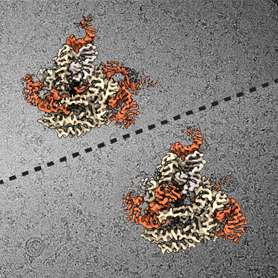 |
Cryo-EM micrographs for the DRT2 reverse transcriptase / ncRNA complex in resting and elongating states [multiple data sets in TIFF format] | Wilkinson ME, Li D, Gao A, Macrae RK, Zhang F [Pubmed: 39208082] [DOI: 10.1126/science.adq3977] |
6.2 TB | 2.91 - 3.17 Å |
| 2025-06-02 | 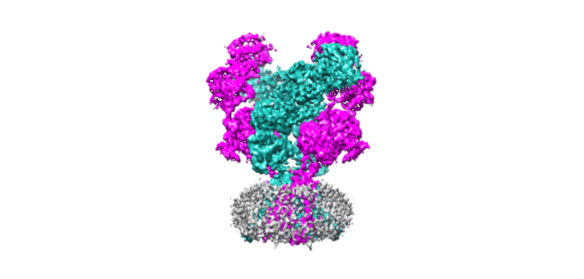 |
Rat GluN1-GluN2B NMDA receptor channel in apo state [8093 multi-frame micrographs composed of 30 frames each in TIFF format] | Chou THC, Furukawa FH [Pubmed: 39085540] [DOI: 10.1038/s41586-024-07742-0] |
1.4 TB | 4.05 Å |
| 2025-06-02 |  |
Cryo-EM Structure of the Full-length hnRNPA1 Amyloid Fibril [5652 multi-frame micrographs composed of 40 frames each in TIFF format] | Sharma K, Schmidt M, Fändrich M [Pubmed: 37481159] [DOI: 10.1016/j.jmb.2023.168211] |
928.0 GB | 3.32 Å |
| 2025-06-02 |  |
Cryo electron micrographs of GSK3β phosphorylated tau fibrils [multiple data sets in MRC and MRCS formats] | Chakraborty P, Ibáñez de Opakua A, Purslow JA, Fromm SA, Chatterjee D, Zachrdla M, Zhuang S, Puri S, Wolozin B, Zweckstetter M [Pubmed: 39693350] [DOI: 10.1073/pnas.2414176121] |
699.4 GB | — |
| 2025-05-30 | 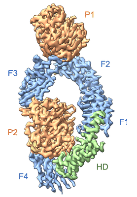 |
Single-particle cryo-EM unaligned micrographs of Sevenless extracellular domain (pH 8) [12010 multi-frame micrographs composed of 50 frames each in TIFF format] | Cerutti G [Pubmed: 39510067] [DOI: 10.1016/j.molcel.2024.10.017] |
2.8 TB | 2.78 Å |
| 2025-05-30 | 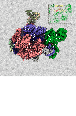 |
Structural insights into transcriptional regulation by the helicase RECQL5 [12100 multi-frame micrographs composed of 50 frames each in TIFF format] | Florez Ariza AJ, Lue NZ, Grob P, Kaeser B, Fang J, Kassube SA, Nogales E [Pubmed: 39975028] [DOI: 10.1101/2025.01.29.634372] |
3.2 TB | 3.2 Å |
| 2025-05-30 | 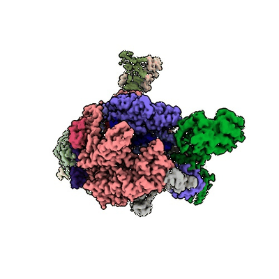 |
Structural insights into transcriptional regulation by the helicase RECQL5 [9048 multi-frame micrographs composed of 50 frames each in TIFF format] | Florez Ariza AJ, Lue NZ, Grob P, Kaeser B, Fang J, Kassube SA, Nogales E [Pubmed: 39975028] [DOI: 10.1101/2025.01.29.634372] |
2.5 TB | 3.7 Å |
| 2025-05-27 | 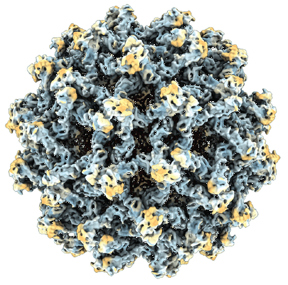 |
CryoEM of purified immature tick borne encephalitis virus particles, strain Kuutsalo-14 [multiple data sets in TIFF format] | Anastasina M, Domanska A, Pulkkinen LIA, Butcher SJ [Pubmed: 38959313] [DOI: 10.1126/sciadv.adl1888] |
26.7 TB | 3.9 - 7.1 Å |
| 2025-05-23 | 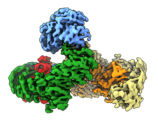 |
AP5βtrunk:SPG11-SPG15 [5724 micrographs in MRC format] | Mai X., Su M.-Y., Stjepanovic G. [Pubmed: 40175557] [DOI: 10.1038/s41594-025-01500-0] |
251.3 GB | 3.26 Å |
| 2025-05-23 |  |
Cryo-EM structure of the 55 kDa nucleoplasmin domain of AtFKBP53 [8687 multi-frame micrographs composed of 594 frames each in EER format] | Bharambe N, Basak S [Pubmed: 40262726] [DOI: 10.1016/j.jsb.2025.108203] |
2.2 TB | 2.0 Å |
| 2025-05-22 |  |
Cryo-ET dataset of dormant microsporidian spores from Encephalitozoon hellem [multiple data sets in MRCS and MRC formats] | Kelley K, Bhabha G, Potter CS, Carragher B, Noble AJ [Pubmed: 40067903] [DOI: 10.1073/pnas.2415233122] |
252.1 GB | — |
| 2025-05-22 |  |
Cryo-ET datasets (1 and 2) of dormant microporidian spores from Encephalitozoon intestinalis [multiple data sets in TIFF and MRC formats] | Usmani M, Coudray N, Raghu R, Ramchandani H, Bobe D, Kopylov M, Zhong ED, Ekiert DC, Bhabha G [Pubmed: 39026755] [DOI: 10.1101/2024.07.13.603322] |
1.3 TB | 44.0 - 70.0 Å |
| 2025-05-22 |  |
Cryo-ET dataset (3) of dormant microporidian spores from Encephalitozoon intestinalis [multiple data sets in TIFF and MRC formats] | Usmani M, Coudray N, Raghu R, Ramchandani H, Bobe D, Kopylov M, Zhong ED, Ekiert DC, Bhabha G [Pubmed: 39026755] [DOI: 10.1101/2024.07.13.603322] |
1.1 TB | 20.0 - 36.0 Å |
| 2025-05-22 | 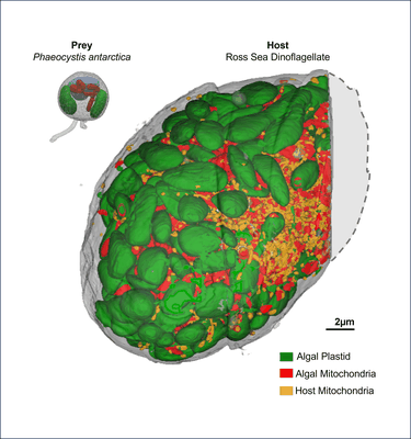 |
Fib-SEM stacks for Prey (Phaeocystis antarctica) and Host (Ross Sea Dinoflagellate; RSD) [multiple data sets in TIFF format] | Rao AK, Gallet B, Jouneau PH, Decelle J [Pubmed: 40250433] [DOI: 10.1101/2024.10.20.619283] |
272.6 GB | — |
| 2025-05-21 |  |
Cryo-EM structure of human GLUT9 bound to urate [multiple data sets in TIFF format] | Matsushita D, Lee Y, Nishizawa T [Pubmed: 40186864] [DOI: 10.1016/j.celrep.2025.115514] |
2.3 TB | 3.39 Å |
| 2025-05-17 |  |
Structural mechanism of FusB-mediated rescue from fusidic acid inhibition of protein synthesis [multiple data sets in TIFF and EER formats] | Gonzalez-Lopez A, Larsson DSD, Selmer M [Pubmed: 40251147] [DOI: 10.1038/s41467-025-58902-3] |
28.2 TB | 1.87 - 2.79 Å |
| 2025-05-14 |  |
Single-particle cryo-EM of Staphylococcus aureus ribosomes inhibited by fusidic acid and fusidic acid cyclopentane [multiple data sets in EER, TIFF and MRC formats] | González-López A, Larsson DSD, Selmer M [Pubmed: 38902339] [DOI: 10.1038/s41598-024-64868-x] |
11.1 TB | 2.02 - 2.49 Å |
