Electron Microscopy Public Image Archive
Electron Microscopy Public Image Archive
Due to a storage failure, some data files are currently inaccessible (Markted: Incomplete dataset).
We are currently working to restore, but we are accepting priority requests.(email, inquiry).
We are restoring lost files from backups in the following order:
We apologize for the inconvenience and appreciate your understanding.
오사카 대학의 EMPIAR-PDBj 팀은, 아시아의 EM 연구자가 용량이 큰 EM 이미지를 EMPIAR 데이터베이스에 전송하는 것을 돕고 있습니다. 인터넷을 통하여 EBI (UK)에 직>접 데이터를 전송하는 대신, 이용자는 우편이나 택배를 통하여 하드 디스크를 오사카 대학으로 보내실 수 있습니다. 혹은 인터넷을 이용하여 오사카 대학의 서버로 전>송 하실 수 있습니다. 오사카 대학에 데이터 전송 서비스를 희망하시는 분은 데이터를 보내시기 전에 먼저 이메일 통하여 등록하시고 싶은 EM데이터에 관하여 상담하십시오.
| Release date | Imageset | Title | Authors and references | Size | Resolution |
|---|---|---|---|---|---|
| 2024-09-03 |  |
ATTRG47E amyloid fibrils from hereditary ATTR amloidosis [12067 multi-frame micrographs composed of 2226 frames each in EER format] | Steinebrei M, Schmidt M, Fändrich M [Pubmed: 37993462] [DOI: 10.1038/s41467-023-43301-3] |
6.8 TB | 2.3673 Å |
| 2024-09-03 |  |
CryoEM Structure of the human Trace Amine-Associated Receptor 1 bound to Ro5256390 [7618 micrographs in MRC format] | Zilberg G, Parpounas AK, Warren AL, Yang S, Wacker D [Pubmed: 38168118] [DOI: 10.1038/s41467-023-44601-4] |
668.9 GB | 3.35 Å |
| 2024-09-03 |  |
AL amyloid fibril from the FOR103 light chain [2788 multi-frame micrographs composed of 40 frames each in TIFF format] | Pfeiffer PB, Karimi-Farsijani S, Schmidt M, Fändrich M [Pubmed: 38879609] [DOI: 10.1038/s41467-024-49520-6] |
504.3 GB | 2.92 Å |
| 2024-09-03 |  |
Volume correlative light X-ray and electron microscopy (CLXEM) of mouse cremaster muscle endothelial cells [multiple data sets in TIFF format] | Reglero-Real N, Pérez-Gutiérrez L, Saleeb R, Nourshargh S, Collinson L, Yoshimura A [DOI: 10.1016/j.xpro.2024.103257] |
79.2 GB | — |
| 2024-09-03 |  |
CryoEM movies of human tau amyloid fibrils extracted from an AD patient brain (in association with in situ cryoET) [10860 multi-frame micrographs composed of 38 frames each in TIFF format] | Gilbert MAG, Fatima N, Jenkins J, O'Sullivan TJ, Schertel A, Halfon Y, Wilkinson M, Morrema THJ, Geibel M, Read RJ, Ranson NA, Radford SE, Hoozemans JJM, Frank RAW [Pubmed: 38987603] [DOI: 10.1038/s41586-024-07680-x] |
1.4 TB | 3.0 Å |
| 2024-09-03 |  |
Structure of Aquifex aeolicus lumazine synthase by cryo-electron microscopy to 1.42 Å resolution [12657 multi-frame micrographs composed of 918 frames each in EER format] | Savva CG, Sobhy MA, De Biasio A, Hamdan SM [Pubmed: 38965901] [DOI: 10.1107/S2052252524005530] |
2.5 TB | 1.42 Å |
| 2024-09-03 |  |
Cryo-EM structure of 1.51 Angstroms mouse heavy-chain apoferritin in 290 nm gold foil holes (HexAuFoil) [multiple data sets in TIFF and MRC formats] | Wang CH, Wu KP, Chang YC | 3.4 TB | 1.51 Å |
| 2024-09-02 |  |
Cryo-ET of in vitro vaccinia core [38 tilt series in MRC format] | Liu J, Turoňová B [Pubmed: 38316878] [DOI: 10.1038/s41594-024-01218-5] |
42.9 GB | 7.7 - 13.4 Å |
| 2024-09-02 |  |
Cryo electron micrographs of 70S Escherichia coli ribosomes [4388 multi-frame micrographs composed of 35 frames each in TIFF format] | Gersteuer FG, Morici MM, Wilson DNW [Pubmed: 38503753] [DOI: 10.1038/s41467-024-46762-2] |
2.0 TB | 2.0 - 2.6 Å |
| 2024-08-29 |  |
Human calcium homeostasis modulator 1 (CALHM1) channel [3285 multi-frame micrographs composed of 30 frames each in TIFF format] | Syrjanen JL, Furukawa H [Pubmed: 37380652] [DOI: 10.1038/s41467-023-39388-3] |
553.5 GB | 3.76 Å |
| 2024-08-29 |  |
Cryo-EM micrographs of human DNA polymerase θ helicase domain bound to inhibitor AB25583 [4500 multi-frame micrographs composed of 2051 frames each in EER format] | Ito F, Li Z, Minakhin L, Chandramouly G, Tyagi M, Betsch R, Krais JJ, Tiberi B, Johnson N, Chen XS, Pomerantz RT [Pubmed: 39143110] [DOI: 10.1038/s41467-024-51351-4] |
4.9 TB | 3.21 Å |
| 2024-08-29 | 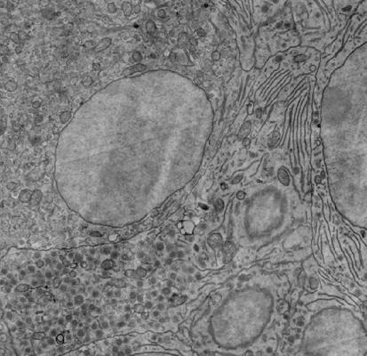 |
FAST-EM array tomography data of rat pancreas prepared with rOTO protocol and stained with neodymium acetate [688 micrographs in TIFF format] | Kievits AJ, Duinkerken BHP, Lane R, de Heus C, van Beijeren Bergen en Henegouwen D, Höppener T, Wolters AHG, Liv N, Giepmans BNG, Hoogenboom JP [Pubmed: 39119255] [DOI: 10.1515/mim-2024-0005] |
69.7 GB | — |
| 2024-08-29 |  |
SPOT-RASTR - a cryo-EM specimen preparation technique that overcomes problems with preferred orientation and the air/water interface [stack of 230000 particles in MRCS format] | Ghazi Esfahani BGE, Randolph PR, Peng RP, Stroupe MES, Grant TG, Stagg SMS [Pubmed: 39108302] [DOI: 10.1093/pnasnexus/pgae284] |
304.6 GB | 3.58 Å |
| 2024-08-29 | 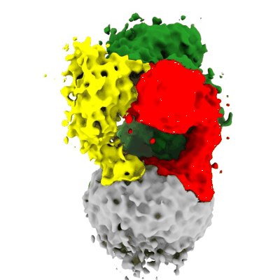 |
The Prothrombin-Prothrombinase Interaction [2390 micrographs in MRC format] | Stojanovski BM, Mohammed BM, Ruben EA, Summers B, Rau MJ, Fitzpatrick JAJ, Di Cera E [Pubmed: 35427420] [DOI: 10.1182/blood.2022015807] |
149.4 GB | 5.3 - 6.47 Å |
| 2024-08-29 | 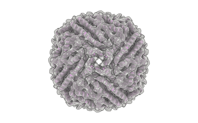 |
Low-dose cryo-electron ptychography of Apoferritin at 4.0 mrad [90 diffraction images in BIG DATA VIEWER HDF5 format] | Küçükoğlu BK, Mohammed IM, Guerrero-Ferreira RCG, Kube MK, Radecke JR, Ribet SR, Varnavides GV, Leidl MLL, Lau KL, Nazarov SN, Myasnikov AM, Sachse CS, Müller-Caspary KMC, Ophus CO, Stahlberg HS [DOI: 10.1101/2024.02.12.579607] |
83.8 GB | 5.8 - 12.3 Å |
| 2024-08-28 |  |
CryoEM micrographs of HK97 small terminase in complex with DNA (2 datasets:550 and 1874 multi-frame micrographs composed of 50 frames in tif format) [multiple data sets in TIFF format] | Antson AA, Chechik M, Greive SJ, Jenkins HT [Pubmed: 39116131] [DOI: 10.1073/pnas.2406138121] |
549.9 GB | 2.92 - 3.0 Å |
| 2024-08-28 |  |
Human calcium homeostasis modulator 1 (CALHM1) channel, I109W point mutation [8664 multi-frame micrographs composed of 30 frames each in TIFF format] | Syrjanen JL, Furukawa H [Pubmed: 37380652] [DOI: 10.1038/s41467-023-39388-3] |
1.8 TB | 2.8 Å |
| 2024-08-28 |  |
Human calcium homeostasis modulator 1 (CALHM1) channel, I109W point mutation in complex with ruthenium red [multiple data sets in TIFF format] | Syrjanen JL, Furukawa H [Pubmed: 37380652] [DOI: 10.1038/s41467-023-39388-3] |
4.6 TB | 3.91 - 4.73 Å |
| 2024-08-28 |  |
Cryoelectron microscopy of IL-21/IL-21R/common gamma signaling complex [3684 multi-frame micrographs composed of 51 frames each in TIFF format] | Abhiraman G.C., Jude K.M., Caveney N.A., Garcia K.C. [Pubmed: 37339051] [DOI: 10.1016/j.celrep.2023.112657] |
4.2 TB | 3.7 Å |
| 2024-08-28 |  |
streptavidin on GSAMs [2144 multi-frame micrographs composed of 32 frames each in MRC format] | Liu N, Xu J, Wang HW | 525.5 GB | 2.56 Å |
| 2024-08-28 |  |
Cryo electron micrographs of CCP5 decorated microtubules [4643 multi-frame micrographs composed of 22 frames each in TIFF format] | Chen J, Zehr EA, Gruschus JM, Szyk A, Liu Y, Tanner ME, Tjandra N, Roll-Mecak A [Pubmed: 39020174] [DOI: 10.1038/s41586-024-07699-0] |
1.3 TB | 2.9 - 3.6 Å |
| 2024-08-28 |  |
Full-length mouse 5-HT3A receptor in complex with SMP100, pre-activated and open-like [multiple data sets in TIFF format] | Felt KCF, Chakrapani SC [Pubmed: 38177669] [DOI: 10.1038/s41594-023-01140-2] |
6.3 TB | 2.7 - 3.79 Å |
| 2024-08-27 |  |
Cryo-EM micrographs of anti-CRISPR associated protein Aca2 bound to its RNA [11232 multi-frame micrographs composed of 40 frames each in TIFF format] | Wilkinson ME, Birkholz N, Kimanius D, Fineran PC [Pubmed: 38987591] [DOI: 10.1038/s41586-024-07644-1] |
1.9 TB | 2.61 Å |
| 2024-08-27 |  |
CryoEM of near-native eisosome/Membrane Compartment containing Can1 (MCC) membrane microdomain scaffolded by Pil1/Lsp1 [2827 multi-frame micrographs composed of 40 frames each in TIFF format] | Kefauver JM, Zou L, Loewith R [Pubmed: 39048819] [DOI: 10.1038/s41586-024-07720-6] |
559.9 GB | 3.2 - 3.67 Å |
| 2024-08-26 |  |
Cryo-EM structure of human pseudouridine synthase 3 [8211 micrographs in TIFF format] | Lin T.-Y., Glatt S. [Pubmed: 38996458] [DOI: 10.1016/j.molcel.2024.06.013] |
3.4 TB | 6.5 Å |
