Electron Microscopy Public Image Archive
Electron Microscopy Public Image Archive
The EMPIAR-PDBj team at Osaka University assists Asian EM researchers with the transfer of big EM image data to EMPIAR. Instead of sending the data directly to the EBI (UK) via the internet, hard drives can also be sent to Osaka University by postal mail or via a courier service. As an alternative, internet transfer to our server in Osaka is also available. If you would like to take advantage of our submission services, please contact us first by e-mail before sending the data to us.
| Release date | Imageset | Title | Authors and references | Size | Resolution |
|---|---|---|---|---|---|
| 2025-05-04 | 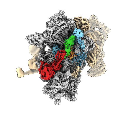 |
Structural basis for translational control by the human 48S initiation complex [multiple data sets in MRC and MRCS formats] | Petrychenko V, Yi SH, Liedtke D, Peng BZ, Rodnina MV, Fischer N [Pubmed: 39289545] [DOI: 10.1038/s41594-024-01378-4] |
— | 2.9 - 3.7 Å |
| 2022-03-28 | 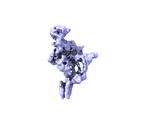 |
Single particle Cryo-EM data set for study the structural basis of Phosphatidylinositol 3-kinase type 2α (PI3KC2α) [multiple data sets in MRC format] | Lo WT, Zhang Y, Vadas O, Roske Y, Gulluni F, De Santis MC, Zagar AV, Stephanowitz H, Hirsch E, Liu F, Daumke O, Kudryashev M, Haucke V [Pubmed: 35256802] [DOI: 10.1038/s41594-022-00730-w] |
0.0 B | 4.4 Å |
| 2019-04-05 | 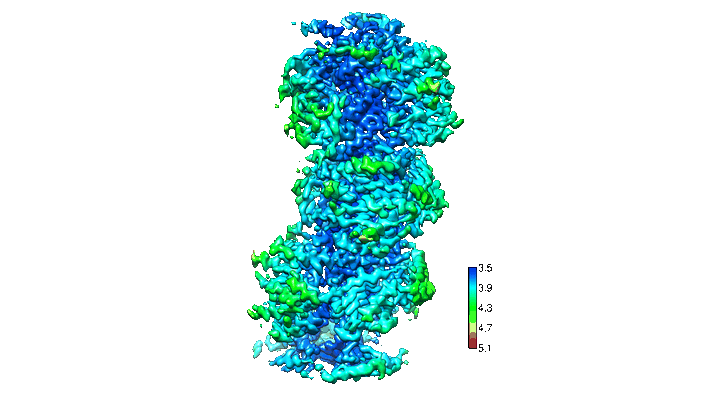 |
mouse MDA5-dsRNA Filaments in presence of 1mM AMPPNP [multiple data sets in MRC format] | Yu Q, Qu K, Modis YE [Pubmed: 30449722] [DOI: 10.1016/j.molcel.2018.10.012] |
0.0 B | 3.68 - 3.93 Å |
| 2021-05-28 | 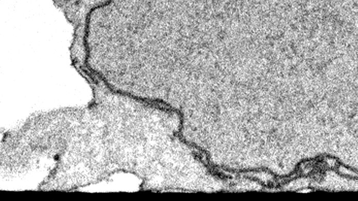 |
CLEM/FIB-SEM Imaging of T Cells after the Formation of Signaling Microclusters at the Immunological Synapse [160 micrographs in TIFF format] | Narayan K | 26.6 MB | — |
| 2021-04-14 |  |
DNA origami signposts for identifying proteins on cell membranes by electron cryotomography [2 tilt series in MRC format] | Silvester E, Vollmer B, Pražák V, Vasishtan D, Machala EA, Whittle C, Black S, Bath J, Turberfield AJ, Grünewald K, Baker LA [Pubmed: 33606980] [DOI: 10.1016/j.cell.2021.01.033] |
38.8 MB | 36.44 Å |
| 2022-01-12 |  |
Cryo-FIB-SEM volume in a Sum159 human cell line [20 micrographs in TIFF format] | Klumpe S, Fung HKH, Goetz SK, Zagoriy I, Hampoelz B, Zhang X, Erdmann PS, Baumbach J, Müller CW, Beck M, Plitzko JM, Mahamid J [Pubmed: 34951584] [DOI: 10.7554/elife.70506] |
120.1 MB | — |
| 2021-02-12 |  |
Ultra-high voltage electron microscope tomography tilt series of neurite section [1 tilt series in MRC format] | Nishida T, Yoshimura R, Nishi R, Imoto Y, Endo Y [Pubmed: 32133640] [DOI: 10.1111/jmi.12885] |
120.3 MB | — |
| 2021-02-12 |  |
Ultra-high voltage electron microscope tomography tilt series of 0.7-μm-thick neurite section acquired at 6,000× magnification at an accelerating voltage of 1 MV [1 tilt series in MRC format] | Nishida T, Yoshimura R, Nishi R, Imoto Y, Endo Y [Pubmed: 32133640] [DOI: 10.1111/jmi.12885] |
120.3 MB | — |
| 2021-02-12 |  |
Ultra-high voltage electron microscope tomography tilt series of 0.7-μm-thick neurite section acquired at 15,000× magnification at an accelerating voltage of 1 MV [1 tilt series in MRC format] | Nishida T, Yoshimura R, Nishi R, Imoto Y, Endo Y [Pubmed: 32133640] [DOI: 10.1111/jmi.12885] |
120.3 MB | — |
| 2020-05-27 |  |
Ultra-high voltage electron microscope tomography tilt series of 0.7-μm-thick neurite section acquired at 20,000× magnification at an accelerating voltage of 1 MV [1 tilt series in MRC format] | Nishida T, Yoshimura R, Nishi R, Imoto Y, Endo Y [Pubmed: 32133640] [DOI: 10.1111/jmi.12885] |
120.3 MB | — |
| 2021-09-01 |  |
Lysosomes as Fiducials for SXT and SIM Correlation [166 tilt series in TIFF format] | Okolo CA, Kounatidis I, Groen J, Nahas KL, Balint S, Fish T, Koronfel MA, López-Cortajarena A, Dobbie I, Pereiro E, Harkiolaki M | 162.4 MB | — |
| 2025-07-03 |  |
Dunaliella tertiolecta PSI-LHCI supercomplex [10090 multi-frame micrographs composed of 50 frames each in TIFF format] | Liu HW, Khera R, Iwai M, Merchant SS [Pubmed: 40523173] [DOI: 10.1073/pnas.2500621122] |
209.0 MB | 2.1 Å |
| 2016-01-20 | 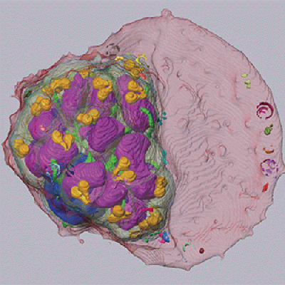 |
SBF-SEM of late schizont-stage malaria parasite infected red blood cell [90 micrographs in MRC format] | Sakaguchi M, Miyazaki N, Fujioka H, Kaneko O, Murata K [Pubmed: 26772147] [DOI: 10.1016/j.jsb.2016.01.003] |
219.7 MB | — |
| 2016-01-20 | 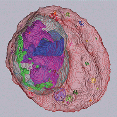 |
SBF-SEM of early schizont-stage malaria parasite infected red blood cell [80 micrographs in MRC format] | Sakaguchi M, Miyazaki N, Fujioka H, Kaneko O, Murata K [Pubmed: 26772147] [DOI: 10.1016/j.jsb.2016.01.003] |
222.1 MB | — |
| 2015-10-09 | 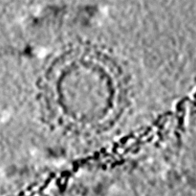 |
Cryo-electron tomography and subtomogram averaging of Rous-Sarcoma-Virus deltaMBD virus-like particles [1 class averages in MRC format] | Schur FKM, Dick RA, Hagen WJH, Vogt VM, Briggs JAG [Pubmed: 26223638] [DOI: 10.1128/JVI.01502-15] |
248.1 MB | — |
| 2021-11-08 | 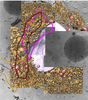 |
Monitoring reversion of hepatitis C virus-induced cellular alterations by Direct-Acting Antivirals using cryo Soft X-ray Tomography and Infrared Microscopy [7 reconstructed volumes in TIFF format] | Perez-Berna AJ, Benseny-Cases N, Rodríguez MJ, Valcarcel R, Carrascosa JL, Gastaminzab P, Pereiroa E [Pubmed: 34726165] [DOI: 10.1107/S2059798321009955] |
260.2 MB | — |
| 2025-03-19 | 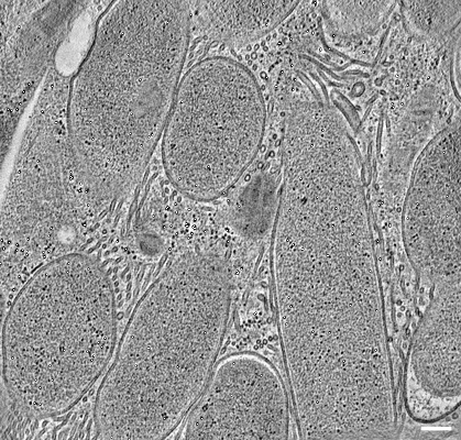 |
Electron tomography of endosymbiotic bacteria inside a bacteriocyte of the cereal weevil Sitophilus oryzae [120 reconstructed volumes in MRC format] | Santarella-Mellwig RSM, Balmand SB, Schwab YS, Zaidman-Remy AZR | 260.8 MB | — |
| 2023-06-20 | 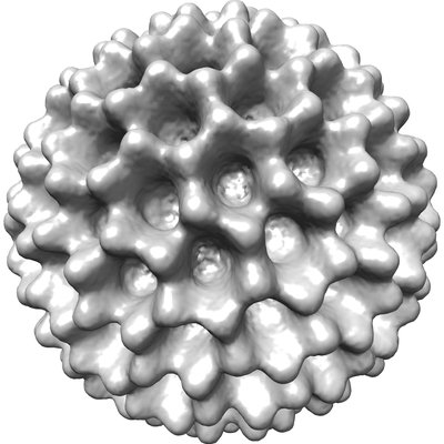 |
Cryo-EPty SPA at CSA of 1.03 mrad [29 micrographs in MRC format] | Pei X, Zhou L, Huang C, Boyce M, Kim JS, Liberti E, Hu Y, Sasaki T, Nellist PD, Zhang P, Stuart DI, Kirkland AI, Wang P [Pubmed: 37230988] [DOI: 10.1038/s41467-023-38268-0] |
262.0 MB | 37.2 Å |
| 2025-07-03 | 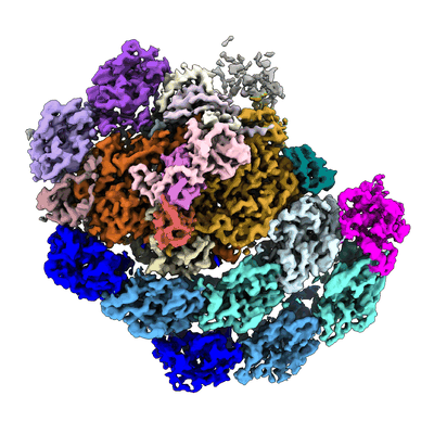 |
Dunaliella salina PSI-LHCI-TIDI1 supercomplex [12541 multi-frame micrographs composed of 50 frames each in TIFF format] | Liu HW, Khera R, Iwai M, Merchant SS [Pubmed: 40523173] [DOI: 10.1073/pnas.2500621122] |
262.0 MB | 3.0 Å |
| 2018-01-17 | 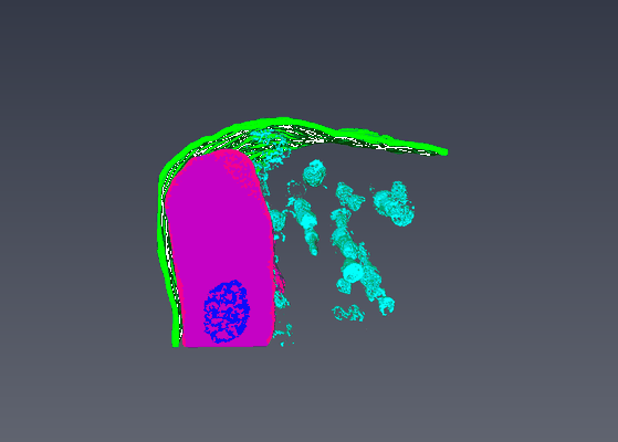 |
Serial Block Face Scanning Electron Micrscopy dataset of fetal day 64 guinea pig psoas muscle in transverse [93 micrographs in TIFF format] | Cocks ET [DOI: 10.1111/jmi.12676] |
264.4 MB | — |
| 2016-01-20 | 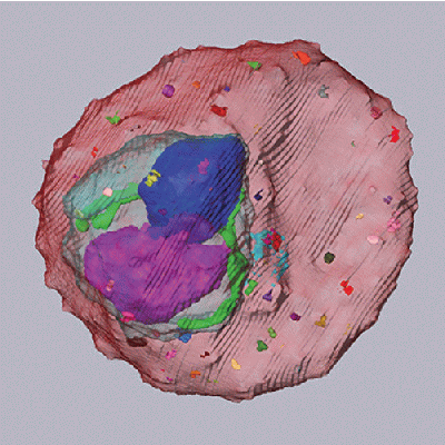 |
SBF-SEM of trophozoite-stage malaria parasite infected red blood cell [110 micrographs in MRC format] | Sakaguchi M, Miyazaki N, Fujioka H, Kaneko O, Murata K [Pubmed: 26772147] [DOI: 10.1016/j.jsb.2016.01.003] |
268.6 MB | — |
| 2017-03-15 | 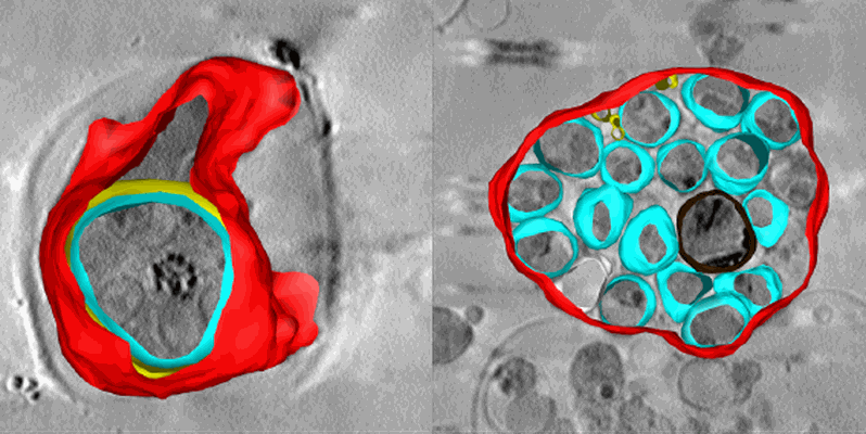 |
Soft X-ray tomography of Plasmodium falciparum infected human erythrocytes stalled in egress by the inhibitors Compound 2 and E64 [1 Soft X-ray tomograms in MRC format] | Hale VL, Saibil HR, Duke E, Fleck RA, Blackman MJ [Pubmed: 28292906] [DOI: 10.1073/pnas.1619441114] |
280.6 MB | — |
| 2016-01-20 | 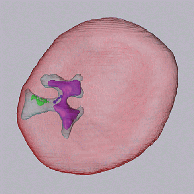 |
SBF-SEM of ring-stage malaria parasite infected red blood cell [120 micrographs in MRC format] | Sakaguchi M, Miyazaki N, Fujioka H, Kaneko O, Murata K [Pubmed: 26772147] [DOI: 10.1016/j.jsb.2016.01.003] |
293.0 MB | — |
| 2018-02-08 |  |
Tilt-series of salmonella enterica wild-type bacterial flagellar motor [1 tilt series in MRC format] | Beeby M, Ribardo DA, Brennan CA, Ruby EG, Jensen GJ, Hendrixson DR [Pubmed: 26976588] [DOI: 10.1073/pnas.1518952113] |
328.0 MB | 69.4 Å |
| 2024-02-13 |  |
Plant SBF-SEM - Tobacco Leaf Chloroplast [130 micrographs in TIFF format] | Wickramanayake JS, Czymmek KJ [Pubmed: 37451777] [DOI: 10.1016/bs.mcb.2023.04.008] |
544.4 MB | — |
