Electron Microscopy Public Image Archive
Electron Microscopy Public Image Archive
The EMPIAR-PDBj team at Osaka University assists Asian EM researchers with the transfer of big EM image data to EMPIAR. Instead of sending the data directly to the EBI (UK) via the internet, hard drives can also be sent to Osaka University by postal mail or via a courier service. As an alternative, internet transfer to our server in Osaka is also available. If you would like to take advantage of our submission services, please contact us first by e-mail before sending the data to us.
| Release date | Imageset | Title | Authors and references | Size | Resolution |
|---|---|---|---|---|---|
| 2023-10-31 | 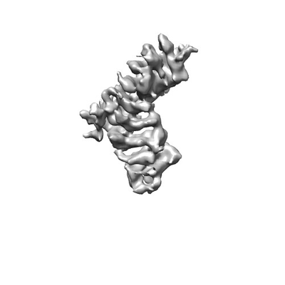 |
Cryogenic electron microscopy structure of human plakophilin-3 [multiple data sets in TIFF and MRC formats] | Gupta J, Rangarajan ES, Izard T [Pubmed: 37298410] [DOI: 10.3390/ijms24119458] |
8.5 TB | 5.03 Å |
| 2022-04-29 | 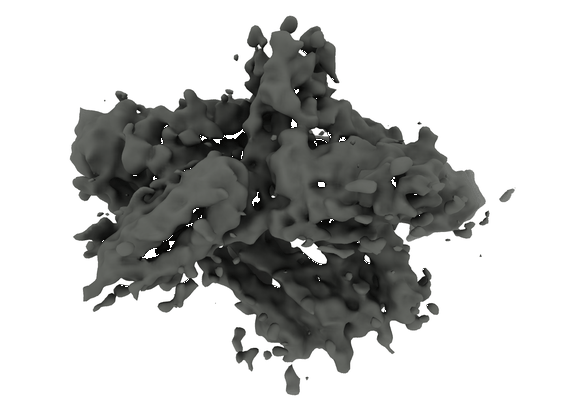 |
Cryogenic electron microscopy structure of full length human meta vinculin [3005 multi-frame micrographs composed of 40 frames each in TIFF format] | Izard T, Rangarajan ES [Pubmed: 33440717] [DOI: 10.3390/ijms22020645] |
744.8 GB | 4.15 - 4.5 Å |
| 2023-10-23 | 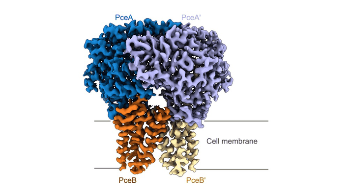 |
Cryogenic electron microscopy spa datset of a membrane-bound menaquinol:organohalide oxidoreductase [13783 micrographs in MRC format] | Ekundayo BE, Ni DC [DOI: 10.1101/2023.07.04.547610] |
861.5 GB | 2.83 Å |
| 2022-03-14 | 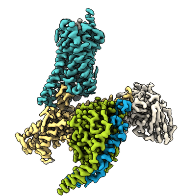 |
Cryogenic electron microscopy of somatostatin receptor 2/SST14/Gi3 complex [9740 multi-frame micrographs composed of 50 frames each in TIFF format] | Robertson MJ [Pubmed: 35210615] [DOI: 10.1038/s41594-022-00727-5] |
4.5 TB | 2.5 Å |
| 2022-03-14 | 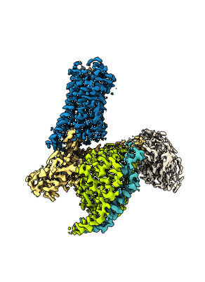 |
Cryogenic electron microscopy of somatostatin receptor 2/Octreotide/Gi3 complex [7576 multi-frame micrographs composed of 55 frames each in TIFF format] | Robertson MJ, Panova O [Pubmed: 35210615] [DOI: 10.1038/s41594-022-00727-5] |
4.2 TB | 2.7 Å |
| 2025-02-03 |  |
Cryogenic electron microscopy of chameleon-plunged acetyl-CoA decarbonylase/synthase [13892 multi-frame micrographs composed of 40 frames each in TIFF format] | Biester A, Drennan CL [Pubmed: 39361653] [DOI: 10.1073/pnas.2410995121] |
1.8 TB | 2.8 - 3.2 Å |
| 2025-01-23 |  |
Cryogenic electron microscopy of blot-plunged acetyl-CoA decarbonylase/synthase [14078 multi-frame micrographs composed of 21 frames each in TIFF format] | Biester A, Drennan CL [Pubmed: 39361653] [DOI: 10.1073/pnas.2410995121] |
1.8 TB | 3.3 Å |
| 2022-12-02 | 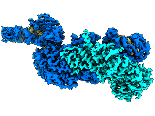 |
Cryogenic electron microscopy SPA of CRISPR Craspase complex Cas7-11 [18968 micrographs in MRC format] | Ekundayo B, Ni DC, Stahlberg H, Torre D, Beckert B, Nazarov S, Myasnikov A [Pubmed: 36471056] [DOI: 10.1038/s41594-022-00894-5] |
1.2 TB | 3.03 - 3.2 Å |
| 2024-08-28 | 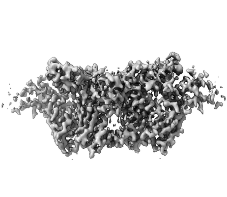 |
Cryoelectron microscopy of IL-21/IL-21R/common gamma signaling complex [3684 multi-frame micrographs composed of 51 frames each in TIFF format] | Abhiraman G.C., Jude K.M., Caveney N.A., Garcia K.C. [Pubmed: 37339051] [DOI: 10.1016/j.celrep.2023.112657] |
4.2 TB | 3.7 Å |
| 2025-06-12 | 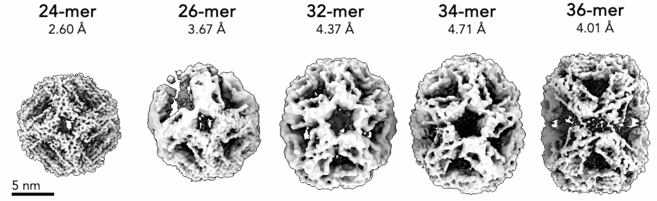 |
Cryoelectron microscopy dataset of apo-state mjHSP16.5/lysozyme at 12:1 monomer ratio (post 75C, 2 hr) [13267 multi-frame micrographs composed of 50 frames each in TIFF format] | Miller AP, Reichow SL [Pubmed: 40240363] [DOI: 10.1038/s41467-025-58964-3] |
7.1 TB | 2.6 - 4.71 Å |
| 2019-07-29 | 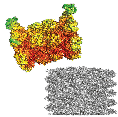 |
CryoWriter: 3.5 Å structure of human 20S proteasome with bound Fabs from microfluidic protein isolation, and 1.9 Å TMV structure [523 multi-frame micrographs composed of 30 frames each in TIFF format] | Schmidli C, Albiez S, Rima L, Righetto R, Mohammed I, Oliva P, Kovacik L, Stahlberg H, Braun T [Pubmed: 31292253] [DOI: 10.1073/pnas.1907214116] |
136.5 GB | 3.5 Å |
| 2023-02-01 | 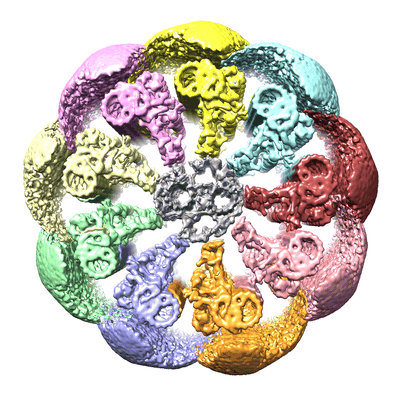 |
CryoET tilt series of mouse sperm flagella after FIB-SEM milling [69 tilt series in MRC format] | Chen Z, Greenan GA, Shiozaki M, Liu Y, Skinner WM, Zhao X, Zhao S, Yan R, Guo C, Yu Z, Lishko PV, Agard DA, Vale RD [Pubmed: 36593309] [DOI: 10.1038/s41594-022-00861-0] |
194.0 GB | 25.0 Å |
| 2017-12-15 | 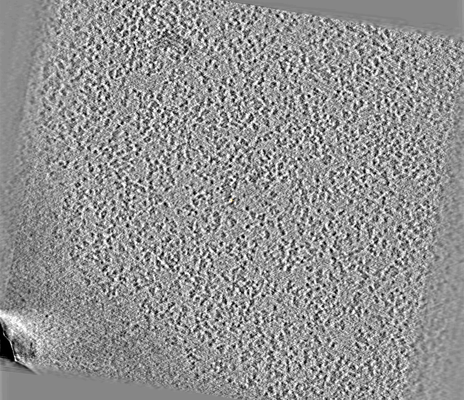 |
CryoET of rabbit muscle aldolase single particle super-res [multiple data sets in MRC format] | Noble AJ, Dandey VP, Wei H, Brasch J, Chase J, Acharya P, Tan YZ, Zhang Z, Kim LY, Scapin G, Rapp M, Eng ET, Rice WJ, Cheng A, Negro CJ, Shapiro L, Kwong PD, Jeruzalmi D, des Georges A, Potter CS, Carragher B [Pubmed: 29809143] [DOI: 10.7554/eLife.34257] |
998.3 GB | — |
| 2017-12-15 | 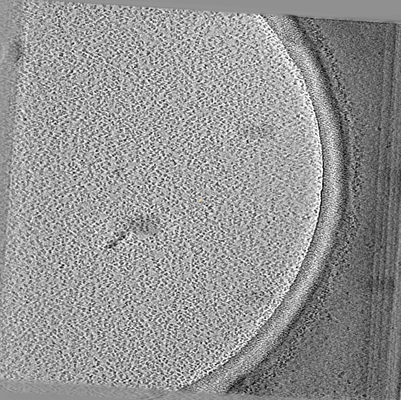 |
CryoET of rabbit muscle aldolase single particle [multiple data sets in MRC format] | Noble AJ, Dandey VP, Wei H, Brasch J, Chase J, Acharya P, Tan YZ, Zhang Z, Kim LY, Scapin G, Rapp M, Eng ET, Rice WJ, Cheng A, Negro CJ, Shapiro L, Kwong PD, Jeruzalmi D, des Georges A, Potter CS, Carragher B [Pubmed: 29809143] [DOI: 10.7554/eLife.34257] |
43.5 GB | — |
| 2022-01-28 |  |
CryoET of presequence protease single particle [multiple data sets in TIFF and MRC formats] | Liang WG, Wijaya J, Wei H, Noble AJ, Mancl JM, Mo S, Lee D, Lin King JV, Pan M, Liu C, Koehler CM, Zhao M, Potter CS, Carragher B, Li S, Tang WJ [Pubmed: 35383169] [DOI: 10.1038/s41467-022-29322-4] |
25.4 GB | — |
| 2019-04-18 | 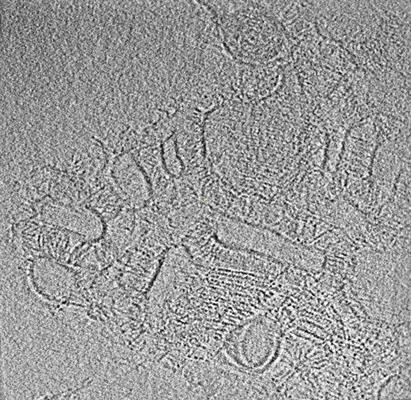 |
CryoET of mouse protocadherin gamma B6 on membranes (without energy filter) [multiple data sets in MRC format] | Brasch J, Goodman KM, Noble AJ, Mannepalli S, Bahna F, Rapp M, Dandey VP, Bepler T, Berger B, Maniatis T, Potter CS, Carragher B, Honig B, Shapiro L [Pubmed: 30971825] [DOI: 10.1038/s41586-019-1089-3] |
363.4 GB | — |
| 2019-04-18 | 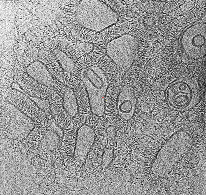 |
CryoET of mouse protocadherin gamma B6 on membranes (with energy filter) [multiple data sets in MRC format] | Brasch J, Goodman KM, Noble AJ, Mannepalli S, Bahna F, Rapp M, Dandey VP, Bepler T, Berger B, Maniatis T, Carragher B [Pubmed: 30971825] [DOI: 10.1038/s41586-019-1089-3] |
411.3 GB | — |
| 2018-04-05 |  |
CryoET of insulin-bound insulin receptor single particle with spot-to-plunge time of 600ms [multiple data sets in MRC format] | Noble AJ, Wei H, Dandey VP, Zhang Z, Potter CS, Carragher B [Pubmed: 30250056] [DOI: 10.1038/s41592-018-0139-3] |
79.4 GB | — |
| 2018-04-05 |  |
CryoET of insulin-bound insulin receptor single particle with spot-to-plunge time of 200ms [multiple data sets in MRC format] | Noble AJ, Wei H, Dandey VP, Zhang Z, Potter CS, Carragher B [Pubmed: 30250056] [DOI: 10.1038/s41592-018-0139-3] |
40.4 GB | — |
| 2017-12-18 |  |
CryoET of hemagglutinin single particle with spot-to-plunge time of 800ms [multiple data sets in MRC format] | Noble AJ, Dandey VP, Wei H, Brasch J, Chase J, Acharya P, Tan YZ, Zhang Z, Kim LY, Scapin G, Rapp M, Eng ET, Rice WJ, Cheng A, Negro CJ, Shapiro L, Kwong PD, Jeruzalmi D, des Georges A, Potter CS, Carragher B [Pubmed: 29809143] [DOI: 10.7554/eLife.34257] |
34.0 GB | — |
| 2018-04-05 |  |
CryoET of hemagglutinin single particle with spot-to-plunge time of 100ms [multiple data sets in MRC format] | Noble AJ, Wei H, Dandey VP, Zhang Z, Potter CS, Carragher B [Pubmed: 30250056] [DOI: 10.1038/s41592-018-0139-3] |
21.9 GB | — |
| 2017-12-15 |  |
CryoET of glutamate dehydrogenase single particle [multiple data sets in MRC format] | Noble AJ, Dandey VP, Wei H, Brasch J, Chase J, Acharya P, Tan YZ, Zhang Z, Kim LY, Scapin G, Rapp M, Eng ET, Rice WJ, Cheng A, Negro CJ, Shapiro L, Kwong PD, Jeruzalmi D, des Georges A, Potter CS, Carragher B [Pubmed: 29809143] [DOI: 10.7554/eLife.34257] |
133.1 GB | — |
| 2017-12-15 |  |
CryoET of glutamate dehydrogenase single particle [multiple data sets in MRC format] | Noble AJ, Dandey VP, Wei H, Brasch J, Chase J, Acharya P, Tan YZ, Zhang Z, Kim LY, Scapin G, Rapp M, Eng ET, Rice WJ, Cheng A, Negro CJ, Shapiro L, Kwong PD, Jeruzalmi D, des Georges A, Potter CS, Carragher B [Pubmed: 29809143] [DOI: 10.7554/eLife.34257] |
37.1 GB | — |
| 2017-12-15 |  |
CryoET of glutamate dehydrogenase + 0.001% DDM single particle [multiple data sets in MRC format] | Noble AJ, Dandey VP, Wei H, Brasch J, Chase J, Acharya P, Tan YZ, Zhang Z, Kim LY, Scapin G, Rapp M, Eng ET, Rice WJ, Cheng A, Negro CJ, Shapiro L, Kwong PD, Jeruzalmi D, des Georges A, Potter CS, Carragher B [Pubmed: 29809143] [DOI: 10.7554/eLife.34257] |
37.1 GB | — |
| 2019-04-18 |  |
CryoET of cis-mutated mouse protocadherin gamma B6 on membranes [multiple data sets in MRC format] | Brasch J, Bepler T, Berger B, Maniatis T, Potter CS, Carragher B, Honig B, Shapiro L [Pubmed: 30971825] [DOI: 10.1038/s41586-019-1089-3] |
379.7 GB | — |
