Electron Microscopy Public Image Archive
Electron Microscopy Public Image Archive
The EMPIAR-PDBj team at Osaka University assists Asian EM researchers with the transfer of big EM image data to EMPIAR. Instead of sending the data directly to the EBI (UK) via the internet, hard drives can also be sent to Osaka University by postal mail or via a courier service. As an alternative, internet transfer to our server in Osaka is also available. If you would like to take advantage of our submission services, please contact us first by e-mail before sending the data to us.
| Release date | Imageset | Title | Authors and references | Size | Resolution |
|---|---|---|---|---|---|
| 2019-06-07 | 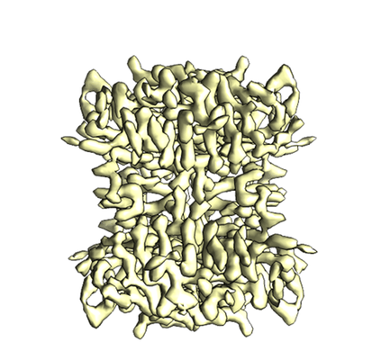 |
Single particle reconstruction of 52 kDa biotin-bound state streptavidin at 3.2 Angstrom resolution [3309 multi-frame micrographs composed of 32 frames each in MRC format] | Fan X, Wang J, Zhang X, Yang Z, Zhang JC, Zhao L, Peng HL, Lei J, Wang HW [Pubmed: 31160591] [DOI: 10.1038/s41467-019-10368-w] |
5.5 TB | 3.2 Å |
| 2019-06-07 | 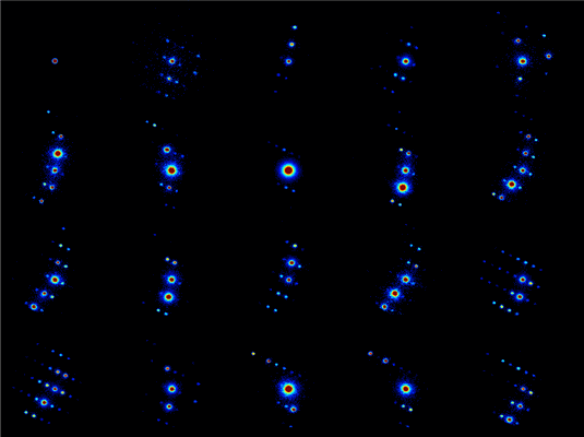 |
Scanning electron diffraction data collected from peptide microcrystals [30000 micrographs in DM4 format] | Gallagher-Jones M, Rodriguez JA, Bustillo K, Ophus C [Pubmed: 30675524] [DOI: 10.1038/s42003-018-0263-8] |
144.3 GB | — |
| 2019-06-19 | 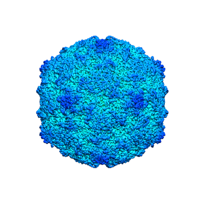 |
Extracellular albumin and endosomal ions prime enterovirus particles for uncoating that can be prevented by fatty acid saturation [multiple data sets in MRC format] | Domanska A, Ruokolainen VP, Pelliccia M, Laajala MA, Marjomäki VS, Butcher SJ [Pubmed: 31189702] [DOI: 10.1128/JVI.00599-19] |
2.4 TB | 3.5 - 3.6 Å |
| 2019-06-21 | 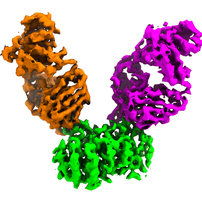 |
LAT1-CD98hc bound to HBJ127 Fab and MEM-108 Fab [1869 multi-frame micrographs composed of 40 frames each in TIFF format] | Lee Y, Wiriyasermkul P, Jin C, Quan L, Ohgaki R, Okuda S, Kusakizako T, Nishizawa T, Oda K, Ishitani R, Yokoyama T, Nakane T, Shirouzu M, Endou H, Nagamori S, Kanai Y, Nureki O [Pubmed: 31160781] [DOI: 10.1038/s41594-019-0237-7] |
969.9 GB | 4.1 Å |
| 2019-06-21 | 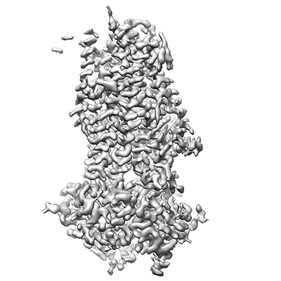 |
Cryo electron micrographs of human cystic fibrosis transmembrane conductance regulator (CFTR) in complex with GLPG [3335 multi-frame micrographs composed of 50 frames each in TIFF format] | Zhang ZZ, Liu FL, Chen JC [Pubmed: 31221859] [DOI: 10.1126/science.aaw7611] |
1.4 TB | 3.2 Å |
| 2019-07-25 | 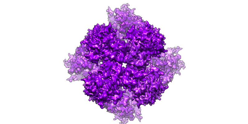 |
Open state structure of the full-length TRPV2 cation channel with a resolved pore turret domain [2447 multi-frame micrographs composed of 50 frames each in MRCS format] | Dosey TL, Wang Z, Fan G [Pubmed: 30598551] [DOI: 10.1038/s41594-018-0168-8] |
934.8 GB | 3.6 Å |
| 2019-07-25 |  |
TRPV2 ion channel gating through allosteric domain coupling revealed by cryo-EM [1969 multi-frame micrographs composed of 35 frames each in MRCS format] | Serysheva II, Chiu W, Wensel TG [Pubmed: 30598551] [DOI: 10.1038/s41594-018-0168-8] |
3.6 TB | 4.0 Å |
| 2019-07-25 | 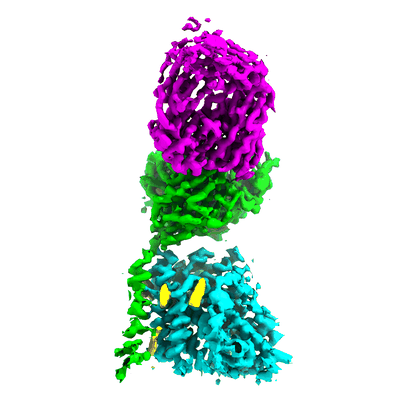 |
LAT1-CD98hc bound to MEM-108 Fab [multiple data sets in TIFF and MRCS formats] | Lee Y, Wiriyasermkul P, Jin C, Quan L, Ohgaki R, Okuda S, Kusakizako T, Nishizawa T, Oda K, Ishitani R, Yokoyama T, Nakane T, Shirouzu M, Endou H, Nagamori S, Kanai Y, Nureki O [Pubmed: 31160781] [DOI: 10.1038/s41594-019-0237-7] |
4.1 TB | 3.31 Å |
| 2019-07-29 | 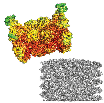 |
CryoWriter: 3.5 Å structure of human 20S proteasome with bound Fabs from microfluidic protein isolation, and 1.9 Å TMV structure [523 multi-frame micrographs composed of 30 frames each in TIFF format] | Schmidli C, Albiez S, Rima L, Righetto R, Mohammed I, Oliva P, Kovacik L, Stahlberg H, Braun T [Pubmed: 31292253] [DOI: 10.1073/pnas.1907214116] |
136.5 GB | 3.5 Å |
| 2019-07-29 | 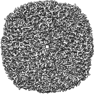 |
Movies of horse spleen apoferritin on multifunctional ultrastable graphene supports for electron cryomicroscopy [480 multi-frame micrographs composed of 38 frames each in MRCS format] | Naydenova K, Peet MJ, Russo CJ [Pubmed: 31127045] [DOI: 10.1073/pnas.1904766116] |
570.0 GB | 2.1 Å |
| 2019-08-07 | 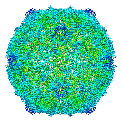 |
Identification of a druggable VP1-VP3 interprotomer pocket in the capsid of enteroviruses [2120 micrographs in MRC format] | Geraets JA, Flatt JW, Domanska A, Butcher SJ [Pubmed: 31185007] [DOI: 10.1371/journal.pbio.3000281] |
2.2 TB | 4.0 Å |
| 2019-08-16 | 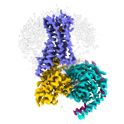 |
Cryo electron microscopy of Cannabinoid Receptor 1-G Protein Complex [2756 multi-frame micrographs composed of 40 frames each in TIFF format] | Krishna Kumar K, Shalev-Benami M, Kobilka BK, Skiniotis G [Pubmed: 30639101] [DOI: 10.1016/j.cell.2018.11.040] |
476.0 GB | 3.0 Å |
| 2019-08-27 | 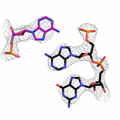 |
Yeast postcatalytic spliceosome, two cryoEM data sets at different magnifications [multiple data sets in MRC format] | Wilkinson ME, Nagai K [Pubmed: 31478901] [DOI: 10.1107/S2059798319010519] |
6.3 TB | 3.3 Å |
| 2019-08-28 | 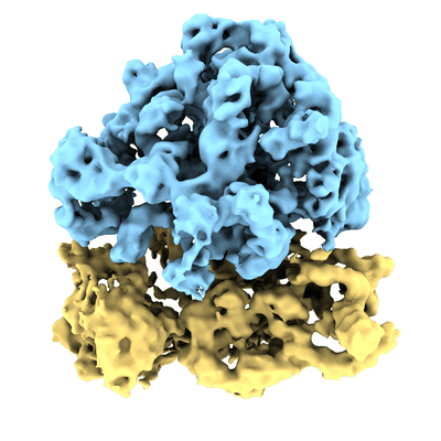 |
Improved applicability and robustness of fast cryo-electron tomography data acquisition [12 tilt series in MRC format] | Eisenstein F, Danev R, Pilhofer M [Pubmed: 31425790] [DOI: 10.1016/j.jsb.2019.08.006] |
21.6 GB | 9.0 Å |
| 2019-08-30 | 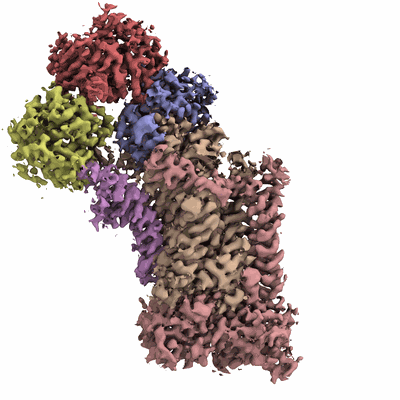 |
Cryo-EM structures of human P4-ATPase flippase [multiple data sets in TIFF format] | Hiraizumi M, Yamashita K, Nishizawa T, Nureki O [Pubmed: 31416931] [DOI: 10.1126/science.aay3353] |
11.0 TB | 2.63 - 3.42 Å |
| 2019-09-27 | 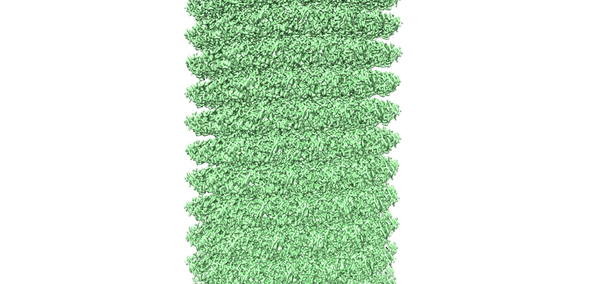 |
Cryo-EM structure of TMV in water [62 multi-frame micrographs composed of 20 frames each in TIFF format] | Weis F, Beckers M, von der Hocht I, Sachse C [Pubmed: 31535454] [DOI: 10.15252/embr.201948451] |
6.8 GB | 1.9 Å |
| 2019-09-27 | 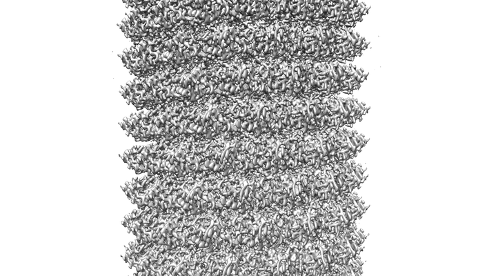 |
Cryo-EM structure of TMV with Ca2+ at low pH [197 multi-frame micrographs composed of 40 frames each in TIFF format] | Weis F, Beckers M, von der Hocht I, Sachse C [Pubmed: 31535454] [DOI: 10.15252/embr.201948451] |
22.0 GB | 2.0 Å |
| 2019-09-30 | 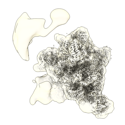 |
Human pre-B spliceosome and U4/U6.U5 tri-snRNP [multiple data sets in MRC and MRCS formats] | Charenton C, Wilkinson ME, Nagai K [Pubmed: 30975767] [DOI: 10.1126/science.aax3289] |
2.3 TB | 2.9 - 28.0 Å |
| 2019-10-04 | 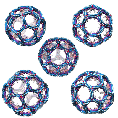 |
Single particle cryo-EM dataset of clathrin cages with phase flipping suitable for refinement [stack of 12785 particles in MRCS format] | Morris KL, Jones JR, Halebian M, Wu S, Baker M, Armache JP, Avila Ibarra A, Sessions RB, Cameron AD, Cheng Y, Smith CJ [Pubmed: 31582853] [DOI: 10.1038/s41594-019-0292-0] |
11.9 GB | 9.07 - 23.68 Å |
| 2019-10-04 | 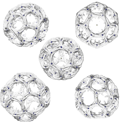 |
Single particle cryo-EM dataset of clathrin cages suitable for subparticle extraction [multiple data sets in MRCS format] | Morris KL, Jones JR, Halebian M, Wu S, Baker M, Armache JP, Avila Ibarra A, Sessions RB, Cameron AD, Cheng Y, Smith CJ [Pubmed: 31582853] [DOI: 10.1038/s41594-019-0292-0] |
19.9 GB | 9.07 - 23.68 Å |
| 2019-10-07 | 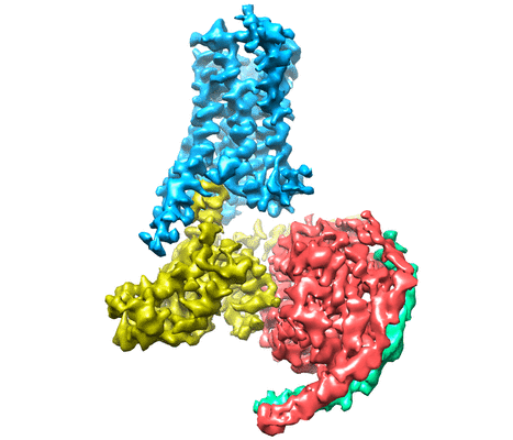 |
Cryo-EM structure of the serotonin 5-HT1B receptor coupled to heterotrimeric Go [multiple data sets in TIFF and MRCS formats] | Garcia-Nafria J, Nehme R, Edwards PC, Tate CG [Pubmed: 29925951] [DOI: 10.1038/s41586-018-0241-9] |
8.0 TB | 3.78 Å |
| 2019-10-07 | 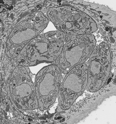 |
Processed FIB SEM images of a parasitophorous vacuole containing Toxoplasma gondii ∆CAP parasites. [1 multi-frame micrographs composed of 1 frames each in MRC format] | Hunt A, Russell MRG, Wagener J, Kent R, Carmeille R, Peddie CJ, Collinson L, Heaslip A, Ward GE, Treeck M [Pubmed: 31577230] [DOI: 10.7554/elife.50598] |
898.1 MB | — |
| 2019-10-07 |  |
Raw FIB SEM images of a parasitophorous vacuole containing Toxoplasma gondii ∆CAP parasites, complemented with CAP. [4529 micrographs in TIFF format] | Hunt A, Russell MRG, Wagener J, Kent R, Carmeille R, Peddie CJ, Collinson L, Heaslip A, Ward GE, Treeck M [Pubmed: 31577230] [DOI: 10.7554/eLife.50598] |
605.2 GB | — |
| 2019-10-07 |  |
Raw FIB SEM images of a parasitophorous vacuole containing Toxoplasma gondii ∆CAP parasites. [4490 micrographs in TIFF format] | Hunt A, Russell MRG, Wagener J, Kent R, Carmeille R, Peddie CJ, Collinson L, Heaslip A, Ward GE, Treeck M [Pubmed: 31577230] [DOI: 10.7554/eLife.50598] |
273.8 GB | — |
| 2019-10-07 | 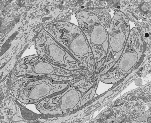 |
Processed FIB SEM images of a parasitophorous vacuole containing Toxoplasma gondii ∆CAP parasites, complemented with CAP. [1 multi-frame micrographs composed of 1 frames each in MRC format] | Hunt A, Russell MRG, Wagener J, Kent R, Carmeille R, Peddie CJ, Collinson L, Heaslip A, Ward GE, Treeck M [Pubmed: 31577230] [DOI: 10.7554/elife.50598] |
583.8 MB | — |
