Electron Microscopy Public Image Archive
Electron Microscopy Public Image Archive
The EMPIAR-PDBj team at Osaka University assists Asian EM researchers with the transfer of big EM image data to EMPIAR. Instead of sending the data directly to the EBI (UK) via the internet, hard drives can also be sent to Osaka University by postal mail or via a courier service. As an alternative, internet transfer to our server in Osaka is also available. If you would like to take advantage of our submission services, please contact us first by e-mail before sending the data to us.
| Release date | Imageset | Title | Authors and references | Size | Resolution |
|---|---|---|---|---|---|
| 2020-06-30 | 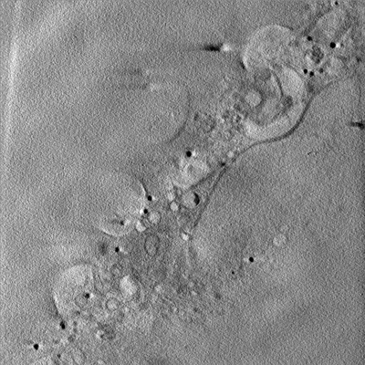 |
Soft X-ray Tomography of mock-infected U2OS cells [1 tilt series in MRC format] | Kounatidis I, Stanifer ML, Phillips MA, Paul-Gilloteaux P, Heiligenstein X, Wang H, Okolo CA, Fish TM, Spink MC, Stuart DI, Davis I, Boulant S, Grimes JM, Dobbie IM, Harkiolaki M [Pubmed: 32610083] [DOI: 10.1016/j.cell.2020.05.051] |
1.2 GB | — |
| 2020-06-30 | 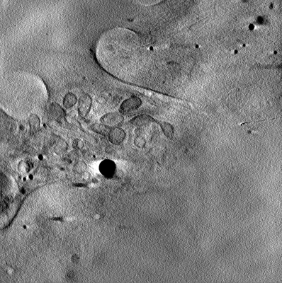 |
Soft X-ray Tomography of mock-infected U2OS cells [1 tilt series in MRC format] | Kounatidis I, Stanifer ML, Phillips MA, Paul-Gilloteaux P, Heiligenstein X, Wang H, Okolo CA, Fish TM, Spink MC, Stuart DI, Davis I, Boulant S, Grimes JM, Dobbie IM, Harkiolaki M [Pubmed: 32610083] [DOI: 10.1016/j.cell.2020.05.051] |
1.2 GB | — |
| 2017-03-15 | 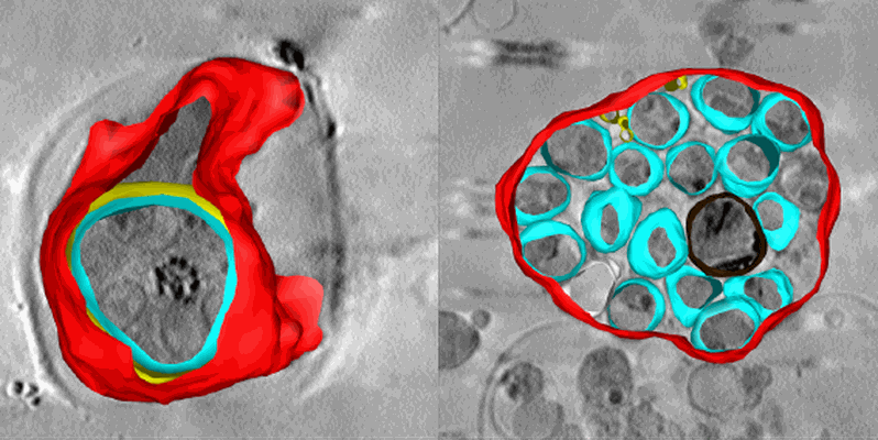 |
Soft X-ray tomography of Plasmodium falciparum infected human erythrocytes stalled in egress by the inhibitors Compound 2 and E64 [1 Soft X-ray tomograms in MRC format] | Hale VL, Saibil HR, Duke E, Fleck RA, Blackman MJ [Pubmed: 28292906] [DOI: 10.1073/pnas.1619441114] |
280.6 MB | — |
| 2023-12-11 | 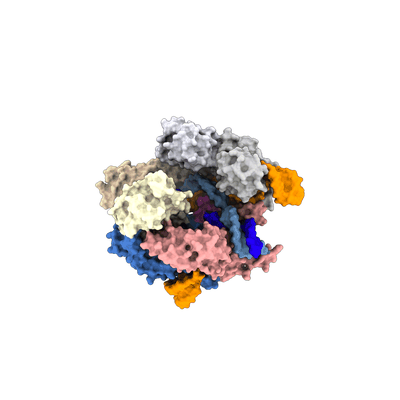 |
SpCas9 bound to 10 nucleotide complementary DNA substrate [multiple data sets in TIFF and DM4 formats] | Pacesa M, Loeff L, Querques I, Muckenfuss LM, Sawicka M, Jinek M [Pubmed: 36002571] [DOI: 10.1038/s41586-022-05114-0] |
157.5 GB | 3.81 Å |
| 2023-12-11 |  |
SpCas9 bound to 12 nucleotide complementary DNA substrate [multiple data sets in TIFF and DM4 formats] | Pacesa M, Loeff L, Querques I, Muckenfuss LM, Sawicka M, Jinek M [Pubmed: 36002571] [DOI: 10.1038/s41586-022-05114-0] |
1.1 TB | 3.64 Å |
| 2023-12-11 |  |
SpCas9 bound to 14 nucleotide complementary DNA substrate [multiple data sets in TIFF and DM4 formats] | Pacesa M, Loeff L, Querques I, Muckenfuss LM, Sawicka M, Jinek M [Pubmed: 36002571] [DOI: 10.1038/s41586-022-05114-0] |
2.1 TB | 3.49 Å |
| 2023-12-11 |  |
SpCas9 bound to 16 nucleotide complementary DNA substrate [multiple data sets in TIFF and DM4 formats] | Pacesa M, Loeff L, Querques I, Muckenfuss LM, Sawicka M, Jinek M [Pubmed: 36002571] [DOI: 10.1038/s41586-022-05114-0] |
1.3 TB | 3.12 Å |
| 2023-12-11 |  |
SpCas9 bound to 18 nucleotide complementary DNA substrate in the catalytic state [multiple data sets in TIFF and DM4 formats] | Pacesa M, Loeff L, Querques I, Muckenfuss LM, Sawicka M, Jinek M [Pubmed: 36002571] [DOI: 10.1038/s41586-022-05114-0] |
1.8 TB | 2.99 Å |
| 2024-02-06 |  |
SpCas9 bound to 18 nucleotide complementary DNA substrate in the checkpoint state [multiple data sets in EER format] | Pacesa M, Loeff L, Querques I, Muckenfuss LM, Sawicka M, Jinek M [Pubmed: 36002571] [DOI: 10.1038/s41586-022-05114-0] |
7.7 TB | 2.54 Å |
| 2024-01-23 |  |
SpCas9 bound to 6 nucleotide complementary DNA substrate [multiple data sets in TIFF and DM4 formats] | Pacesa M, Loeff L, Querques I, Muckenfuss LM, Sawicka M, Jinek M [Pubmed: 36002571] [DOI: 10.1038/s41586-022-05114-0] |
1.5 TB | 3.87 Å |
| 2023-12-11 |  |
SpCas9 bound to 8 nucleotide complementary DNA substrate [multiple data sets in TIFF and DM4 formats] | Pacesa M, Loeff L, Querques I, Muckenfuss LM, Sawicka M, Jinek M [Pubmed: 36002571] [DOI: 10.1038/s41586-022-05114-0] |
1.4 TB | 4.14 Å |
| 2020-09-11 |  |
Spatial Intra- and Intercellular Alignment of Respiratory Cilia and its Relation to Function [16 multi-frame micrographs composed of 100 frames each in MRC format] | Schneiter M, Halm S, Odriozola A, Mogel H, Rička J, Stoffel MH, Zuber B, Frenz M, Tschanz SA [DOI: 10.1101/735332] |
67.5 GB | — |
| 2024-04-17 |  |
Spatial mapping of hepatic ER and mitochondria architecture reveals zonated remodeling in fasting and obesity [multiple data sets in TIFF format] | Parlakgul G | 2.9 TB | — |
| 2023-02-13 |  |
Spinacia oleracea endogenous cytochrome b6f complex [7651 multi-frame micrographs composed of 40 frames each in TIFF format] | Jaciuk M, Koziej L, Pintscher S, Indyka P, Rawski M, Glatt S [Pubmed: 36638176] [DOI: 10.1126/sciadv.add9688] |
2.9 TB | 2.13 Å |
| 2023-09-19 |  |
Spraguea lophii 100S ribosome [multiple data sets in TIFF format] | McLaren MJ, Gil Diez P, Isupov M, Conners R, Gambelli L, Gold V, Connell S, Walter A, Williams B, Daum B [Pubmed: 37709902] [DOI: 10.1038/s41564-023-01469-w] |
9.8 TB | 2.79 Å |
| 2022-07-18 |  |
Staphylococcal self-loading helicases couple the staircase mechanism 1 with inter domain high flexibility [3345 multi-frame micrographs composed of 40 frames each in MRC format] | Qiao C [Pubmed: 35871290] [DOI: 10.1093/nar/gkac625] |
1.6 TB | 3.9 - 3.96 Å |
| 2022-08-12 |  |
Staphylococcal self-loading helicases couple the staircase mechanism with inter domain high flexibility [3121 multi-frame micrographs composed of 40 frames each in TIFF format] | Qiao C, Debiasi-Anders G, Mir-Sanchis I [Pubmed: 35871290] [DOI: 10.1093/nar/gkac625] |
395.0 GB | 3.14 Å |
| 2022-07-18 |  |
Staphylococcal self-loading helicases couple the staircase mechanism with inter domain high flexibility [3765 multi-frame micrographs composed of 40 frames each in TIFF format] | Qiao QCC, Mir Sanchis IMS [Pubmed: 35871290] [DOI: 10.1093/nar/gkac625] |
467.6 GB | 3.1 - 3.3 Å |
| 2022-01-21 |  |
Structural Insights of Transcriptionally Active, Full-Length Androgen Receptor Coactivator Complexes [1958 multi-frame micrographs composed of 50 frames each in MRC format] | Yu X, Yi P [Pubmed: 32668201] [DOI: 10.1016/j.molcel.2020.06.031] |
5.1 TB | 13.0 Å |
| 2022-01-24 |  |
Structural Insights of Transcriptionally Active, Full-Length Androgen Receptor Coactivator Complexes [multiple data sets in MRC and TIFF formats] | Yu X, Yi P [Pubmed: 32668201] [DOI: 10.1016/j.molcel.2020.06.031] |
2.0 TB | 20.0 Å |
| 2021-09-10 |  |
Structural analysis of receptors and actin polarity in platelet protrusions [3 reconstructed volumes in EM format] | Sorrentino S, Conesa JJ, Cuervo A, Melero R, Martins B, Fernandez-Gimenez E, de Isidro-Gomez FP, de la Morena J, Studt JD, Sorzano COS, Eibauer M, Carazo JM, Medalia O [Pubmed: 34504018] [DOI: 10.1073/pnas.2105004118] |
6.0 GB | 26.6 Å |
| 2022-06-13 |  |
Structural and functional analyses of the tridomain-nonribosomal peptide synthetase FmoA3 for 4-methyloxazoline ring formation [1886 multi-frame micrographs composed of 39 frames each in TIFF format] | Katsuyama Y, Sone K, Harada A, Kawai S, Urano N, Adachi N, Moriya T, Kawasaki M, Shin-Ya K, Senda T, Ohnishi Y [Pubmed: 33783097] [DOI: 10.1002/anie.202102760] |
1.4 TB | 3.55 Å |
| 2024-04-17 |  |
Structural and quantum chemical basis for OCP-mediated quenching of phycobilisomes [16285 multi-frame micrographs composed of 1176 frames each in EER format] | Sauer PV, Kotecha A, Koh AF [Pubmed: 38578996] [DOI: 10.1126/sciadv.adk7535] |
6.1 TB | 1.63 - 2.2 Å |
| 2023-12-04 |  |
Structural architecture of the acidic region of the B domain of coagulation factor V [8429 micrographs in MRC format] | Mohammed BM, Basore K, Summers B, Pelc LA, Di Cera E [Pubmed: 33684942] [DOI: 10.1182/blood.2021010684] |
447.1 GB | 3.05 - 3.3 Å |
| 2021-07-30 | 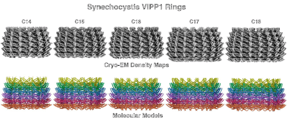 |
Structural basis for VIPP1 oligomerization and maintenance of thylakoid membrane integrity [8120 multi-frame micrographs composed of 80 frames each in TIFF format] | Gupta TK, Klumpe S, Gries K, Strauss M, Rudack T, Schuller JM, Schroda M, Engel BD [Pubmed: 34166613] [DOI: 10.1016/j.cell.2021.05.011] |
2.0 TB | 3.8 Å |
