Electron Microscopy Public Image Archive
Electron Microscopy Public Image Archive
The EMPIAR-PDBj team at Osaka University assists Asian EM researchers with the transfer of big EM image data to EMPIAR. Instead of sending the data directly to the EBI (UK) via the internet, hard drives can also be sent to Osaka University by postal mail or via a courier service. As an alternative, internet transfer to our server in Osaka is also available. If you would like to take advantage of our submission services, please contact us first by e-mail before sending the data to us.
| Release date | Imageset | Title | Authors and references | Size | Resolution |
|---|---|---|---|---|---|
| 2022-01-28 |  |
CryoET of presequence protease single particle [multiple data sets in TIFF and MRC formats] | Liang WG, Wijaya J, Wei H, Noble AJ, Mancl JM, Mo S, Lee D, Lin King JV, Pan M, Liu C, Koehler CM, Zhao M, Potter CS, Carragher B, Li S, Tang WJ [Pubmed: 35383169] [DOI: 10.1038/s41467-022-29322-4] |
25.4 GB | — |
| 2022-03-07 | 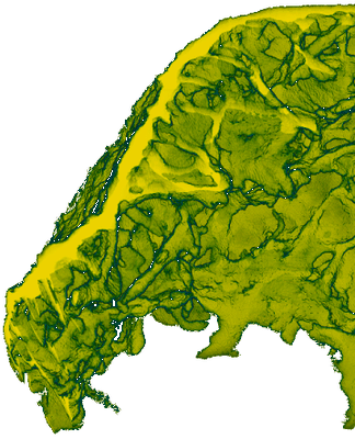 |
A biological nano-foam: the wall of coniferous bisaccate pollen (data sets) [multiple data sets in TIFF and JPEG formats] | Cojocaru R, Mannix O, Capron M, Miller CG, Jouneau PH, Gallet B, Falconet D, Pacureanu A, Stukins S [Pubmed: 35138906] [DOI: 10.1126/sciadv.abd0892] |
25.2 GB | — |
| 2021-02-12 | 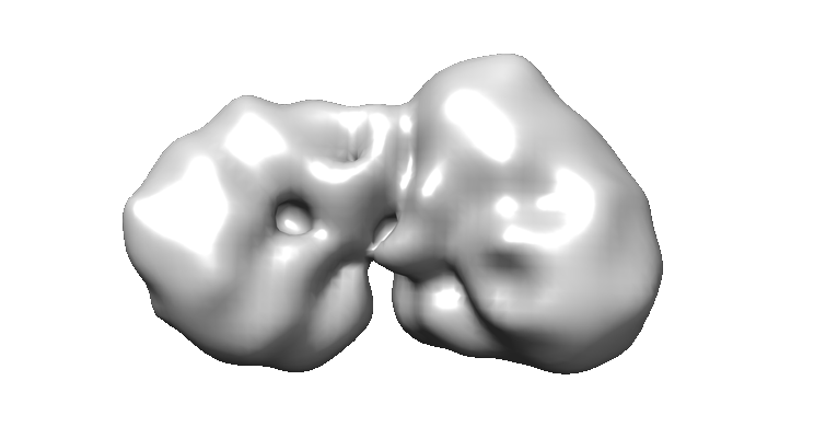 |
Negative-stained transmission electron microscopy of the nucleosome and deacetylase complex purified from murine erythroleukemia cells. [400 micrographs in MRC format] | Mackay JP, Landsberg MJ, Jackman MJ [Pubmed: 33264611] [DOI: 10.1016/j.celrep.2020.108450] |
25.0 GB | 14.0 Å |
| 2021-11-09 | 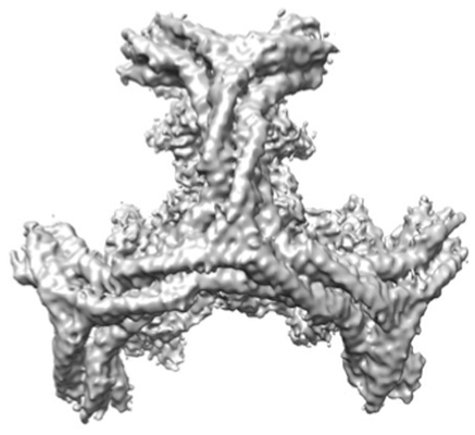 |
Multi-modal adaptor-clathrin contacts drive coated vesicle assembly [stack of 54488 particles in MRCS format] | Smith SM, Larocque G, Wood KM, Morris KL, Roseman AM, Sessions RB, Royle SJ, Smith CJ [Pubmed: 34487371] [DOI: 10.15252/embj.2021108795] |
25.0 GB | 10.5 Å |
| 2023-01-30 | 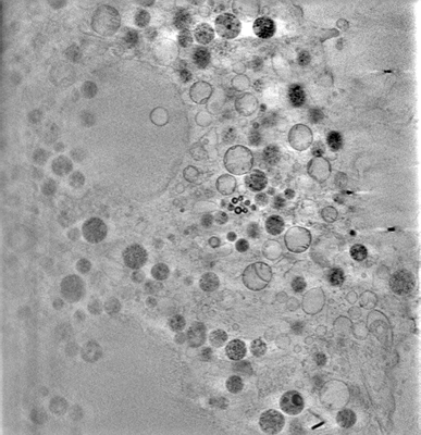 |
3D-surface reconstruction of cellular cryo-soft X-ray microscopy tomograms using semi-supervised deep learning [7 reconstructed volumes in MRC format] | Dyhr MCA, Sadeghi M, Moynova R, Knappe C, Kepsutlu Çakmak B, Werner S, Schneider G, McNally J, Noe F, Ewers H [DOI: 10.1101/2022.05.16.492055] |
23.5 GB | — |
| 2020-03-23 | 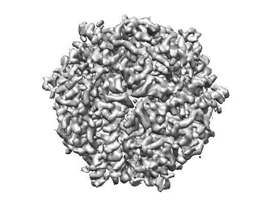 |
Micrographs of DPS collected at 100 keV using a hybrid pixel direct electron detector [739 multi-frame micrographs composed of 32 frames each in MRCS format] | Naydenova K, McMullan G, Peet MJ, Lee Y, Edwards PC, Chen S, Leahy E, Scotcher S, Henderson R, Russo CJ [Pubmed: 31709064] [DOI: 10.1107/S2052252519012612] |
23.3 GB | 3.4 Å |
| 2017-12-18 | 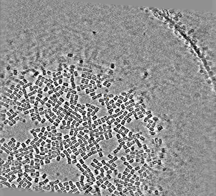 |
Phase plate cryoET of T20S proteasome single particle [multiple data sets in MRC format] | Noble AJ, Dandey VP, Wei H, Brasch J, Chase J, Acharya P, Tan YZ, Zhang Z, Kim LY, Scapin G, Rapp M, Eng ET, Rice MJ, Cheng A, Negro CJ, Shapiro L, Kwong PD, Jeruzalmi D, des Georges A, Potter CS, Carragher B [Pubmed: 29809143] [DOI: 10.7554/eLife.34257] |
23.3 GB | — |
| 2014-09-09 | 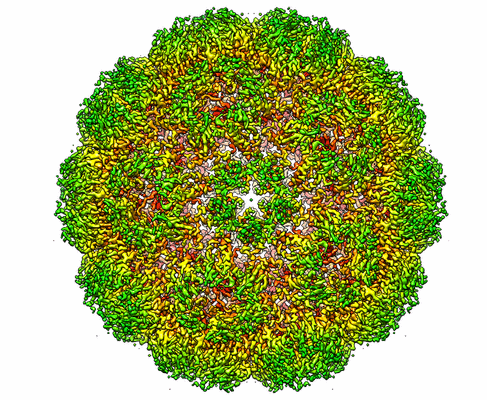 |
Full virus map of Brome Mosaic Virus (picked particles) [stack of 35142 particles in IMAGIC format] | Wang Z, Hryc C, Bammes B, Afonine P, Jakana J, Chen DH, Liu XA, Baker M, Kao C, Ludtke S, Schmid M, Adams P, Chiu W [Pubmed: 25185801] [DOI: 10.1038/ncomms5808] |
23.1 GB | 3.8 Å |
| 2017-12-18 | 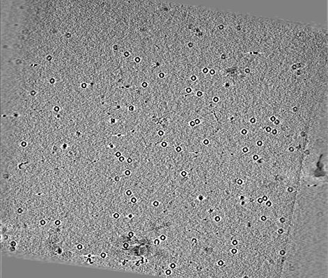 |
CryoET of apoferritin single particle [multiple data sets in MRC format] | Noble AJ, Dandey VP, Wei H, Brasch J, Chase J, Acharya P, Tan YZ, Zhang Z, Kim LY, Scapin G, Rapp M, Eng ET, Rice MJ, Cheng A, Negro CJ, Shapiro L, Kwong PD, Jeruzalmi D, des Georges A, Potter CS, Carragher B [Pubmed: 29809143] [DOI: 10.7554/eLife.34257] |
23.0 GB | — |
| 2022-12-13 | 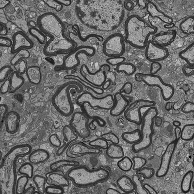 |
Focused ion beam-scanning electron microscopy links pathological myelin outfoldings to axonal changes in mice lacking Plp1 or Mag [1290 micrographs in TIFF format] | Steyer AM, Möbius W [Pubmed: 36354016] [DOI: 10.1002/glia.24290] |
22.4 GB | — |
| 2022-12-13 | 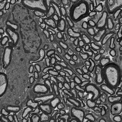 |
Focused ion beam-scanning electron microscopy links pathological myelin outfoldings to axonal changes in mice lacking Plp1 or Mag [1553 micrographs in TIFF format] | Steyer AM, Möbius W [Pubmed: 36354016] [DOI: 10.1002/glia.24290] |
22.3 GB | — |
| 2019-09-27 | 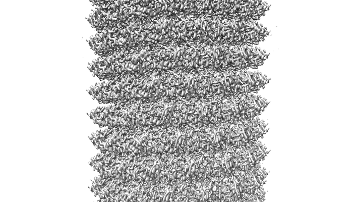 |
Cryo-EM structure of TMV with Ca2+ at low pH [197 multi-frame micrographs composed of 40 frames each in TIFF format] | Weis F, Beckers M, von der Hocht I, Sachse C [Pubmed: 31535454] [DOI: 10.15252/embr.201948451] |
22.0 GB | 2.0 Å |
| 2018-04-05 | 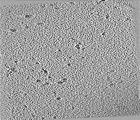 |
CryoET of hemagglutinin single particle with spot-to-plunge time of 100ms [multiple data sets in MRC format] | Noble AJ, Wei H, Dandey VP, Zhang Z, Potter CS, Carragher B [Pubmed: 30250056] [DOI: 10.1038/s41592-018-0139-3] |
21.9 GB | — |
| 2019-08-28 | 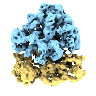 |
Improved applicability and robustness of fast cryo-electron tomography data acquisition [12 tilt series in MRC format] | Eisenstein F, Danev R, Pilhofer M [Pubmed: 31425790] [DOI: 10.1016/j.jsb.2019.08.006] |
21.6 GB | 9.0 Å |
| 2022-01-12 | 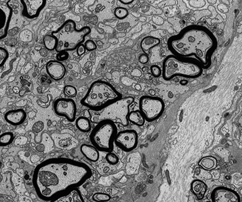 |
FIB-SEM of mouse optic nerve of an inducible conditional Mbp knock-out 26 weeks after induction [747 micrographs in TIFF format] | Meschkat M, Steyer AM, Ruhwedel T, Möbius W [DOI: 10.1101/2020.09.02.279612] |
21.5 GB | — |
| 2020-11-18 | 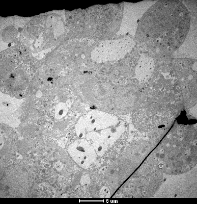 |
SARS-CoV-2 infection in human adult lung alveolar stem cells [multiple data sets in TIFF format] | Youk J, Kim T, Evans KV, Jeong YI, Hur Y, Hong SP, Kim JH, Yi K, Kim SY, Na KJ, Bleazard T, Kim HM, Fellows M, Mahbubani KT, Saeb-Parsy K, Kim SY, Kim YT, Koh GY, Choi BS, Ju YS, Lee JH [Pubmed: 33142113] [DOI: 10.1016/j.stem.2020.10.004] |
20.8 GB | — |
| 2021-10-01 | 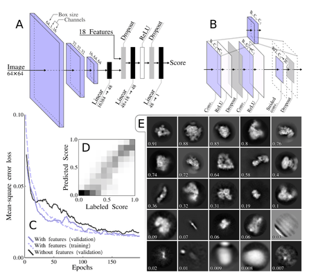 |
Training data set for automated 2D class selection [18051 class averages in MRCS format] | Kimanius D, Dong L, Sharov G, Nakane T, Scheres SHW [Pubmed: 34783343] [DOI: 10.1042/bcj20210708] |
20.7 GB | — |
| 2021-02-12 |  |
Negative-stained transmission electron microscopy of the MTA-HDAC-MBD core complex purified from EXPI cells [325 micrographs in MRC format] | Mackay JP, Landsberg MJ, Jackman MJ [Pubmed: 33264611] [DOI: 10.1016/j.celrep.2020.108450] |
20.3 GB | 29.0 Å |
| 2024-02-19 | 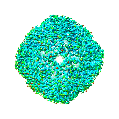 |
Graphene-sandwiched apoferritin [55 multi-frame micrographs composed of 32 frames each in TIFF format] | Liu N, Xu J, Wang HW [Pubmed: 38252835] [DOI: 10.1073/pnas.2309384121] |
20.2 GB | 3.1 Å |
| 2019-10-04 | 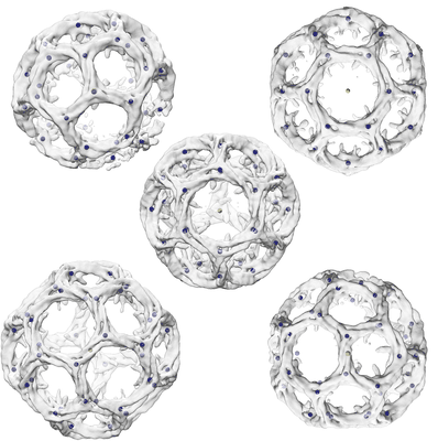 |
Single particle cryo-EM dataset of clathrin cages suitable for subparticle extraction [multiple data sets in MRCS format] | Morris KL, Jones JR, Halebian M, Wu S, Baker M, Armache JP, Avila Ibarra A, Sessions RB, Cameron AD, Cheng Y, Smith CJ [Pubmed: 31582853] [DOI: 10.1038/s41594-019-0292-0] |
19.9 GB | 9.07 - 23.68 Å |
| 2022-12-19 | 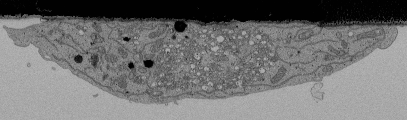 |
CLEMSite, a software for automated phenotypic screens using light microscopy and FIB-SEM. [multiple data sets in TIFF format] | Lleti JMSL, Steyer AMS, Schwab YS | 19.7 GB | — |
| 2018-10-23 | 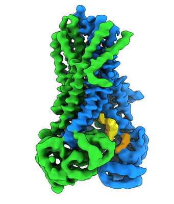 |
Cryo-electron microscopy data of thermostabilized avian CFTR [stack of 2 particles in MRCS format] | Fay JF [Pubmed: 30281975] [DOI: 10.1021/acs.biochem.8b00763] |
19.3 GB | 4.3 - 6.6 Å |
| 2023-02-28 | 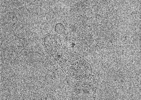 |
Cryo-ET tilt series of HPIV3 clinical isolates in the presence of an F neutralizing Fab [6 tilt series in MRC format] | Marcink T, Cheng W, Porotto M, des Georges A, Moscona A [Pubmed: 36763666] [DOI: 10.1126/sciadv.ade2727] |
19.1 GB | 16.5 Å |
| 2022-01-12 | 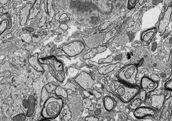 |
FIB-SEM of an Mbp-deficient shiverer mouse optic nerve [857 micrographs in TIFF format] | Meschkat M, Steyer AM, Ruhwedel T, Möbius W [Pubmed: 35246535] [DOI: 10.1038/s41467-022-28720-y] |
18.8 GB | — |
| 2023-02-28 | 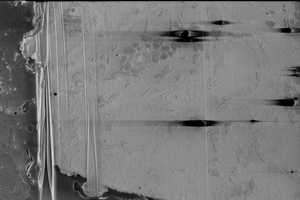 |
Cryo serial FIB/SEM of mouse heart tissue [136 micrographs in TIFF format] | Dumoux M, Glen T, Smith JLR, Ho EML, Perdigão LMA, Pennington A, Klumpe S, Yee NBY, Farmer DA, Lai PYA, Bowles W, Kelley R, Plitzko JM, Wu L, Basham M, Clare DK, Siebert CA, Darrow MC, Naismith JH, Grange M [Pubmed: 36805107] [DOI: 10.7554/elife.83623] |
18.0 GB | — |
