Electron Microscopy Public Image Archive
Electron Microscopy Public Image Archive
The EMPIAR-PDBj team at Osaka University assists Asian EM researchers with the transfer of big EM image data to EMPIAR. Instead of sending the data directly to the EBI (UK) via the internet, hard drives can also be sent to Osaka University by postal mail or via a courier service. As an alternative, internet transfer to our server in Osaka is also available. If you would like to take advantage of our submission services, please contact us first by e-mail before sending the data to us.
| Release date | Imageset | Title | Authors and references | Size | Resolution |
|---|---|---|---|---|---|
| 2018-01-22 | 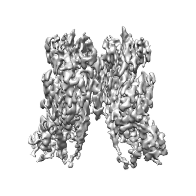 |
Cryo-EM structure of the TMEM16A in LMNG [multiple data sets in MRCS format] | Dang S, Cheng Y [Pubmed: 29236684] [DOI: 10.1038/nature25024] |
244.2 GB | 3.8 Å |
| 2018-01-22 | 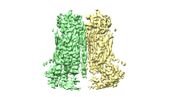 |
Cryo-EM structure of the TMEM16A in Nanodisc [stack of 3149 particles in MRCS format] | Dang S, Cheng Y [Pubmed: 29236684] [DOI: 10.1038/nature25024] |
226.7 GB | 3.8 Å |
| 2017-10-23 | 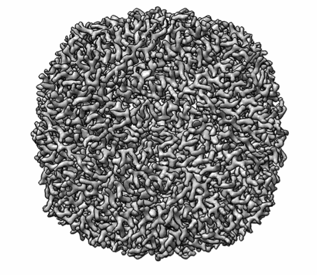 |
Mixed under-/over-focused data collected by VPP-Cs-corrector coupled EM. [1 micrographs in MRC format] | Fan X, Wang HW [Pubmed: 28943337] [DOI: 10.1016/j.str.2017.08.008] |
34.4 GB | 3.0 Å |
| 2018-02-26 | 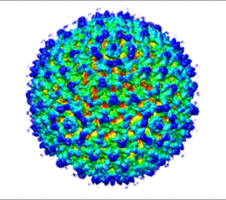 |
Cryo-EM reconstruction of a modified human Adenovirus C5 [231 multi-frame micrographs composed of 50 frames each in MRC format] | Schmid M, Ernst P, Honegger A, Suomalainen M, Zimmermann M, Braun L, Stauffer S, Thom C, Dreier B, Eibauer M, Kipar A, Vogel V, Greber UF, Medalia O, Plückthun A [Pubmed: 29386504] [DOI: 10.1038/s41467-017-02707-6] |
612.7 GB | 7.4 Å |
| 2018-02-07 | 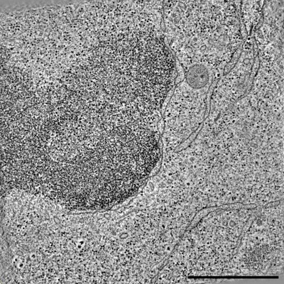 |
Raw 2d tomographic tilt series of a dividing cell [65 tilt series in ST format] | Otsuka S [Pubmed: 29323269] [DOI: 10.1038/s41594-017-0001-9] |
237.8 GB | — |
| 2018-01-24 | 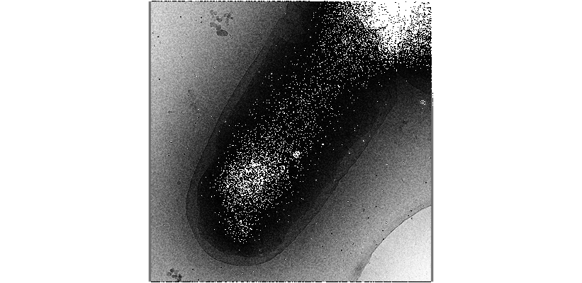 |
Tilt-series of e. coli carrying the ple7 plasmid carrying YFP-MreB hyper-overexpressed by induction with 1uM IPTG [5 tilt series in MRC format] | Swulius MT, Jensen GJ [Pubmed: 22904287] [DOI: 10.1128/JB.00505-12] |
5.0 GB | — |
| 2018-01-24 | 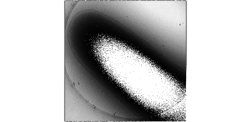 |
Tilt-series of e. coli carrying the ple7 plasmid carrying YFP-MreB induced with 20 uM IPTG [7 tilt series in MRC format] | Swulius MT, Jensen GJ [Pubmed: 22904287] [DOI: 10.1128/JB.00505-12] |
6.6 GB | — |
| 2018-01-24 | 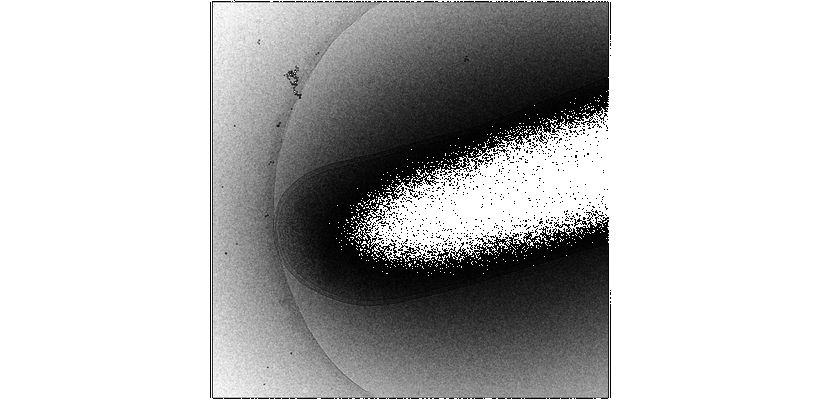 |
Tilt-series of e. coli carrying empty ple6 plasmid induced with 20 uM IPTG [4 tilt series in MRC format] | Swulius MT, Jensen GJ [Pubmed: 22904287] [DOI: 10.1128/JB.00505-12] |
3.8 GB | — |
| 2018-02-08 |  |
Tilt-series of salmonella enterica wild-type bacterial flagellar motor [1 tilt series in MRC format] | Beeby M, Ribardo DA, Brennan CA, Ruby EG, Jensen GJ, Hendrixson DR [Pubmed: 26976588] [DOI: 10.1073/pnas.1518952113] |
328.0 MB | 69.4 Å |
| 2017-07-14 | 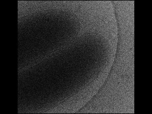 |
Tilt-series for the electron cryotomogram of Vibrio cholerae O395 N1 [16 tilt series in MRC format] | Chang YW, Kjaer A, Ortega DR, Kovacikova G, Sutherland JA, Rettberg LA, Taylor RK, Jensen GJ [Pubmed: 28165453] [DOI: 10.1038/nmicrobiol.2016.269] |
51.3 GB | 50.0 Å |
| 2017-11-30 | 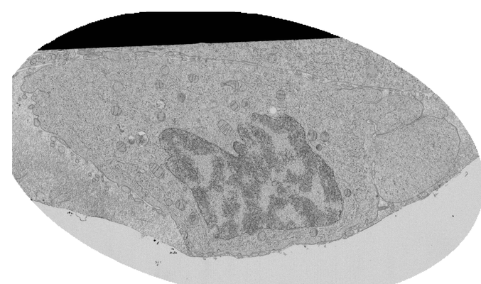 |
FIB-SEM of a dividing cell at 11.2 min after anaphase onset [777 multi-frame micrographs composed of 1 frames each in TIFF format] | Otsuka S, Steyer AM, Schorb M, Heriche JK, Hossain MJ, Sethi S, Kueblbeck M, Schwab Y, Beck M, Ellenberg J [DOI: 10.1038/s41594-017-0001-9] |
11.0 GB | — |
| 2017-06-01 | 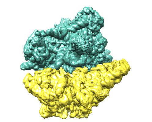 |
The Structure of the Yeast Mitochondrial Ribosome [stack of 2524 particles in MRCS format] | Desai N, Brown A, Amunts A, Ramakrishnan V [Pubmed: 28154081] [DOI: 10.1126/science.aal2415] |
138.6 GB | 3.2 - 4.97 Å |
| 2019-05-21 | 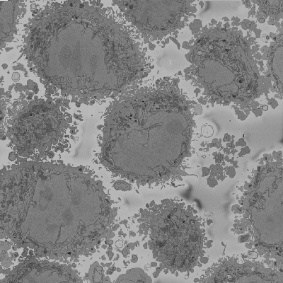 |
Serial Block Face SEM of HeLa cell pellet with 5 nm pixels and 50 nm slices (benchmark dataset) [518 micrographs in DM4 format] | Peddie CP, Jones ML, Collinson LM | 129.8 GB | — |
| 2017-11-28 | 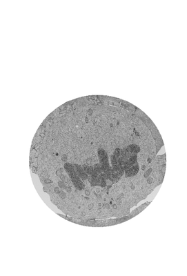 |
FIB-SEM of a dividing cell at 5.7 min after anaphase onset [2620 multi-frame micrographs composed of 1 frames each in TIFF format] | Otsuka S, Steyer AM, Schorb M, Hériché JK, Hossain MJ, Sethi S, Kueblbeck M, Schwab Y, Beck M, Ellenberg J [Pubmed: 29323269] [DOI: 10.1038/s41594-017-0001-9] |
63.0 GB | — |
| 2017-11-30 | 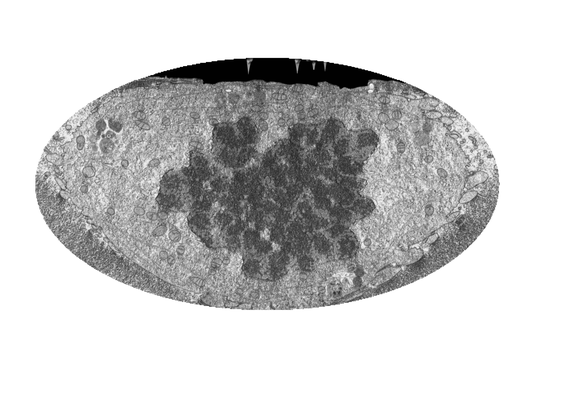 |
FIB-SEM of a dividing cell at 5.3 min after anaphase onset [1998 multi-frame micrographs composed of 1 frames each in TIFF format] | Otsuka S, Steyer AM, Schorb M, Hériché JK, Hossain MJ, Sethi S, Kueblbeck M, Schwab Y, Beck M, Ellenberg J [Pubmed: 29323269] [DOI: 10.1038/s41594-017-0001-9] |
31.2 GB | — |
| 2017-11-30 | 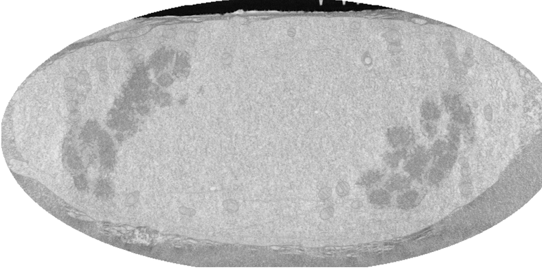 |
FIB-SEM of a dividing cell at 4.3 min after anaphase onset [1358 multi-frame micrographs composed of 1 frames each in TIFF format] | Otsuka S, Steyer AM, Schorb M, Hériché JK, Hossain MJ, Sethi S, Kueblbeck M, Schwab Y, Beck M, Ellenberg J [Pubmed: 29323269] [DOI: 10.1038/s41594-017-0001-9] |
14.0 GB | — |
| 2017-11-28 | 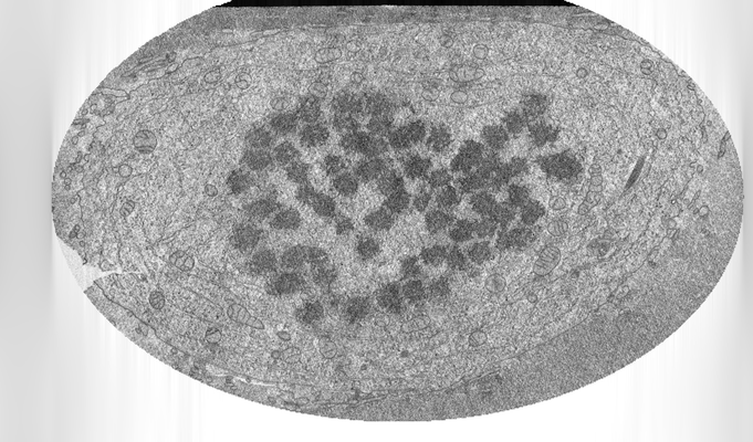 |
FIB-SEM of a dividing cell at 3.9 min after anaphase onset [2293 multi-frame micrographs composed of 1 frames each in TIFF format] | Otsuka S, Steyer AM, Schorb M, Hériché JK, Hossain MJ, Sethi S, Kueblbeck M, Schwab Y, Beck M, Ellenberg J [Pubmed: 29323269] [DOI: 10.1038/s41594-017-0001-9] |
26.8 GB | — |
| 2017-11-30 | 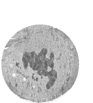 |
FIB-SEM of a dividing cell at 6.3 min after anaphase onset [3206 multi-frame micrographs composed of 1 frames each in TIFF format] | Otsuka S, Steyer AM, Schorb M, Hériché JK, Hossain MJ, Sethi S, Kueblbeck M, Schwab Y, Beck M, Ellenberg J [Pubmed: 29323269] [DOI: 10.1038/s41594-017-0001-9] |
83.2 GB | — |
| 2017-11-28 | 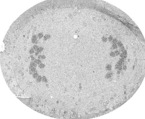 |
FIB-SEM of a dividing cell at 3.1 min after anaphase onset [1652 multi-frame micrographs composed of 1 frames each in TIFF format] | Otsuka S, Steyer AM, Schorb M, Hériché JK, Hossain MJ, Sethi S, Kueblbeck M, Schwab Y, Beck M, Ellenberg J [Pubmed: 29323269] [DOI: 10.1038/s41594-017-0001-9] |
27.0 GB | — |
| 2017-08-29 | 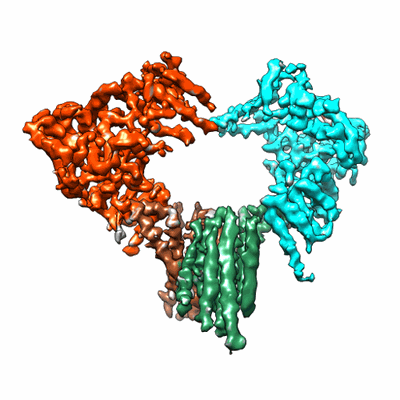 |
Cryo-EM structure of Hrd1 and Hrd3 complex [multiple data sets in MRC format] | Mi WM, Schoebel SS, Stein AS, Rapoport TAR, Liao ML [Pubmed: 28682307] [DOI: 10.1038/nature23314] |
708.3 GB | 4.7 Å |
| 2018-05-30 |  |
Cryo-ET of natural chromatin from Ostreococcus tauri and Saccharyomyces cerevisiae [25 class averages in MRC format] | Cai S, Song Y, Chen C, Shi J [Pubmed: 29742050] [DOI: 10.1091/mbc.E17-07-0449] |
40.1 GB | — |
| 2017-08-16 | 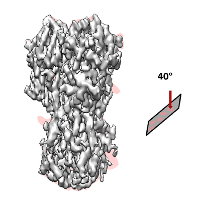 |
40 Degree Tilted Single-Particle CryoEM of Highly Preferred Orientated Influenza Hemagglutinin Trimer [multiple data sets in MRC format] | Tan YZ, Lyumkis D [Pubmed: 28671674] [DOI: 10.1038/nmeth.4347] |
1.8 TB | 4.2 Å |
| 2017-05-15 | 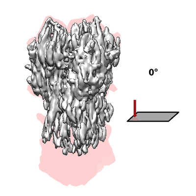 |
Untilted Single-Particle CryoEM of Highly Preferred Orientated Influenza Hemagglutinin Trimer [multiple data sets in MRC format] | Tan YZ, Lyumkis D [Pubmed: 28671674] [DOI: 10.1038/nmeth.4347] |
1.2 TB | 4.2 Å |
| 2018-01-03 |  |
Structure of the Z-disk isolated from the indirect flight muscle of the honey bee [96 class averages in MRC format] | Rusu M, Hu Z, Taylor KA, Trinick J [Pubmed: 28733815] [DOI: 10.1007/s10974-017-9477-5] |
3.0 GB | 60.0 Å |
| 2019-05-21 | 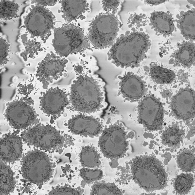 |
Serial Block Face SEM of HeLa cell pellet with 10 nm pixels and 50 nm slices (benchmark dataset) [518 micrographs in DM4 format] | Peddie CP, Jones ML, Collinson LM | 129.8 GB | — |
