EMPIAR-10109
FIB-SEM of a dividing cell at 11.2 min after anaphase onset [777 multi-frame micrographs composed of 1 frames each in TIFF format]
Publication:

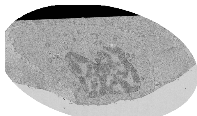
Postmitotic nuclear pore assembly proceeds by radial dilation of small ER membrane openings

Otsuka S, Steyer AM, Schorb M, Heriche JK, Hossain MJ, Sethi S, Kueblbeck M, Schwab Y, Beck M, Ellenberg J

Nature Structural & Molecular Biology (2017)

Related EMDB entry:
Deposited:
2017-06-17
Released:
2017-11-30
Last modified:
2017-11-30
Imageset size:
10.96 GB
Imageset DOI:
Experimental metadata:
Download xml json
Contains:
  • micrographs - multiframe
1. FIB-SEM images of a dividing cell at 11.2 min after anaphase onset
Category:
micrographs - multiframe
Image format:
TIFF
No. of images or tilt series:
777
Image size:
(5064, 2992)
Pixel type:
UNSIGNED BYTE
Pixel spacing:
(5.0, 8.0)
Details:
The pixel size in xy was set to be 5 nm and the FIB-milling was done every 8 nm. The acquired raw FIB-SEM images were aligned using TrackEM2 (Cardona et al., PloS one, e38011, 2012) in ImageJ (http://rsbweb.nih.gov/ij/). After alignment, the stack was cropped, gray levels were inverted and the images were smoothened with a mean filter (kernel size: 15 × 15 nm). A small part of the nucleus is missing.
Files:
Loading...
Files:
Loading...

Ito F, Alvarez-Cabrera AL, Liu S, Yang H, Shiriaeva A, Zhou ZH, Chen XS. (2023)
Rigden DJ, Fernández XM. (2023)
Iudin A, Korir PK, Somasundharam S, Weyand S, Cattavitello C, Fonseca N, Salih O, Kleywegt GJ, Patwardhan A. (2023)
Serra Lleti JM, Steyer AM, Schieber NL, Neumann B, Tischer C, Hilsenstein V, Holtstrom M, Unrau D, Kirmse R, Lucocq JM, Pepperkok R, Schwab Y. (2023)
Caldwell BJ, Norris AS, Karbowski CF, Wiegand AM, Wysocki VH, Bell CE. (2022)