Electron Microscopy Public Image Archive
Electron Microscopy Public Image Archive
Our EMPIAR-PDBj will suspend the following operations from August 10, 2024 (Saturday) to August 18, 2024 (Sunday):
Web servers and data downloads will remain available during this period.
Additionally, please be aware that EMPIAR-EBI will also be closed for annotation operations from July 25, 2024 (Thursday) to August 18, 2024 (Sunday).
Thank you.
The EMPIAR-PDBj team at Osaka University assists Asian EM researchers with the transfer of big EM image data to EMPIAR. Instead of sending the data directly to the EBI (UK) via the internet, hard drives can also be sent to Osaka University by postal mail or via a courier service. As an alternative, internet transfer to our server in Osaka is also available. If you would like to take advantage of our submission services, please contact us first by e-mail before sending the data to us.
| Release date | Imageset | Title | Authors and references | Size | Resolution |
|---|---|---|---|---|---|
| 2024-07-18 |  |
Rhodobacter microvirus Ebor attached to B10 host cell, single particle data [2750 multi-frame micrographs composed of 25 frames each in TIFF format] | Bardy P, MacDonald CIW, Kirchberger PC, Jenkins HT, Botka T, Byrom L, Alim NTB, Traore DAK, Konig HC, Nicholas TR, Chechik M, Hart SJ, Turkenburg JP, Blaza JN, Beatty JT, Fogg PCM, Antson AA [Pubmed: 38915634] [DOI: 10.1101/2024.06.11.598214] |
498.5 GB | 7.84 Å |
| 2024-07-09 |  |
Empty particles of Rhodobacter microvirus Ebor [1099 multi-frame micrographs composed of 2639 frames each in EER format] | Bardy P, MacDonald CIW, Kirchberger PC, Jenkins HT, Botka T, Byrom L, Alim NTB, Traore DAK, Konig HC, Nicholas TR, Chechik M, Hart SJ, Turkenburg JP, Blaza JN, Beatty JT, Fogg PCM, Antson AA [DOI: 10.1101/2024.06.11.598214] |
1.6 TB | 3.3 Å |
| 2024-07-16 |  |
In vitro-induced genome-releasing intermediate of Rhodobacter microvirus Ebor computed with C5 symmetry [2810 multi-frame micrographs composed of 2765 frames each in EER format] | Bardy P, MacDonald CIW, Kirchberger PC, Jenkins HT, Botka T, Byrom L, Alim NTB, Traore DAK, Konig HC, Nicholas TR, Chechik M, Hart SJ, Turkenburg JP, Blaza JN, Beatty JT, Fogg PCM, Antson AA [Pubmed: 38915634] [DOI: 10.1101/2024.06.11.598214] |
4.3 TB | 25.0 Å |
| 2024-07-09 |  |
Native capsid of Rhodobacter microvirus Ebor computed with I4 symmetry [954 multi-frame micrographs composed of 2870 frames each in EER format] | Bardy P, MacDonald CIW, Kirchberger PC, Jenkins HT, Botka T, Byrom L, Alim NTB, Traore DAK, Konig HC, Nicholas TR, Chechik M, Hart SJ, Turkenburg JP, Blaza JN, Beatty JT, Fogg PCM, Antson AA [DOI: 10.1101/2024.06.11.598214] |
1.5 TB | 3.2 Å |
| 2024-07-08 |  |
Structure of mavacamten-free human cardiac thick filaments within the sarcomere by cryoelectron tomography [multiple data sets in MRC format] | Chen L, Liu J, Rastegarpouyani H, Janssen PML, Pinto JR, Taylor KA [Pubmed: 38386705] [DOI: 10.1073/pnas.2311883121] |
25.6 GB | 16.5 - 26.4 Å |
| 2024-06-27 |  |
Cryo-EM reveals a nearly complete PCNA loading process and unique features of the human alternative clamp loader CTF18-RFC [18776 multi-frame micrographs composed of 50 frames each in TIFF format] | Wang FW [Pubmed: 38669181] [DOI: 10.1073/pnas.2319727121] |
7.9 TB | 2.75 - 3.35 Å |
| 2024-07-16 |  |
Cryo-EM of Caulobacter crescentus FljM flagellar filament [9780 multi-frame micrographs composed of 45 frames each in TIFF format] | Sanchez JC, Montemayor EJ, Ploscariu NT, Parrell D, Baumgardt JK, Yang JE, Sibert B, Cai K, Wright ER [Pubmed: 37503001] [DOI: 10.1101/2023.07.10.548443] |
1.9 TB | 2.11 Å |
| 2024-07-19 |  |
Particle stacks from heart-derived AL amyloid (AL59) cryo-EM data [multiple data sets in MRCS format] | Schulte T, Speranzini V, Chaves-Sanjuan A, Milazzo M, Ricagno S | 130.1 GB | 3.6 Å |
| 2024-06-13 |  |
Cryo-EM structure of guide RNA and target RNA bound Cas7-11 [4177 multi-frame micrographs composed of 60 frames each in TIFF format] | Goswami HN, Rai J, Das A, Li H [Pubmed: 36190192] [DOI: 10.7554/eLife.81678] |
2.0 TB | 2.82 Å |
| 2024-06-13 |  |
Cryo electron microscopy unaligned movie frames for single particle analysis of the Escherichia coli 70S ribosome with Myxovalargin A [8208 multi-frame micrographs composed of 40 frames each in TIFF format] | Koller TO, Wilson DN [Pubmed: 36603206] [DOI: 10.1021/jacs.2c08816] |
946.3 GB | 2.1 - 3.0 Å |
| 2024-06-13 |  |
Cryo-EM micrographs of human RYBP-PRC1 bound to unmodified mononucleosome [14852 micrographs in MRC format] | Ciapponi MC, Karlukova EK, Schkölziger SS, Benda CB, Müller JM [Pubmed: 38528151] [DOI: 10.1038/s41594-024-01258-x] |
1.3 TB | 2.91 Å |
| 2024-07-08 |  |
Particle stack from SARS-CoV-2 (B.1.1.529/Omicron variant) Spike protein in complex with the single chain fragment scFv76-77 single particle cryo-EM data [stack of 3471 particles in MRCS format] | Chaves-Sanjuan A, Berlinguer M, Bolognesi M | 366.7 GB | 3.7 - 4.3 Å |
| 2024-07-08 | 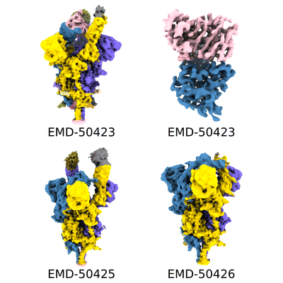 |
Particle stack from SARS-CoV-2 (B.1.1.529/Omicron variant) Spike protein in complex with the single chain fragment scFv76 single particle cryo-EM data [stack of 6020 particles in MRCS format] | Chaves-Sanjuan A, Berlinguer M, Bolognesi M | 758.7 GB | 3.0 - 3.8 Å |
| 2024-07-16 | 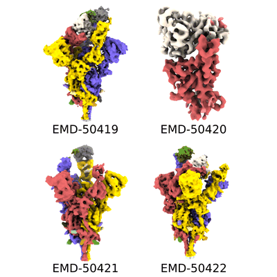 |
Particle stack from SARS-CoV-2 (wuhan variant) Spike protein in complex with the single chain fragment scFv41N single particle cryo-EM data [stack of 4991 particles in MRCS format] | Chaves-Sanjuan A, Berlinguer M, Bolognesi M | 712.4 GB | 3.8 - 4.0 Å |
| 2024-06-13 | 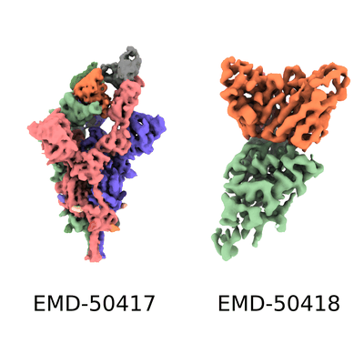 |
Particle stack from SARS-CoV-2 (wuhan variant) Spike protein in complex with the single chain fragment scFv76-77 single particle cryo-EM data [stack of 3774 particles in MRCS format] | Chaves-Sanjuan A, Berlinguer M, Bolognesi M | 304.7 GB | 3.9 - 4.0 Å |
| 2024-07-16 |  |
In situ cryoET of beta-amyloid and tau in post-mortem Alzheimer's disease brain [61 reconstructed volumes in MRC format] | Jenkins J [Pubmed: 38987603] [DOI: 10.1038/s41586-024-07680-x] |
140.6 GB | 8.7 - 33.0 Å |
| 2024-07-16 |  |
In situ cryoET of beta-amyloid and tau in post-mortem Alzheimer's disease brain [61 reconstructed volumes in MRC format] | Jenkins J [Pubmed: 38987603] [DOI: 10.1038/s41586-024-07680-x] |
167.0 GB | 8.7 - 33.0 Å |
| 2024-07-16 |  |
In situ cryoET of beta-amyloid and tau in post-mortem Alzheimer's disease brain [61 reconstructed volumes in MRC format] | Jenkins J [Pubmed: 38987603] [DOI: 10.1038/s41586-024-07680-x] |
127.0 GB | 8.7 - 33.0 Å |
| 2024-06-11 | 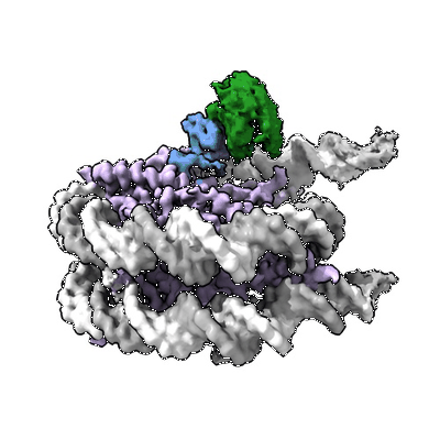 |
Cryo-EM micrographs of human RYBP-PRC1 bound to H2Aub1 mononucleosome [10669 micrographs in MRC format] | Ciapponi MC, Karlukova EK, Schkölziger SS, Benda CB, Müller JM [Pubmed: 38528151] [DOI: 10.1038/s41594-024-01258-x] |
935.6 GB | 3.18 Å |
| 2024-07-16 |  |
In situ cryoET of beta-amyloid and tau in post-mortem Alzheimer's disease brain [61 tilt series in MRC format] | Jenkins J [Pubmed: 38987603] [DOI: 10.1038/s41586-024-07680-x] |
149.4 GB | 8.7 - 33.0 Å |
| 2024-07-16 | 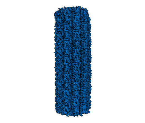 |
Cryo-EM of Caulobacter crescentus FljK flagellar filament [8813 multi-frame micrographs composed of 55 frames each in TIFF format] | Sanchez JC, Montemayor EJ, Ploscariu NT, Parrell D, Baumgardt JK, Yang JE, Sibert B, Cai K, Wright ER [Pubmed: 37503001] [DOI: 10.1101/2023.07.10.548443] |
2.0 TB | 2.71 Å |
| 2024-05-16 |  |
The cryo-electron microscopy structure of a Vitiosangium bacterial gasdermin in an slinky-like oligomeric conformation [8156 multi-frame micrographs composed of 50 frames each in TIFF format] | Johnson AG, Mayer ML, Schaefer SL, McNamara-Bordewick NK, Hummer G, Kranzusch PJ [Pubmed: 38509367] [DOI: 10.1038/s41586-024-07216-3] |
2.1 TB | 3.3 Å |
| 2024-07-16 | 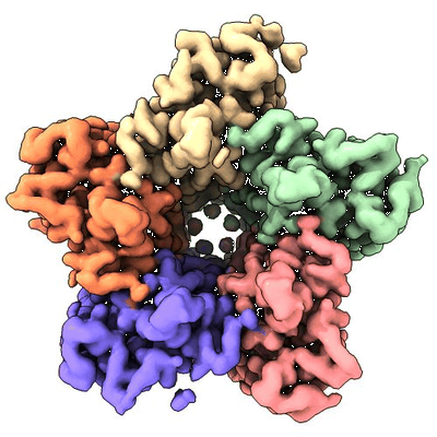 |
Cryo electron micrographs of retinal-free Proteoopsin bound to decanoate [20910 multi-frame micrographs composed of 40 frames each in TIFF format] | Hirschi S, Lemmin T, Fotiadis D | 3.0 TB | 2.97 Å |
| 2024-06-16 | 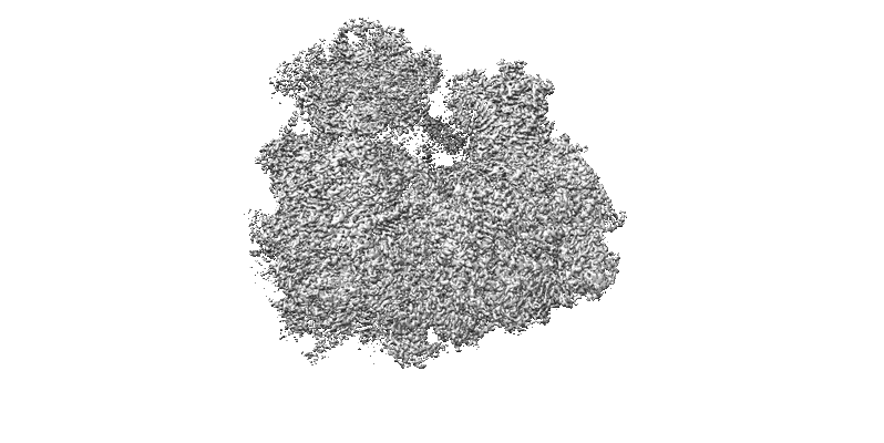 |
Hypopseudouridylated yeast 80S bound with Taura syndrome virus (TSV) internal ribosome entry site (IRES) [ three datasets, in TIF format] [multiple data sets in TIFF format] | Zhao Y, Rai J, Li H [Pubmed: 37595043] [DOI: 10.1126/sciadv.adg8190] |
16.0 TB | 2.2 - 2.87 Å |
| 2024-06-12 | 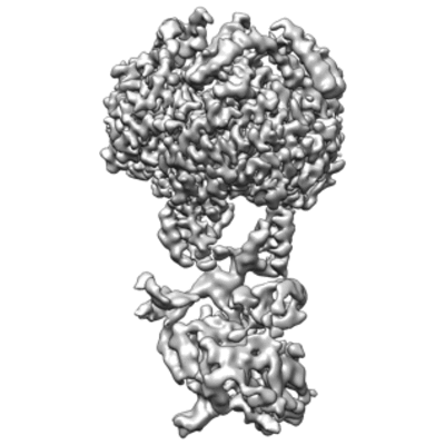 |
Single-particle cryo-EM structures of PNPase in complex with the RNA chaperone Hfq and regulatory RNA [6271 multi-frame micrographs composed of 38 frames each in TIFF format] | Dendooven T, Sinha D, Roeselová A, Cameron TA, De Lay NR, Luisi BF, Bandyra KJ [Pubmed: 34157309] [DOI: 10.1016/j.molcel.2021.05.032] |
6.8 TB | 3.7 Å |
