Electron Microscopy Public Image Archive
Electron Microscopy Public Image Archive
The EMPIAR-PDBj team at Osaka University assists Asian EM researchers with the transfer of big EM image data to EMPIAR. Instead of sending the data directly to the EBI (UK) via the internet, hard drives can also be sent to Osaka University by postal mail or via a courier service. As an alternative, internet transfer to our server in Osaka is also available. If you would like to take advantage of our submission services, please contact us first by e-mail before sending the data to us.
| Release date | Imageset | Title | Authors and references | Size | Resolution |
|---|---|---|---|---|---|
| 2020-07-10 | 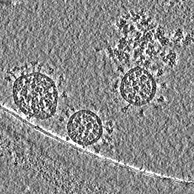 |
Cryo electron tomography - tilt-series of Sars-Cov-2 [266 tilt series in MRC format] | Turoňová B, Sikora M, Schürmann C, Hagen WJH, Welsch S, Blanc FEC, von Bülow S, Gecht M, Bagola K, Hörner C, van Zandbergen G, Landry J, de Azevedo NTD, Mosalaganti S, Schwarz A, Covino R, Mühlebach MD, Hummer G, Krijnse Locker J, Beck M [Pubmed: 32817270] [DOI: 10.1126/science.abd5223] |
469.8 GB | 4.9 - 7.9 Å |
| 2024-05-03 |  |
Initiation factor 3 bound to the 30S ribosomal subunit in an initial step of translation [multiple data sets in MRC and MRCS formats] | Uday AB, Mishra RK, Hussain T [Pubmed: 38148682] [DOI: 10.1002/prot.26655] |
4.8 TB | 4.4 - 4.6 Å |
| 2021-04-14 | 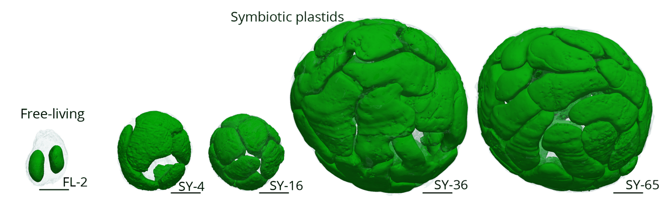 |
Cytoklepty in the plankton: a host strategy to optimize the bioenergetic machinery of endosymbiotic algae [500 multi-frame micrographs composed of 1 frames each in TIFF format] | Uwizeye C, Mars Brisbin M, Gallet B, LeKieffre C, Schieber NL, Wangpraseurt D, Decelle J [Pubmed: bioRxiv] [DOI: 10.1101/2020.12.08.416644] |
2.9 GB | — |
| 2021-10-05 | 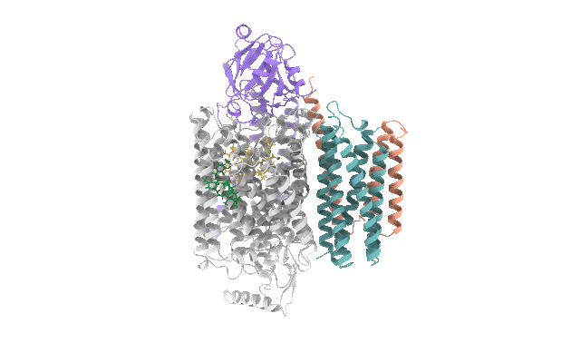 |
Cryo-EM structures of E. coli cytochrome bo3 in MSP Nanodiscs [3446 multi-frame micrographs composed of 50 frames each in TIFF format] | Vallese F [Pubmed: 34417297] [DOI: 10.1073/pnas.2106750118] |
851.4 GB | 2.19 Å |
| 2022-08-08 | 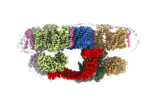 |
Architecture of the human erythrocyte ankyrin-1 complex [multiple data sets in TIFF, MRC and MRCS formats] | Vallese F, Clarke OB [Pubmed: 35835865] [DOI: 10.1038/s41594-022-00792-w] |
5.2 TB | 2.4 Å |
| 2024-04-17 |  |
Data set of tilted cryo-EM images of human N-deacetylase/N-sulfotransferase 1 [multiple data sets in TIFF format] | Vallet SD, Annaval T, Wild R, Lortat-Jacob H [DOI: 10.1002/pgr2.8] |
1.9 TB | 4.5 Å |
| 2023-09-26 |  |
Single particle cryo-EM dataset of human nucleolar and nuclear pre-60S assembly intermediates [multiple data sets in TIFF and MRCS formats] | Vanden Broeck A, Klinge S [Pubmed: 37410842] [DOI: 10.1126/science.adh3892] EMD-29104 EMD-29252 EMD-29105 EMD-29253 EMD-29106 EMD-29254 EMD-29107 EMD-29255 EMD-29108 EMD-29256 EMD-29109 EMD-29257 EMD-29110 EMD-29258 EMD-29111 EMD-29259 EMD-29112 EMD-29260 EMD-29113 EMD-29261 EMD-29114 EMD-29262 EMD-29115 EMD-29263 EMD-29130 EMD-29131 EMD-29132 EMD-29133 EMD-29134 EMD-29135 EMD-29136 EMD-29137 EMD-29138 EMD-29139 EMD-29140 EMD-29141 EMD-29142 EMD-29128 EMD-29129 EMD-29143 EMD-29144 EMD-29145 EMD-29146 EMD-29147 EMD-29148 EMD-29149 EMD-29150 EMD-29151 EMD-29152 EMD-29153 EMD-29154 EMD-29155 EMD-29156 EMD-29157 EMD-29158 EMD-29159 EMD-29160 EMD-29161 EMD-29162 EMD-29163 EMD-29164 EMD-29165 EMD-29166 EMD-29167 EMD-29168 EMD-29169 EMD-29170 EMD-29171 EMD-29173 EMD-29174 EMD-29175 EMD-29176 EMD-29177 EMD-29178 EMD-29179 EMD-29180 EMD-29181 EMD-29182 EMD-29183 EMD-29184 EMD-29185 EMD-29186 EMD-29187 EMD-29188 EMD-29189 EMD-29192 EMD-29194 EMD-29193 EMD-29116 EMD-29265 EMD-29117 EMD-29266 EMD-29118 EMD-29267 EMD-29119 EMD-29268 EMD-29120 EMD-29269 EMD-29121 EMD-29271 EMD-29122 EMD-29272 EMD-29123 EMD-29273 EMD-29124 EMD-29274 EMD-29125 EMD-29275 EMD-29126 EMD-29276 EMD-29127 EMD-29277 EMD-29195 EMD-29196 EMD-29197 EMD-29198 EMD-29199 EMD-29200 EMD-29201 EMD-29202 EMD-29204 EMD-29205 EMD-29206 |
140.5 TB | 2.33 - 3.75 Å |
| 2023-05-10 |  |
Microtubule depolymerization contributes to spontaneous neurotransmitter release [multiple data sets in MRC format] | Velasco C, Santarella-Mellwig R, Schorb M, Gao L, Thorn-Seshold O, Llobet A [Pubmed: 37147475] [DOI: 10.1038/s42003-023-04779-1] |
123.8 GB | — |
| 2020-12-04 |  |
Structure of the class D GPCR Ste2 dimer coupled to two G proteins [multiple data sets in MRC and TIFF formats] | Velazhahan V, Ma N, Pándy-Szekeres G, Kooistra AJ, Lee Y, Gloriam DE, Vaidehi N, Tate CG [Pubmed: 33268889] [DOI: 10.1038/s41586-020-2994-1] |
1.3 TB | 3.3 Å |
| 2022-03-21 | 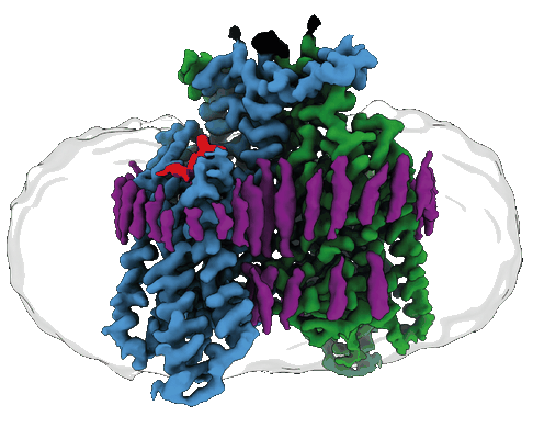 |
Structure of the GPCR dimer Ste2 bound to an antagonist [15751 multi-frame micrographs composed of 59 frames each in TIFF format] | Velazhahan V, Tate CG [Pubmed: 35296853] [DOI: 10.1038/s41586-022-04498-3] |
4.1 TB | 2.7 Å |
| 2022-03-21 | 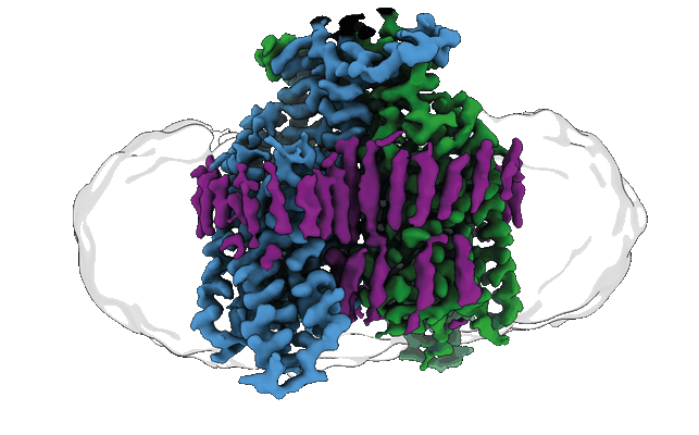 |
Structure of the ligand-free GPCR dimer Ste2 [9369 multi-frame micrographs composed of 53 frames each in EER format] | Velazhahan V, Tate CG [Pubmed: 35296853] [DOI: 10.1038/s41586-022-04498-3] |
8.2 TB | 3.1 Å |
| 2022-03-21 | 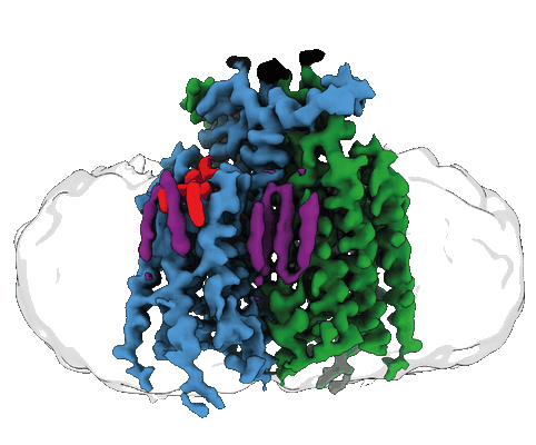 |
Structure of the agonist-bound GPCR dimer Ste2 [6944 multi-frame micrographs composed of 50 frames each in MRC format] | Velazhahan V, Tate CG [Pubmed: 35296853] [DOI: 10.1038/s41586-022-04498-3] |
1.3 TB | 3.46 - 3.53 Å |
| 2023-03-01 | 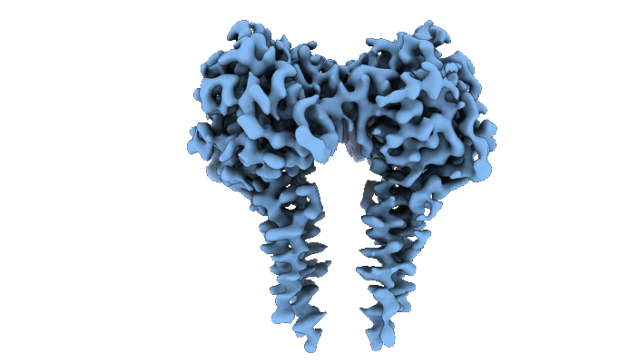 |
CryoEM structure of full-length dimeric ClbP [3888 multi-frame micrographs composed of 50 frames each in TIFF format] | Velilla JA, Walsh RM, Gaudet R [Pubmed: 36253550] [DOI: 10.1038/s41589-022-01142-z] |
1.1 TB | 3.73 Å |
| 2016-08-18 |  |
Structure and Dynamics of Single-isoform Recombinant Neuronal Human Tubulin [304 multi-frame micrographs composed of 23 frames each in MRC format] | Vemu A, Atherton J, Spector JO, Szyk A, Moores CA, Roll-Mecak A [Pubmed: 27129203] [DOI: 10.1074/jbc.C116.731133] |
487.7 GB | 4.0 Å |
| 2016-06-28 |  |
Designer nanoscale DNA assemblies programmed from the top down [50 micrographs in MRC format] | Veneziano R, Ratanalert S, Zhang K, Zhang F, Yan H, Chiu W, Bathe M [Pubmed: 27229143] [DOI: 10.1126/science.aaf4388] |
3.7 GB | 22.0 Å |
| 2016-06-28 |  |
Designer nanoscale DNA assemblies programmed from the top down [100 micrographs in MRC format] | Veneziano R, Ratanalert S, Zhang K, Zhang F, Yan H, Chiu W, Bathe M [Pubmed: 27229143] [DOI: 10.1126/science.aaf4388] |
7.3 GB | 25.0 Å |
| 2016-06-28 |  |
Designer nanoscale DNA assemblies programmed from the top down [177 micrographs in MRC format] | Veneziano R, Ratanalert S, Zhang K, Zhang F, Yan H, Chiu W, Bathe M [Pubmed: 27229143] [DOI: 10.1126/science.aaf4388] |
13.0 GB | 20.0 Å |
| 2023-10-03 |  |
Cryo electron tomography of Cytochalasin D-induced protrusions of Drosophila S2 cells treated with thapsigargin or MG132 [multiple data sets in TIFF and MRC formats] | Ventura Santos C, Carter AP [Pubmed: 37702953] [DOI: 10.15252/embr.202357264] |
107.6 GB | — |
| 2023-07-10 |  |
Cryo electron tomography of Cytochalasin D-induced protrusions of Drosophila S2 cells - Datasets 1 - 4 [multiple data sets in TIFF and MRC formats] | Ventura Santos C, Carter AP, Rogers SL [Pubmed: 37034688] [DOI: 10.1101/2023.03.31.535077] |
446.0 GB | — |
| 2023-07-10 |  |
Cryo electron tomography of Cytochalasin D-induced protrusions of Drosophila S2 cells treated with DMSO or thapsigargin - Datasets 5 - 7 [multiple data sets in TIFF and MRC formats] | Ventura Santos C, Carter AP, Rogers SL [Pubmed: 37034688] [DOI: 10.1101/2023.03.31.535077] |
373.1 GB | — |
| 2023-07-10 |  |
Cryo electron tomography of Cytochalasin D-induced protrusions of Drosophila S2 alpha-tubulin acetyltransferase knock-out (dTAT KO) cells - Dataset 8 [multiple data sets in TIFF and MRC formats] | Ventura Santos C, Carter AP, Rogers SL [Pubmed: 37034688] [DOI: 10.1101/2023.03.31.535077] |
231.5 GB | — |
| 2023-07-10 |  |
Cryo electron tomography of induced protrusions of cofilin or control knock-down Drosophila S2 cells - Datasets 9 - 12 [multiple data sets in TIFF and MRC formats] | Ventura Santos C, Carter AP, Rogers SL [Pubmed: 37034688] [DOI: 10.1101/2023.03.31.535077] |
491.9 GB | — |
| 2022-11-29 |  |
1.42 Angstrom Apoferritin structure determined using G1 Titan krios S-FEG operated at 300kV, zero loss imaging using Gatan BioQuantum energy filter operated at 10eV slit width and imaged using K2 camera. [multiple data sets in MRC format] | Venugopal H | 668.6 GB | 1.42 Å |
| 2021-11-02 |  |
CryoEM single particle dataset for NanR dimer-DNA hetero-complex. [3465 multi-frame micrographs composed of 32 frames each in MRC format] | Venugopal H, Horne CR, Ramm G, Dobson RCJ [Pubmed: 33790291] [DOI: 10.1038/s41467-021-22253-6] |
364.8 GB | 3.9 Å |
| 2022-06-22 |  |
single particle cryo-EM of red blood cell lysate (hemolysate, hemoglobin reduced, filtered by SEC) [6608 multi-frame micrographs composed of 20 frames each in TIFF format] | Verbeke EJ [Pubmed: 35858567] [DOI: 10.1016/j.celrep.2022.111103] |
1.0 TB | 3.4 Å |
