Electron Microscopy Public Image Archive
Electron Microscopy Public Image Archive
The EMPIAR-PDBj team at Osaka University assists Asian EM researchers with the transfer of big EM image data to EMPIAR. Instead of sending the data directly to the EBI (UK) via the internet, hard drives can also be sent to Osaka University by postal mail or via a courier service. As an alternative, internet transfer to our server in Osaka is also available. If you would like to take advantage of our submission services, please contact us first by e-mail before sending the data to us.
| Release date | Imageset | Title | Authors and references | Size | Resolution |
|---|---|---|---|---|---|
| 2020-10-23 | 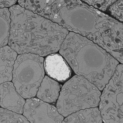 |
170314, Five-day-old Col-0 Arabidopsis thaliana root, phloem pole unloading zone (339-381 um from the root tip) [multiple data sets in MRC format] | Paterlini A, Belevich I, Jokitalo E, Helariutta Y [Pubmed: 31182845] [DOI: 10.1038/s41477-019-0429-5] |
43.2 GB | — |
| 2020-06-19 | 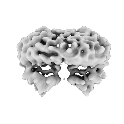 |
Tetrameric SARS-CoV-2 ORF3a in a lipid nanodisc [7092 multi-frame micrographs composed of 50 frames each in TIFF format] | Kern DM, Sorum B, Mali SS, Hoel CM, Sridharan S, Remis JP, Toso DB, Kotecha A, Bautista DM, Brohawn SG [Pubmed: 34158638] [DOI: 10.1038/s41594-021-00619-0] |
4.8 TB | 6.5 Å |
| 2020-06-19 | 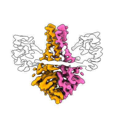 |
SARS-CoV-2 ORF3a dimer with added Emodin in an MSP1E3D1 lipid nanodisc [6750 multi-frame micrographs composed of 50 frames each in TIFF format] | Kern DM, Sorum B, Mali SS, Hoel CM, Sridharan S, Remis JP, Toso DB, Kotecha A, Bautista DM, Brohawn SG [Pubmed: 34158638] [DOI: 10.1038/s41594-021-00619-0] |
4.5 TB | 3.7 Å |
| 2020-06-19 | 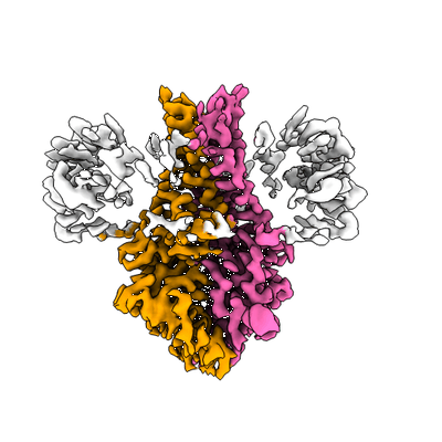 |
SARS-CoV-2 ORF3a dimer in an MSP1E3D1 lipid nanodisc [6309 multi-frame micrographs composed of 50 frames each in TIFF format] | Kern DM, Sorum B, Mali SS, Hoel CM, Sridharan S, Remis JP, Toso DB, Kotecha A, Bautista DM, Brohawn SG [Pubmed: 34158638] [DOI: 10.1038/s41594-021-00619-0] |
4.4 TB | 2.9 Å |
| 2020-09-28 | 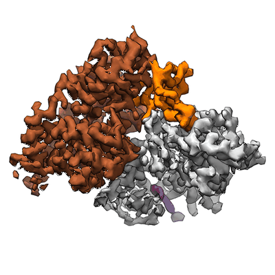 |
Single-particle cryo-EM of the human CDK-activating kinase in complex with THZ1 [4453 multi-frame micrographs composed of 69 frames each in TIFF format] | Greber BJ, Perez-Bertoldi JM, Lim K, Iavarone AT, Toso DB, Nogales E [Pubmed: 32855301] [DOI: 10.1073/pnas.2009627117] |
2.4 TB | 3.3 Å |
| 2021-11-08 | 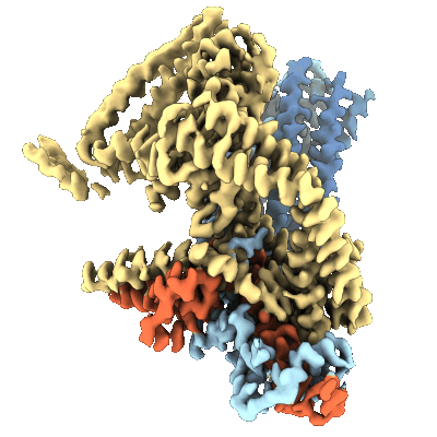 |
Single-particle cryo-EM of the full-length merozoite surface protein 1 from Plasmodium falciparum [multiple data sets in MRC and MRCS formats] | Dijkman PM, Marzluf T, Zhang Y, Chang SYS, Helm D, Lanzer M, Bujard H, Kudryashev M [Pubmed: 34078606] [DOI: 10.1126/sciadv.abg0465] |
4.3 TB | 3.1 - 3.6 Å |
| 2021-10-26 | 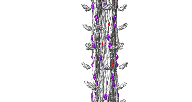 |
The CryoEM Structure of Drosophila Flight Muscle Thick Filaments at 7Å Resolution [1510 multi-frame micrographs composed of 44 frames each in MRC format] | Daneshparvar N, Taylor DW, O'Leary TS, Rahmani H, Abbasiyeganeh F, Previs MJ, Taylor KA [Pubmed: 32718994] [DOI: 10.1101/2020.06.05.136580] |
8.7 TB | 7.0 - 8.0 Å |
| 2022-01-21 | 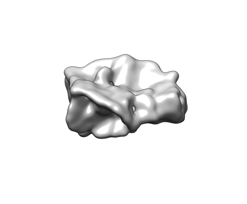 |
Structural Insights of Transcriptionally Active, Full-Length Androgen Receptor Coactivator Complexes [1958 multi-frame micrographs composed of 50 frames each in MRC format] | Yu X, Yi P [Pubmed: 32668201] [DOI: 10.1016/j.molcel.2020.06.031] |
5.1 TB | 13.0 Å |
| 2020-08-12 | 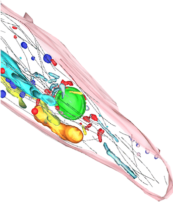 |
TEM tomograms of Drosophila tracheal terminal cells during subcellular tube formation [multiple data sets in TIFF and MRC formats] | Mathew R, Rios-Barrera LD, Machado P, Schwab Y, Leptin M [Pubmed: 32657472] [DOI: 10.15252/embj.2020105332] |
366.7 GB | — |
| 2020-06-16 | 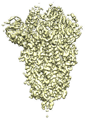 |
Characterization of the SARS-CoV-2 S Protein: Biophysical, Biochemical, Structural, and Antigenic Analysis [multiple data sets in TIFF and MRC formats] | Herrera NG, Morano NC, Celikgil A, Georgiev GI, Malonis R, Lee JH, Tong K, Vergnolle O, Massimi A, Yen LY, Noble AJ, Kopylov M, Bonanno JB, Garrett-Thomson SC, Hayes DB, Bortz R, Wirchnianski A, Florez C, Laudermilch E, Haslwanter D, Fels J, Dieterle M, Jangra R, Barnhill J, Mengotto A, Kimmel D, Daily J, Pirofski L, Chandran K, Brenowitz M, Garforth S, Eng E, Lai JR, Almo SC [Pubmed: 32587972] [DOI: 10.1101/2020.06.14.150607] |
484.3 GB | 3.22 Å |
| 2020-10-02 | 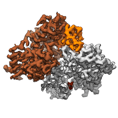 |
Single-particle cryo-EM of the human CDK-activating kinase in complex with ATP-gamma-S [multiple data sets in TIFF format] | Greber BJ, Perez-Bertoldi JM, Lim K, Iavarone AT, Toso DB, Nogales E [Pubmed: 32855301] [DOI: 10.1073/pnas.2009627117] |
3.5 TB | 2.8 Å |
| 2020-07-06 | 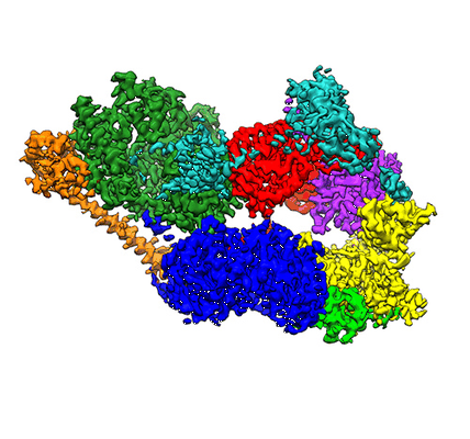 |
Phase-plate cryo-EM of human TFIIH [multiple data sets in TIFF format] | Greber BJ, Toso DB, Fang J, Nogales E [Pubmed: 30860024] [DOI: 10.7554/eLife.44771] |
9.2 TB | 3.7 Å |
| 2020-07-14 | 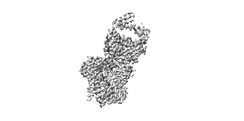 |
3 Å resolution single particle reconstruction of glucosyltransferase ALG6 in nanodisc [multiple data sets in MRC and TIFF formats] | Bloch JS, Pesciullesi G, Boilevin J, Nosol K, Irobalieva RN, Darbre T, Aebi M, Kossiakoff AA, Reymond JL, Locher KP [Pubmed: 32103179] [DOI: 10.1038/s41586-020-2044-z] |
2.8 TB | 3.0 Å |
| 2020-08-06 | 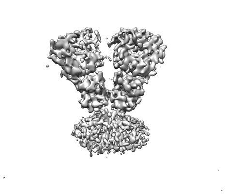 |
Cryo-EM Structure of GluD1-Orphan Delta Receptor Reveals a Novel Architecture in the Ionotropic Glutamate Receptor Family [3938 multi-frame micrographs composed of 40 frames each in MRC format] | Burada AP, Vinnakota R, Kumar J [Pubmed: 31925409] [DOI: 10.1038/s41594-019-0359-y] |
6.5 TB | 8.1 Å |
| 2021-05-14 | 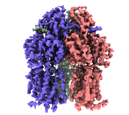 |
3.9 Angstrom reconstruction of E.coli AcrB embedded in the liposome [5757 multi-frame micrographs composed of 32 frames each in MRCS format] | Yao X, Fan X, Yan N [Pubmed: 32680969] [DOI: 10.1073/pnas.2009385117] |
2.4 TB | 3.9 Å |
| 2022-01-18 | 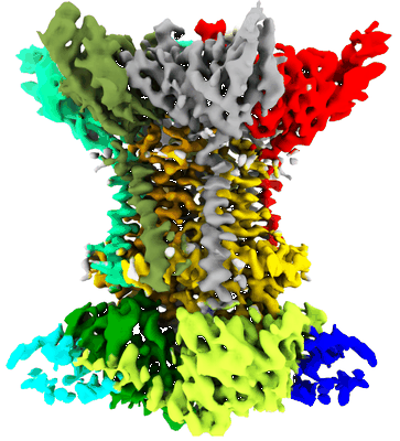 |
Cryo-EM structure of the A.baumannii MlaBDEF complex bound to AppNHp [2557 micrographs in MRC format] | Mann D, Bergeron JRC [Pubmed: 34188171] [DOI: 10.1038/s42003-021-02318-4] |
135.6 GB | 3.9 Å |
| 2020-05-27 | 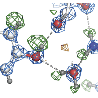 |
Atomic resolution structure of apoferritin [3370 multi-frame micrographs composed of 434 frames each in EER format] | Nakane T, Kotecha A, Sente A, McMullan G, Masiulis S, Brown PMGE, Grigoras IT, Malinauskaite L, Malinauskas T, Miehling J, Uchański T, Yu L, Karia D, Pechnikova EV, de Jong E, Keizer J, Bischoff M, McCormack J, Tiemeijer P, Hardwick SW, Chirgadze DY, Murshudov G, Aricescu AR, Scheres SHW [Pubmed: 33087931] [DOI: 10.1038/s41586-020-2829-0] |
538.1 GB | 1.22 Å |
| 2020-07-21 | 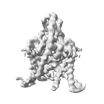 |
TASK2 in MSP1D1 lipid nanodisc at pH6.5 [3024 multi-frame micrographs composed of 50 frames each in TIFF format] | Li B, Brohawn SG [Pubmed: 32999458] [DOI: 10.1038/s41586-020-2770-2] |
2.0 TB | 3.45 Å |
| 2020-07-21 | 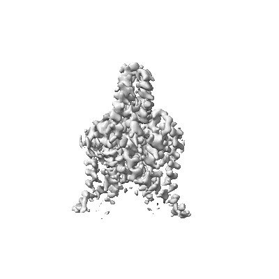 |
TASK2 in MSP1D1 lipid nanodisc at pH8.5 [3470 multi-frame micrographs composed of 50 frames each in TIFF format] | Li B, Brohawn SG [Pubmed: 32999458] [DOI: 10.1038/s41586-020-2770-2] |
2.3 TB | 3.52 Å |
| 2021-05-17 | 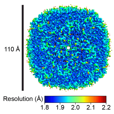 |
Single-Particle CryoEM of Human Apoferritin Light Chain Vitrified Using Back-it-up [multiple data sets in MRCS and MRC formats] | Tan YZ, Rubinstein JL [Pubmed: 33135680] [DOI: 10.1107/S2059798320012474] |
2.1 TB | 2.0 Å |
| 2020-07-30 | 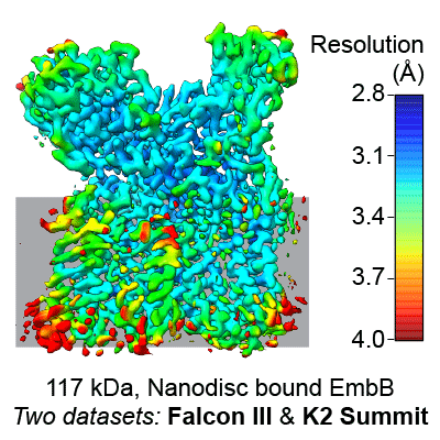 |
Single-Particle Cryo-EM of Arabinosyltransferase EmbB from Mycobacterium smegmatis, Collected using both Falcon III and K2 Detectors [multiple data sets in MRCS, TIFF and MRC formats] | Tan YZ, Rodrigues J, Keener JE, Zheng RB, Brunton R, Kloss B, Giacometti SI, Rosário AL, Zhang L, Niederweis M, Clarke OB, Lowary TL, Marty MT, Archer M, Potter CS, Carragher B, Mancia F [Pubmed: 32636380] [DOI: 10.1038/s41467-020-17202-8] |
4.5 TB | 3.3 Å |
| 2020-06-30 |  |
Soft X-ray Tomography of mock-infected U2OS cells [1 tilt series in MRC format] | Kounatidis I, Stanifer ML, Phillips MA, Paul-Gilloteaux P, Heiligenstein X, Wang H, Okolo CA, Fish TM, Spink MC, Stuart DI, Davis I, Boulant S, Grimes JM, Dobbie IM, Harkiolaki M [Pubmed: 32610083] [DOI: 10.1016/j.cell.2020.05.051] |
1.2 GB | — |
| 2020-06-30 | 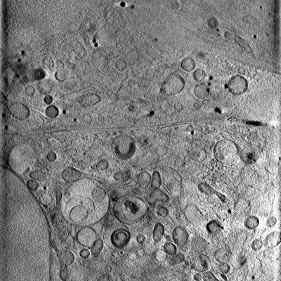 |
Soft X-ray Tomography of mock-infected U2OS cells [1 tilt series in MRC format] | Kounatidis I, Stanifer ML, Phillips MA, Paul-Gilloteaux P, Heiligenstein X, Wang H, Okolo CA, Fish TM, Spink MC, Stuart DI, Davis I, Boulant S, Grimes JM, Dobbie IM, Harkiolaki M [Pubmed: 32610083] [DOI: 10.1016/j.cell.2020.05.051] |
1.2 GB | — |
