EMPIAR-10442
170314, Five-day-old Col-0 Arabidopsis thaliana root, phloem pole unloading zone (339-381 um from the root tip) [multiple data sets in MRC format]
Publication:

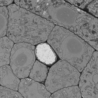
Sphingolipid biosynthesis modulates plasmodesmal ultrastructure and phloem unloading

Yan D, Yadav SR, Paterlini A, Nicolas WJ, Petit JD, Brocard L, Belevich I, Grison MS, Vaten A, Karami L, El-Showk S, Lee JY, Murawska GM, Mortimer J, Knoblauch M, Jokitalo E, Markham JE, Bayer EM, Helariutta Y

Nature plants 5 (2019) 604-615

PMID: 31182845

Related EMDB entry:
Deposited:
2020-06-04
Released:
2020-10-23
Last modified:
2020-10-23
Imageset size:
43.17 GB
Imageset DOI:
Experimental metadata:
Download xml json
Contains:
  • micrographs - multiframe
1. Sieve element cells of Arabidopsis thaliana roots
Category:
micrographs - multiframe
Image format:
MRC
No. of images or tilt series:
1
Image size:
(3874, 3253)
Pixel type:
UNSIGNED BYTE
Pixel spacing:
(70.0, 70.0)
Details:
Processed datasets: image processing was done in Microscopy Image Browser and included adjustment of contrast, alignment and calibration. The bounding box coordinates represent position of the dataset relative to the tip of the root

Dataset: 170314_Col-0_R20_339-381um.am
Voxel size: 0.007 x 0.007 x 0.04 um
BoundingBox (xmin, xmax, ymin, ymax, zmin, zmax) 527.30140 555.40970 -686.83160 -663.23020 339.14530 379.14530
MIB(1704051124): Flip: mode=flipZ
MIB(1704051130): ContrastGamma: Min:12585.2355, Max: 36949.7536, Gamma: 1
MIB(1704051130): Converted to from uint16 to uint8
MIB(1704051135): Aligned using Drift correction; relative to 0
MIB(1704051143): Aligned using Single landmark point; relative to 0
MIB(1704051206): ImCrop: [x1 y1 dx dy z1 dz t1 dt]: [682 418 3874 3253 1 1061 1 1]
Files:
Loading...
2. Sieve element cells of Arabidopsis thaliana roots
Category:
micrographs - multiframe
Image format:
MRC
No. of images or tilt series:
1
Image size:
(3430, 2010)
Pixel type:
UNSIGNED BYTE
Pixel spacing:
(70.0, 70.0)
Details:
Processed datasets: image processing was done in Microscopy Image Browser and included adjustment of contrast, alignment and calibration. The bounding box coordinates represent position of the dataset relative to the tip of the root

Dataset: 170821_Col-0_R01_294-317um.am
Voxel size: 0.007 x 0.007 x 0.04 um
BoundingBox -46.17730 -20.82255 71.14690 86.00187 294.89500 314.89500
MIB(1709011612): Aligned using Single landmark point; relative to 0
MIB(1709011614): Aligned using Drift correction; relative to 0
MIB(1709011623): Aligned using Single landmark point; relative to 0
MIB(1709011623): ImCrop: [x1 y1 dx dy z1 dz t1 dt]: [119 482 3430 2010 1 544 1 1]
MIB(1709011626): Normalize contrast, mode: mask, Ch: 1
MIB(1709011628): ContrastGamma: Min:14265.0396, Max: 23898.9022, Gamma: 1
MIB(1709011628): Converted to from uint16 to uint8
MIB(1709011628): Flip: mode=flipZ
Files:
Loading...
3. Sieve element cells of Arabidopsis thaliana roots
Category:
micrographs - multiframe
Image format:
MRC
No. of images or tilt series:
1
Image size:
(2341, 1689)
Pixel type:
UNSIGNED BYTE
Pixel spacing:
(100.0, 100.0)
Details:
Processed datasets: image processing was done in Microscopy Image Browser and included adjustment of contrast, alignment and calibration. The bounding box coordinates represent position of the dataset relative to the tip of the root

Dataset: 180130_PLM_SE_down_278-307um.am
Voxel size: 0.010 x 0.01 x 0.04 um
BoundingBox -166.42400 -143.02300 -290.90100 -274.02000 278.23500 306.23500
MIB(1802021325): Aligned using Drift correction; relative to 0
MIB(1802021327): Aligned using Single landmark point; relative to 0
MIB(1802021330): Normalize contrast, mode: mask, Ch: 1
MIB(1802021334): Normalize contrast, mode: mask, Ch: 1
MIB(1802021335): ContrastGamma: Min:29058.337, Max: 44837.221, Gamma: 1
MIB(1802021335): Converted to from uint16 to uint8
MIB(1802021336): ImCrop: [x1 y1 dx dy z1 dz t1 dt]: [296 448 2341 1689 1 717 1 1]
Files:
Loading...
4. Sieve element cells of Arabidopsis thaliana roots
Category:
micrographs - multiframe
Image format:
MRC
No. of images or tilt series:
1
Image size:
(2596, 1731)
Pixel type:
UNSIGNED BYTE
Pixel spacing:
(100.0, 100.0)
Details:
Processed datasets: image processing was done in Microscopy Image Browser and included adjustment of contrast, alignment and calibration. The bounding box coordinates represent position of the dataset relative to the tip of the root

Dataset: 180130_PLM_SE_up_278-307um.am
Voxel size: 0.010 x 0.01 x 0.04 um
BoundingBox -161.23100 -135.28000 -327.25700 -309.95700 278.23500 306.23500
MIB(1802021310): Aligned using Drift correction; relative to 0
MIB(1802021312): Aligned using Single landmark point; relative to 0
MIB(1802021316): ImCrop: [x1 y1 dx dy z1 dz t1 dt]: [183 462 2596 1731 1 717 1 1]
MIB(1802021319): Normalize contrast, mode: mask, Ch: 1
MIB(1802021320): ContrastGamma: Min:28724.6413, Max: 45826.6123, Gamma: 1
MIB(1802021320): Converted to from uint16 to uint8
Files:
Loading...
Files:
Loading...

Ito F, Alvarez-Cabrera AL, Liu S, Yang H, Shiriaeva A, Zhou ZH, Chen XS. (2023)
Rigden DJ, Fernández XM. (2023)
Iudin A, Korir PK, Somasundharam S, Weyand S, Cattavitello C, Fonseca N, Salih O, Kleywegt GJ, Patwardhan A. (2023)
Serra Lleti JM, Steyer AM, Schieber NL, Neumann B, Tischer C, Hilsenstein V, Holtstrom M, Unrau D, Kirmse R, Lucocq JM, Pepperkok R, Schwab Y. (2023)
Caldwell BJ, Norris AS, Karbowski CF, Wiegand AM, Wysocki VH, Bell CE. (2022)