Electron Microscopy Public Image Archive
Electron Microscopy Public Image Archive
The EMPIAR-PDBj team at Osaka University assists Asian EM researchers with the transfer of big EM image data to EMPIAR. Instead of sending the data directly to the EBI (UK) via the internet, hard drives can also be sent to Osaka University by postal mail or via a courier service. As an alternative, internet transfer to our server in Osaka is also available. If you would like to take advantage of our submission services, please contact us first by e-mail before sending the data to us.
| Release date | Imageset | Title | Authors and references | Size | Resolution |
|---|---|---|---|---|---|
| 2023-03-17 | 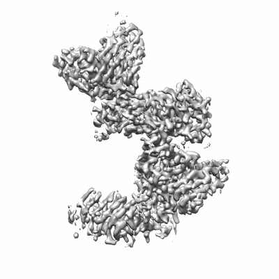 |
FMC63 scFv in complex with soluble CD19 [multiple data sets in TIFF format] | Meyerson JR, He C [Pubmed: 36867678] [DOI: 10.1126/sciimmunol.adf1426] |
1.4 TB | 3.05 Å |
| 2023-03-17 | 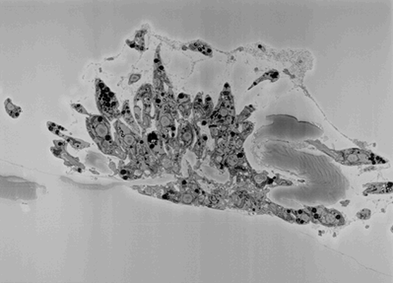 |
SBFSEM imaging of Leishmania haptomonad-like cells attached to plastic [252 multi-frame micrographs composed of 1 frames each in MRC format] | Yanase R, Sunter JD [DOI: 10.1101/2022.10.28.514187] |
33.6 GB | — |
| 2023-03-17 | 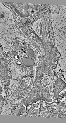 |
Serial section electron tomography of a Leishmania haptomonad on the stomodeal valve in the sand fly [670 multi-frame micrographs composed of 1 frames each in MRC format] | Yanase R, Sunter JD [DOI: 10.1101/2022.10.28.514187] |
6.7 GB | — |
| 2023-03-17 | 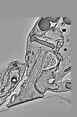 |
Serial section electron tomography of a Leishmania haptomonad-like cell attached to plastic [718 multi-frame micrographs composed of 1 frames each in MRC format] | Yanase R, Sunter JD [DOI: 10.1101/2022.10.28.514187] |
10.8 GB | — |
| 2023-03-22 |  |
Electron cryo-tomography data on the ER-mitochondria encounter structure in cryo-FIB milled yeast cells [multiple data sets in MRC and TIFF formats] | Wozny MR, Di Luca A, Morado DR, Picco A, Khaddaj R, Campomanes P, Ivanovic L, Hoffmann PC, Miller EA, Vanni S, Kukulski W [Pubmed: 37165187] [DOI: 10.1038/s41586-023-06050-3] |
236.6 GB | — |
| 2023-03-27 |  |
Cryo electron microscopy of beta-2-microglobulin amyloid fibrils for the variant deltaN6 (in vitro, pH 6.2) [4095 multi-frame micrographs composed of 28 frames each in TIFF format] | Wilkinson M, Gallardo RU, Martinez RM, Guthertz N, So M, Aubrey LD, Radford SE, Ranson NA [Pubmed: 36864041] [DOI: 10.1038/s41467-023-36791-8] |
616.1 GB | 3.0 - 3.4 Å |
| 2023-03-27 |  |
Cryo electron microscopy of beta-2-microglobulin amyloid fibrils for the variant V27M (in vitro, pH 6.2) [611 multi-frame micrographs composed of 40 frames each in TIFF format] | Wilkinson M, Gallardo RU, Martinez RM, Guthertz N, So M, Aubrey LD, Radford SE, Ranson NA [Pubmed: 36864041] [DOI: 10.1038/s41467-023-36791-8] |
92.7 GB | 2.8 Å |
| 2023-03-27 |  |
Cryo electron microscopy of beta-2-microglobulin amyloid fibrils for the variant D76N (in vitro, pH 6.2) [3849 multi-frame micrographs composed of 36 frames each in TIFF format] | Wilkinson M, Gallardo RU, Martinez RM, Guthertz N, So M, Aubrey LD, Radford SE, Ranson NA [Pubmed: 36864041] [DOI: 10.1038/s41467-023-36791-8] |
575.5 GB | 3.0 - 4.1 Å |
| 2023-03-27 |  |
Cryo-EM structure of the ribosome/Sec61 translocon with a macrocyclic inhibitor KZR-8445 [multiple data sets in TIFF and MRC formats] | Rehan S [DOI: 10.1101/2022.07.03.498529] |
12.9 TB | 3.2 Å |
| 2023-03-27 | 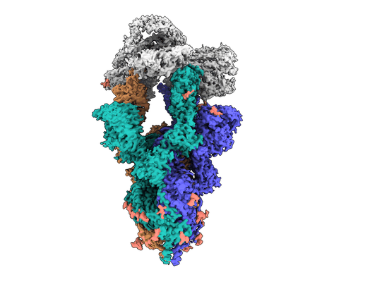 |
mouse ACE2 receptor bound to SARS-CoV-2 variant Omicron BA.2.12.1 Spike [8046 multi-frame micrographs composed of 945 frames each in EER format] | Ni D, Myasnikov A, Stahlberg S, Lau K [DOI: 10.1371/journal.ppat.1011206] |
4.0 TB | 2.89 - 3.03 Å |
| 2023-03-27 | 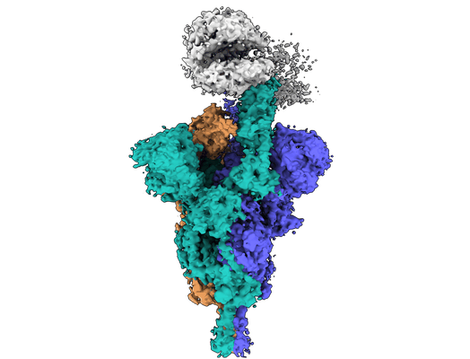 |
mouse ACE2 receptor bound to SARS-CoV-2 variant Beta Spike [5671 multi-frame micrographs composed of 40 frames each in MRC format] | Ni D, Myasnikov A, Stahlberg S, Lau K [DOI: 10.1371/journal.ppat.1011206] |
3.8 TB | 4.4 Å |
| 2023-03-30 | 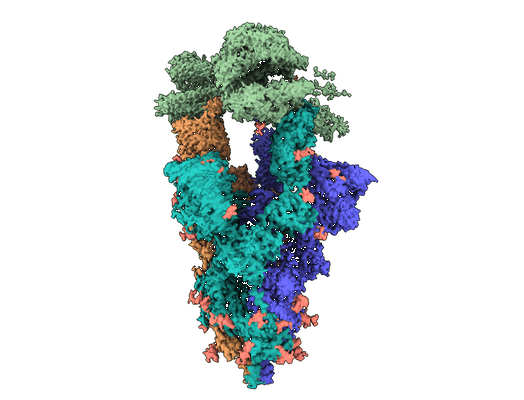 |
human ACE2 receptor bound to SARS-CoV-2 variant Omicron BA.4/5 Spike [7254 multi-frame micrographs composed of 1092 frames each in EER format] | Ni D, Myasnikov A, Stahlberg S, Lau K [DOI: 10.1371/journal.ppat.1011206] |
4.1 TB | 2.92 Å |
| 2023-03-30 | 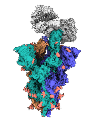 |
mouse ACE2 receptor bound to SARS-CoV-2 variant Omicron BA.4/5 Spike [7989 multi-frame micrographs composed of 1092 frames each in EER format] | Ni D, Myasnikov A, Stahlberg S, Lau K [DOI: 10.1371/journal.ppat.1011206] |
4.4 TB | 2.92 - 3.3 Å |
| 2023-04-11 | 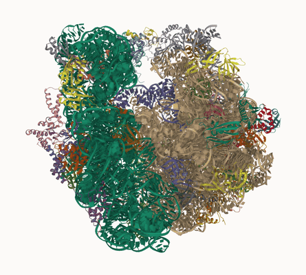 |
Sarecycline inhibits protein translation in Cutibacterium acnes 70S ribosome using a two-site mechanism [multiple data sets in TIFF and MRC formats] | Lomakin IB, Devarkar SC, Patel S, Grada A, Bunick CG [Pubmed: 36864821] [DOI: 10.1093/nar/gkad103] |
6.2 TB | 2.78 Å |
| 2023-04-11 | 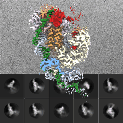 |
Cryo-EM micrographs for the Bombyx mori R2 retrotransposon initiating target-primed reverse transcription [16551 multi-frame micrographs composed of 40 frames each in TIFF format] | Wilkinson ME, Zhang F [Pubmed: 37023171] [DOI: 10.1126/science.adg7883] |
2.3 TB | 3.08 Å |
| 2023-04-11 | 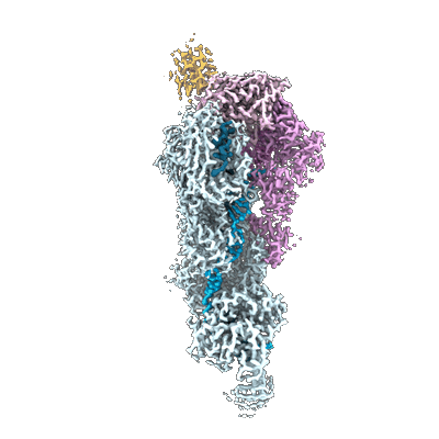 |
Desulfonema ishimotonii Cas7-11 in complex with Csx29 and Csx30 [multiple data sets in TIFF format] | Strecker J, Demircioglu FE, Li D, Faure G, Wilkinson ME, Macrae RK, Zhang F [Pubmed: 36423276] [DOI: 10.1126/science.add7450] |
5.7 TB | 2.53 - 3.15 Å |
| 2023-04-11 | 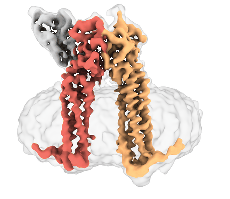 |
Human PRPH2-ROM1 hetero-dimer [10448 multi-frame micrographs composed of 61 frames each in TIFF format] | El Mazouni D [Pubmed: 36351012] [DOI: 10.1126/sciadv.add3677] |
3.4 TB | 3.7 Å |
| 2023-04-11 |  |
Human PRPH2-ROM1 oligomers [multiple data sets in TIFF format] | El Mazouni D [Pubmed: 36351012] [DOI: 10.1126/sciadv.add3677] |
6.0 TB | 7.6 - 8.2 Å |
| 2023-04-11 | 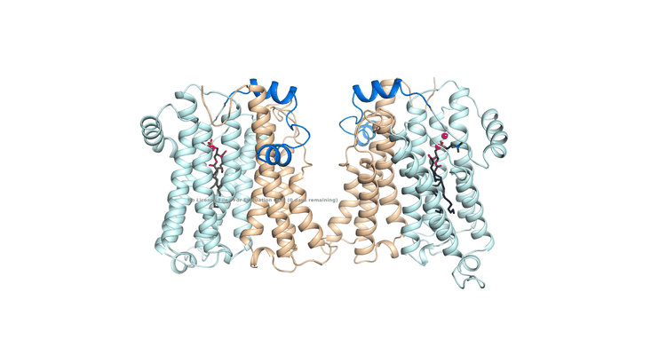 |
Structure of human choline/ethanolamine phosphotransferase [3115 multi-frame micrographs composed of 32 frames each in TIFF format] | Qian H | 1.4 TB | 3.7 Å |
| 2023-04-12 | 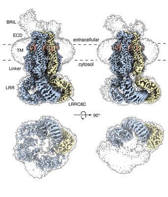 |
LRRC8A-BRIL:C Heteromer in GDN [multiple data sets in TIFF and MRCS formats] | Kern DM, Brohawn SG [Pubmed: 36928458] [DOI: 10.1038/s41594-023-00944-6] |
8.9 TB | 2.95 - 4.16 Å |
| 2023-04-12 | 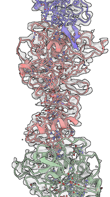 |
Cryo-Electron Microscopy structure of OmcZ nanowires from Geobacter sulfurreducens [14005 multi-frame micrographs composed of 50 frames each in TIFF format] | Gu YG, Srikanth V, Salazar-Morales AI, Samatey FA, Malvankar NS, Guberman-Pfeffer MGP, Shen CS, Gupta KG, Londer YL, Giska FG, Batista VB [Pubmed: 36732469] [DOI: 10.1038/s41564-022-01315-5] |
6.5 TB | 3.4 Å |
| 2023-04-13 | 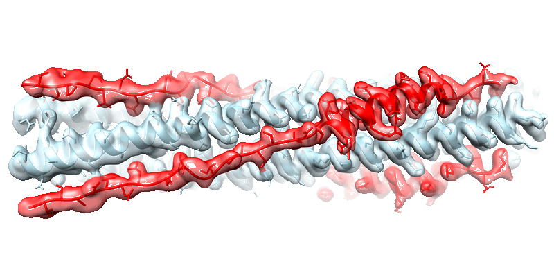 |
Cryo-EM structure of the SARS-CoV-2 HR1HR2 fusion core complex with extended HR2 [18846 multi-frame micrographs composed of 40 frames each in TIFF format] | Yang K, Brunger AT [Pubmed: 36122200] [DOI: 10.1073/pnas.2210990119] |
4.6 TB | 2.22 Å |
| 2023-04-13 | 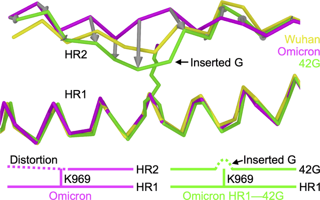 |
Structure-based design of a SARS-CoV-2 Omicron-specific inhibitor [multiple data sets in TIFF format] | Yang K, Brunger AT [Pubmed: 36940324] [DOI: 10.1073/pnas.2300360120] |
13.5 TB | 2.51 Å |
| 2023-04-13 |  |
Cryo electron microscopy of DNA Polymerase alpha - primase bound to SARS-CoV-2 nsp1 virulence factor [2919 multi-frame micrographs composed of 48 frames each in TIFF format] | Kilkenny ML, Pellegrini L [Pubmed: 34719824] [DOI: 10.1002/pro.4220] |
751.3 GB | 4.12 - 4.4 Å |
| 2023-04-14 |  |
Structural insights into the human Prostaglandin E receptor EP3-Gi signaling complex [11241 multi-frame micrographs composed of 70 frames each in TIFF format] | Suno R, Sugita Y, Kobayashi T [Pubmed: 36103815] [DOI: 10.1016/j.celrep.2022.111323] |
3.1 TB | 3.375 Å |
