Electron Microscopy Public Image Archive
Electron Microscopy Public Image Archive
The EMPIAR-PDBj team at Osaka University assists Asian EM researchers with the transfer of big EM image data to EMPIAR. Instead of sending the data directly to the EBI (UK) via the internet, hard drives can also be sent to Osaka University by postal mail or via a courier service. As an alternative, internet transfer to our server in Osaka is also available. If you would like to take advantage of our submission services, please contact us first by e-mail before sending the data to us.
| Release date | Imageset | Title | Authors and references | Size | Resolution |
|---|---|---|---|---|---|
| 2024-02-06 | 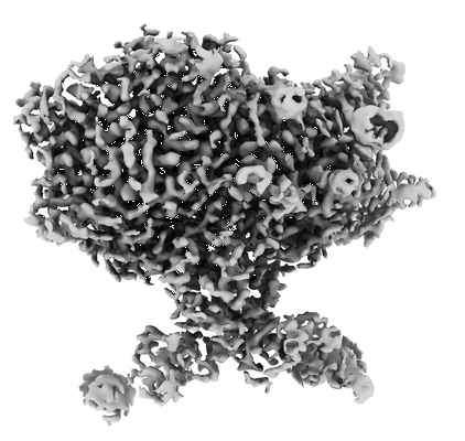 |
CryoEM structures of the human CLC-2-AK42 voltage gated chloride channel reveal a ball and chain gating mechanism [stack of 11498 particles in MRC format] | Xu M, Pintilie G, Liu Y, Chiu W, Maduke M | 1.2 TB | 2.46 Å |
| 2021-04-06 | 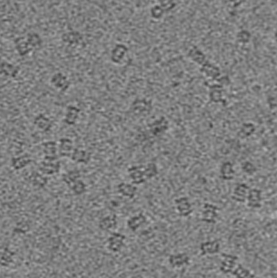 |
CryoET dataset of T20S proteasome for testing motion-aware tilt-series alignment and 3D reconstruction [3 tilt series in MRC format] | Fernandez JJ [Pubmed: 29410148] [DOI: 10.1016/j.jsb.2018.02.001] |
5.1 GB | 9.0 Å |
| 2017-12-15 | 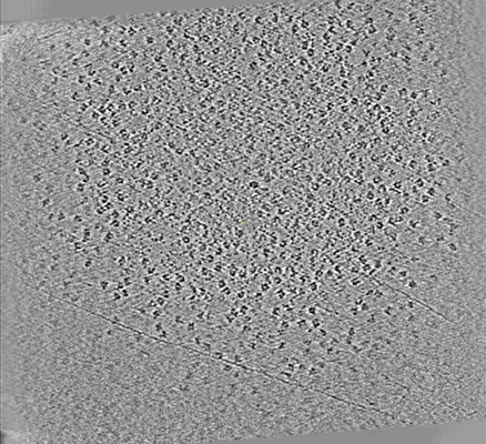 |
CryoET of DNAB helicase-helicase loader single particle [multiple data sets in MRC format] | Noble AJ, Dandey VP, Wei H, Brasch J, Chase J, Acharya P, Tan YZ, Zhang Z, Kim LY, Scapin G, Rapp M, Eng ET, Rice WJ, Cheng A, Negro CJ, Shapiro L, Kwong PD, Jeruzalmi D, des Georges A, Potter CS, Carragher B [Pubmed: 29809143] [DOI: 10.7554/eLife.34257] |
120.1 GB | — |
| 2022-03-21 | 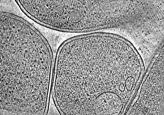 |
CryoET of E. coli prepared with the Waffle Method [1504 multi-frame micrographs composed of 14 frames each in TIFF format] | Kelley K, Raczkowski AM, Klykov O, Jaroenlak P, Bobe D, Kopylov M, Eng ET, Bhabha G, Potter CS, Carragher B, Noble AJ [Pubmed: 35387991] [DOI: 10.1038/s41467-022-29501-3] |
94.0 GB | — |
| 2017-12-18 |  |
CryoET of Mtb 20S proteasome single particle [multiple data sets in MRC format] | Noble AJ, Dandey VP, Wei H, Brasch J, Chase J, Acharya P, Tan YZ, Zhang Z, Kim LY, Scapin G, Rapp M, Eng ET, Rice MJ, Cheng A, Negro CJ, Shapiro L, Kwong PD, Jeruzalmi D, des Georges A, Potter CS, Carragher B [Pubmed: 29809143] [DOI: 10.7554/eLife.34257] |
9.1 GB | — |
| 2017-12-18 |  |
CryoET of T20S proteasome single particle [multiple data sets in MRC format] | Noble AJ, Dandey VP, Wei H, Brasch J, Chase J, Acharya P, Tan YZ, Zhang Z, Kim LY, Scapin G, Rapp M, Eng ET, Rice WJ, Cheng A, Negro CJ, Shapiro L, Kwong PD, Jeruzalmi D, des Georges A, Potter CS, Carragher B [Pubmed: 29809143] [DOI: 10.7554/eLife.34257] |
235.3 GB | — |
| 2017-12-18 |  |
CryoET of T20S proteasome single particle [multiple data sets in MRC format] | Dandey VP, Wei H, Brasch J, Chase J, Acharya P, Tan YZ, Zhang Z, Kim LY, Scapin G, Rapp M, Eng ET, Rice MJ, Cheng A, Negro CJ, Shapiro L, Kwong PD, Jeruzalmi D, des Georges A, Potter CS, Carragher B [Pubmed: 29809143] [DOI: 10.7554/eLife.34257] |
12.4 GB | — |
| 2017-12-18 |  |
CryoET of apoferritin single particle [multiple data sets in MRC format] | Noble AJ, Dandey VP, Wei H, Brasch J, Chase J, Acharya P, Tan YZ, Zhang Z, Kim LY, Scapin G, Rapp M, Eng ET, Rice MJ, Cheng A, Negro CJ, Shapiro L, Kwong PD, Jeruzalmi D, des Georges A, Potter CS, Carragher B [Pubmed: 29809143] [DOI: 10.7554/eLife.34257] |
46.1 GB | — |
| 2017-12-18 | 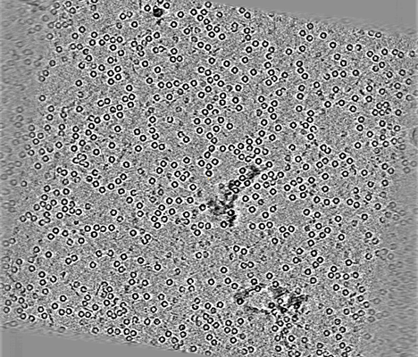 |
CryoET of apoferritin single particle [multiple data sets in MRC format] | Dandey VP, Wei H, Brasch J, Chase J, Acharya P, Tan YZ, Zhang Z, Kim LY, Scapin G, Rapp M, Eng ET, Rice MJ, Cheng A, Negro CJ, Shapiro L, Kwong PD, Jeruzalmi D, des Georges A, Potter CS, Carragher B [Pubmed: 29809143] [DOI: 10.7554/eLife.34257] |
29.2 GB | — |
| 2017-12-18 | 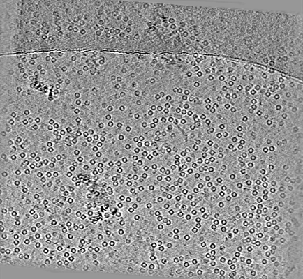 |
CryoET of apoferritin single particle [multiple data sets in MRC format] | Noble AJ, Dandey VP, Wei H, Brasch J, Chase J, Acharya P, Tan YZ, Zhang Z, Kim LY, Scapin G, Rapp M, Eng ET, Rice MJ, Cheng A, Negro CJ, Shapiro L, Kwong PD, Jeruzalmi D, des Georges A, Potter CS, Carragher B [Pubmed: 29809143] [DOI: 10.7554/eLife.34257] |
301.1 GB | — |
| 2017-12-18 | 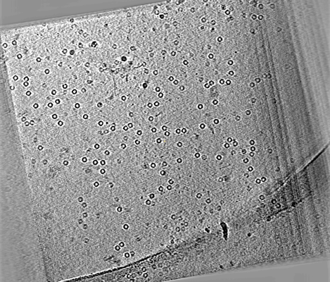 |
CryoET of apoferritin single particle [multiple data sets in MRC format] | Noble AJ, Dandey VP, Wei H, Brasch J, Chase J, Acharya P, Tan YZ, Zhang Z, Kim LY, Scapin G, Rapp M, Eng ET, Rice MJ, Cheng A, Negro CJ, Shapiro L, Kwong PD, Jeruzalmi D, des Georges A, Potter CS, Carragher B [Pubmed: 29809143] [DOI: 10.7554/eLife.34257] |
32.9 GB | — |
| 2017-12-18 | 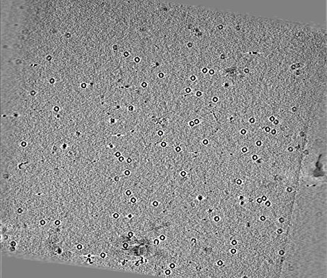 |
CryoET of apoferritin single particle [multiple data sets in MRC format] | Noble AJ, Dandey VP, Wei H, Brasch J, Chase J, Acharya P, Tan YZ, Zhang Z, Kim LY, Scapin G, Rapp M, Eng ET, Rice MJ, Cheng A, Negro CJ, Shapiro L, Kwong PD, Jeruzalmi D, des Georges A, Potter CS, Carragher B [Pubmed: 29809143] [DOI: 10.7554/eLife.34257] |
23.0 GB | — |
| 2018-04-05 | 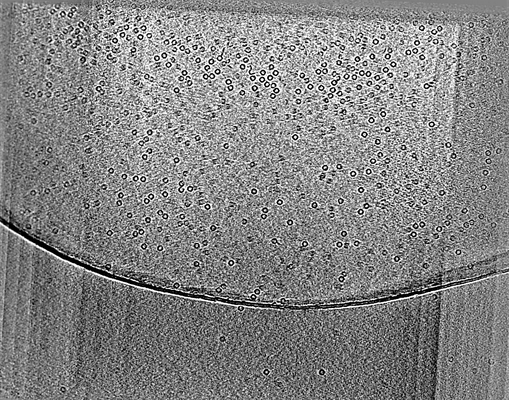 |
CryoET of apoferritin single particle with spot-to-plunge time of 100ms [multiple data sets in MRC and DYNAMO TBL AND OMD FILES formats] | Noble AJ, Wei H, Dandey VP, Zhang Z, Potter CS, Carragher B [Pubmed: 30250056] [DOI: 10.1038/s41592-018-0139-3] |
119.3 GB | — |
| 2018-04-05 | 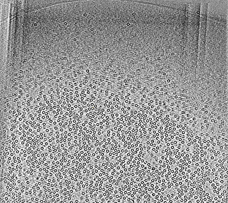 |
CryoET of apoferritin single particle with spot-to-plunge time of 400ms [multiple data sets in MRC and DYNAMO TBL AND OMD FILES formats] | Noble AJ, Wei H, Dandey VP, Zhang Z, Potter CS, Carragher B [Pubmed: 30250056] [DOI: 10.1038/s41592-018-0139-3] |
98.8 GB | — |
| 2018-04-05 | 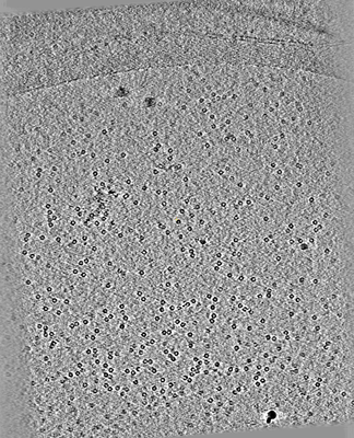 |
CryoET of apoferritin with 0.5 mM TCEP single particle [multiple data sets in MRC format] | Noble AJ, Dandey VP, Wei H, Brasch J, Chase J, Acharya P, Tan YZ, Zhang Z, Kim LY, Scapin G, Rapp M, Eng ET, Rice MJ, Cheng A, Negro CJ, Shapiro L, Kwong PD, Jeruzalmi D, des Georges A, Potter CS, Carragher B [Pubmed: 29809143] [DOI: 10.7554/eLife.34257] |
35.3 GB | — |
| 2018-04-05 | 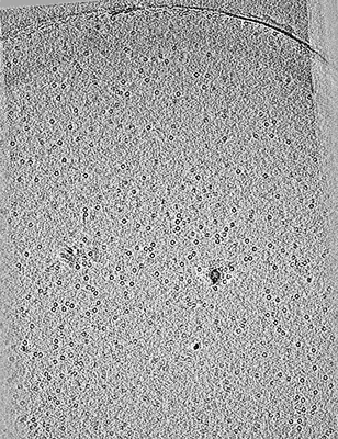 |
CryoET of apoferritin with 0.5 mM TCEP single particle with spot-to-plunge time of 170ms [multiple data sets in MRC and DYNAMO TBL AND OMD FILES formats] | Noble AJ, Wei H, Dandey VP, Zhang Z, Potter CS, Carragher B [Pubmed: 30250056] [DOI: 10.1038/s41592-018-0139-3] |
31.6 GB | — |
| 2018-10-25 |  |
CryoET of bacterial RNA polymerase with several detergents [multiple data sets in MRC and RAW TEXT formats] | Chen J, Noble AJ, Kang JY, Darst SA [DOI: 10.1016/j.yjsbx.2019.100005] |
207.5 GB | — |
| 2019-04-18 |  |
CryoET of cis-mutated mouse protocadherin gamma B6 on membranes [multiple data sets in MRC format] | Brasch J, Bepler T, Berger B, Maniatis T, Potter CS, Carragher B, Honig B, Shapiro L [Pubmed: 30971825] [DOI: 10.1038/s41586-019-1089-3] |
379.7 GB | — |
| 2017-12-15 |  |
CryoET of glutamate dehydrogenase + 0.001% DDM single particle [multiple data sets in MRC format] | Noble AJ, Dandey VP, Wei H, Brasch J, Chase J, Acharya P, Tan YZ, Zhang Z, Kim LY, Scapin G, Rapp M, Eng ET, Rice WJ, Cheng A, Negro CJ, Shapiro L, Kwong PD, Jeruzalmi D, des Georges A, Potter CS, Carragher B [Pubmed: 29809143] [DOI: 10.7554/eLife.34257] |
37.1 GB | — |
| 2017-12-15 |  |
CryoET of glutamate dehydrogenase single particle [multiple data sets in MRC format] | Noble AJ, Dandey VP, Wei H, Brasch J, Chase J, Acharya P, Tan YZ, Zhang Z, Kim LY, Scapin G, Rapp M, Eng ET, Rice WJ, Cheng A, Negro CJ, Shapiro L, Kwong PD, Jeruzalmi D, des Georges A, Potter CS, Carragher B [Pubmed: 29809143] [DOI: 10.7554/eLife.34257] |
133.1 GB | — |
| 2017-12-15 | 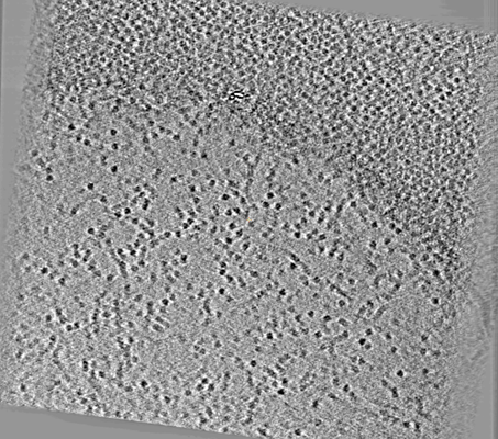 |
CryoET of glutamate dehydrogenase single particle [multiple data sets in MRC format] | Noble AJ, Dandey VP, Wei H, Brasch J, Chase J, Acharya P, Tan YZ, Zhang Z, Kim LY, Scapin G, Rapp M, Eng ET, Rice WJ, Cheng A, Negro CJ, Shapiro L, Kwong PD, Jeruzalmi D, des Georges A, Potter CS, Carragher B [Pubmed: 29809143] [DOI: 10.7554/eLife.34257] |
37.1 GB | — |
| 2018-04-05 | 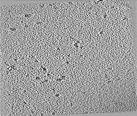 |
CryoET of hemagglutinin single particle with spot-to-plunge time of 100ms [multiple data sets in MRC format] | Noble AJ, Wei H, Dandey VP, Zhang Z, Potter CS, Carragher B [Pubmed: 30250056] [DOI: 10.1038/s41592-018-0139-3] |
21.9 GB | — |
| 2017-12-18 | 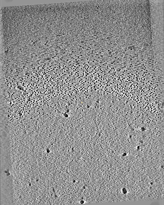 |
CryoET of hemagglutinin single particle with spot-to-plunge time of 800ms [multiple data sets in MRC format] | Noble AJ, Dandey VP, Wei H, Brasch J, Chase J, Acharya P, Tan YZ, Zhang Z, Kim LY, Scapin G, Rapp M, Eng ET, Rice WJ, Cheng A, Negro CJ, Shapiro L, Kwong PD, Jeruzalmi D, des Georges A, Potter CS, Carragher B [Pubmed: 29809143] [DOI: 10.7554/eLife.34257] |
34.0 GB | — |
| 2018-04-05 | 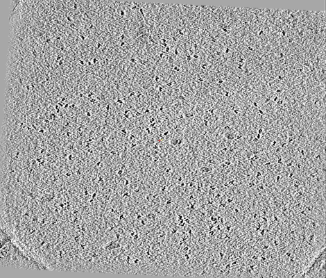 |
CryoET of insulin-bound insulin receptor single particle with spot-to-plunge time of 200ms [multiple data sets in MRC format] | Noble AJ, Wei H, Dandey VP, Zhang Z, Potter CS, Carragher B [Pubmed: 30250056] [DOI: 10.1038/s41592-018-0139-3] |
40.4 GB | — |
| 2018-04-05 |  |
CryoET of insulin-bound insulin receptor single particle with spot-to-plunge time of 600ms [multiple data sets in MRC format] | Noble AJ, Wei H, Dandey VP, Zhang Z, Potter CS, Carragher B [Pubmed: 30250056] [DOI: 10.1038/s41592-018-0139-3] |
79.4 GB | — |
