Electron Microscopy Public Image Archive
Electron Microscopy Public Image Archive
現在、ストレージの障害により、一部データセットがアクセスできないか不完全な状態(Marked: Incomplete dataset)になっています。
以下の順で復旧作業継続中ですが、優先リクエスト承ります(email、inquiry)
ご不便をおかけしますがご理解よろしくお願いします。
大阪大学のEMPIAR-PDBjチームでは、アジアの電顕研究者が、EMPIARデータベースに大きな電顕画像を転送するお手伝いをしています。 ネットワークでイギリスに転送する代わりに、ハードディスク自体を郵送・宅配便で大阪大学に送付、あるいは阪大のサーバまでネットワーク転送していただければ、こちらで仲介して登録サイトへの転送を代行します。 EMPIARへの登録を希望する方で、阪大へのデータ送付・転送をご希望の方は、まず、e-mail で、登録したい電顕画像データについて、ご相談ください。
| Release date | Imageset | Title | Authors and references | Size | Resolution |
|---|---|---|---|---|---|
| 2025-03-10 | 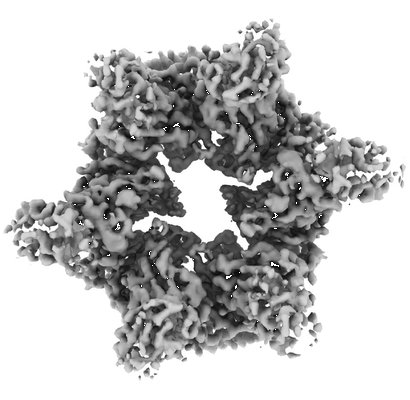 |
TadA with AMPPNP [5108 multi-frame micrographs composed of 40 frames each in TIFF format] | Hohl M, Low HH [Pubmed: 39103374] [DOI: 10.1038/s41467-024-50280-6] |
2.2 TB | 3.8 Å |
| 2025-03-10 | 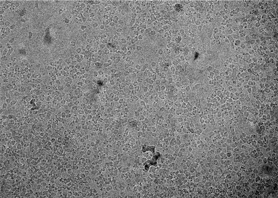 |
Cryo-electron micrographs of Cholinephosphotransferase in complex with diacylglycerol [multiple data sets in TIFF and MRC formats] | Roberts JR [Pubmed: 39747155] [DOI: 10.1038/s41467-024-55673-1] |
1.2 TB | 3.2 Å |
| 2025-03-10 | 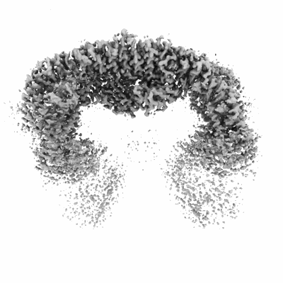 |
Cryo-EM micrographs of purified human BIRC6 in presence of MAP1LC3B [7580 micrographs in MRC format] | Ehrmann JF, Grabarczyk DB, Clausen T [Pubmed: 36758105] [DOI: 10.1126/science.ade8873] |
473.8 GB | 3.3 Å |
| 2025-03-05 |  |
Cryo-EM micrographs of purified human BIRC6 in presence of CASP9 [9289 micrographs in MRC format] | Ehrmann JF, Grabarczyk DB, Clausen T [Pubmed: 36758105] [DOI: 10.1126/science.ade8873] |
580.6 GB | 3.3 Å |
| 2025-03-05 | 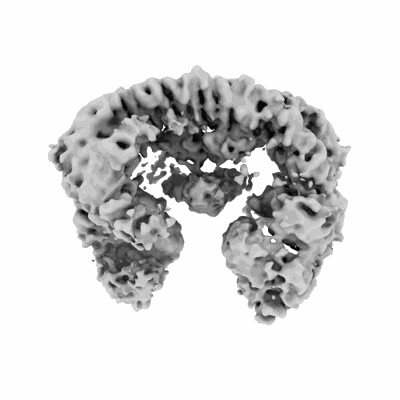 |
Cryo-EM micrographs of purified human BIRC6 in presence of HTRA2 [8842 micrographs in MRC format] | Ehrmann JF, Grabarczyk DB, Clausen T [Pubmed: 36758105] [DOI: 10.1126/science.ade8873] |
552.7 GB | 3.3 - 6.2 Å |
| 2025-03-04 |  |
PSI-FCPI of the diatom Thalassiosira pseudonana CCMP1335 [8950 multi-frame micrographs composed of 50 frames each in TIFF format] | Kato K, Nagao R, Nakajima Y, Shen JR [Pubmed: 39480899] [DOI: 10.7554/eLife.99858] |
1.7 TB | 2.3 Å |
| 2025-02-27 |  |
Single particle cryogenic electron micrographs of isolated mouse mitochondrial supercomplex I2+III2 [multiple data sets in TIFF format] | Letts JA, Padavannil A [Pubmed: 39788125] [DOI: 10.1016/j.cmet.2024.11.011] |
18.4 TB | 3.67 - 4.25 Å |
| 2025-02-27 |  |
EcPKS2 inhibited with Malonyl-CoA [17420 multi-frame micrographs composed of 50 frames each in TIFF format] | Schubert HL [Pubmed: 39913209] [DOI: 10.1073/pnas.2419884122] |
4.6 TB | 2.92 - 3.15 Å |
| 2025-02-24 |  |
Cryo-ET of Dictyostelium discoideum in HL5 medium (control condition) [59 tilt series in MRC format] | Hoffmann PC, Beck M [Pubmed: 39729993] [DOI: 10.1016/j.molcel.2024.11.038] |
386.5 GB | 30.0 Å |
| 2025-02-24 | 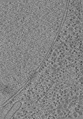 |
Cryo-ET of Dictyostelium discoideum in 0.4 M sorbitol (hyperosmotic stress) [59 tilt series in MRC format] | Hoffmann PC, Beck M [Pubmed: 39729993] [DOI: 10.1016/j.molcel.2024.11.038] |
332.1 GB | 38.0 Å |
| 2025-02-21 | 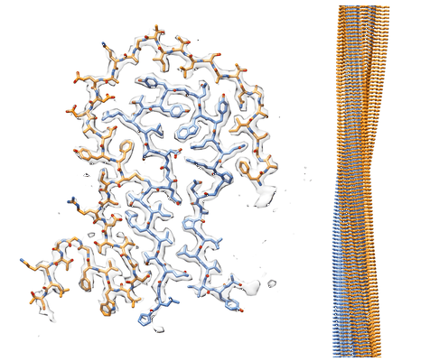 |
AL amyloid fibril from the FOR010 light chain [4008 multi-frame micrographs composed of 963 frames each in EER format] | Pfeiffer PB, Banerjee S, Schmidt M, Fändrich M [Pubmed: 38879609] [DOI: 10.1038/s41467-024-49520-6] |
1.8 TB | 2.25 Å |
| 2025-02-21 | 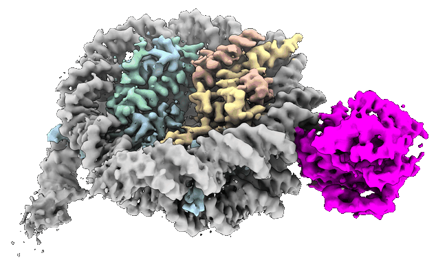 |
Single particle cryoEM dataset of Haspin kinase bound to a nucleosome [9999 multi-frame micrographs composed of 1485 frames each in EER format] | Hicks CW, Wolberger C [Pubmed: 39979508] [DOI: 10.1038/s41594-025-01502-y] |
5.8 TB | 2.99 - 3.64 Å |
| 2025-02-21 |  |
movies for NKCC1 bound with bumetanide [6027 multi-frame micrographs composed of 40 frames each in TIFF format] | Cao E [Pubmed: 39875725] [DOI: 10.1038/s44318-025-00368-6] |
1.5 TB | 2.5 Å |
| 2025-02-13 | 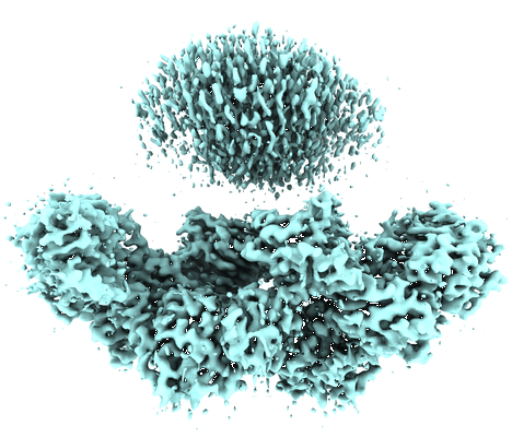 |
Polyketide Synthase EcPKS1 [2913 multi-frame micrographs composed of 36 frames each in TIFF format] | Schubert HL [Pubmed: 39913209] [DOI: 10.1073/pnas.2419884122] |
2.0 TB | 3.5 - 4.3 Å |
| 2025-02-11 | 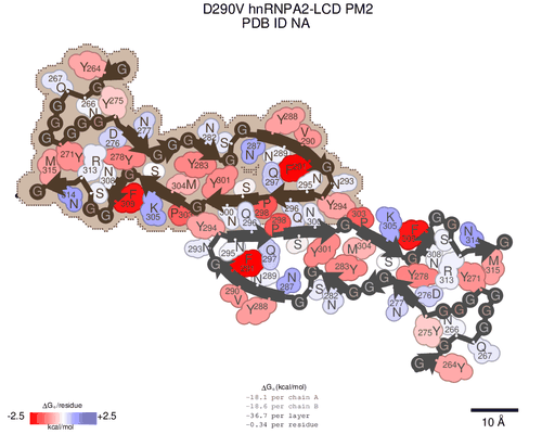 |
CryoEM data for D290V hnRNPA2 LCDs [11900 multi-frame micrographs composed of 50 frames each in TIFF format] | Lu JL [Pubmed: 38072051] [DOI: 10.1016/j.jbc.2023.105531] |
1.9 TB | 3.2 - 3.9 Å |
| 2025-02-11 |  |
EcPKS2 with acylated pPANT and three ACP-docked positions [6244 multi-frame micrographs composed of 40 frames each in TIFF format] | Schubert HL [Pubmed: 39913209] [DOI: 10.1073/pnas.2419884122] |
2.9 TB | 2.92 - 3.11 Å |
| 2025-02-10 |  |
cryo-EM structure of human tRNA(GlnUUG) - PUS4 and PUS7 modified [8610 micrographs in MRC format] | Biela AP, Glatt S [Pubmed: 40301665] [DOI: 10.1038/s44318-025-00443-y] |
756.0 GB | 5.1 Å |
| 2025-02-10 |  |
cryo-EM structure of human tRNA(AspGUC) - PUS4 and PUS7 modified [7511 micrographs in MRC format] | Biela AP, Glatt S [Pubmed: 40301665] [DOI: 10.1038/s44318-025-00443-y] |
659.5 GB | 5.83 Å |
| 2025-02-10 | 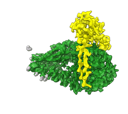 |
Cryo electron structure of the ectodomain of Site-one protease without SPRING [6171 multi-frame micrographs composed of 1 frames each in EER format] | Kober DL [Pubmed: 38977690] [DOI: 10.1038/s41467-024-50068-8] |
3.1 TB | 2.27 Å |
| 2025-02-10 | 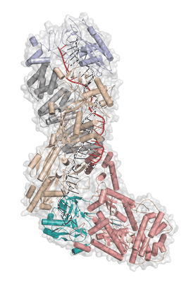 |
Cryo-EM structure of the active Lactococcus lactis Csm bound to target in post-cleavage stage [9775 multi-frame micrographs composed of 74 frames each in TIFF format] | Goswami HN, Wang B, Ahmadizadeh F, Addo-Yobo D, Zhao Y, Whittington AC, Terns MP [Pubmed: 38989619] [DOI: 10.1093/nar/gkae603] |
7.9 TB | 2.58 - 2.79 Å |
| 2025-02-10 |  |
TniQ-capped TnsC-ATP-dsDNA complex [multiple data sets in TIFF and DM4 formats] | Querques I, Schmitz M, Oberli S, Chanez C, Jinek M [Pubmed: 36435179] [DOI: 10.1016/j.cell.2022.11.009] |
2.9 TB | 3.44 Å |
| 2025-02-10 |  |
High resolution C. elegans 80S ribosome single particle cryoEM structure [8337 multi-frame micrographs composed of 51 frames each in TIFF format] | Sehgal E., Balasco Serrao V.H., Arribere J. [Pubmed: 39209556] [DOI: 10.1261/rna.080103.124] |
2.8 TB | 2.59 - 2.63 Å |
| 2025-02-10 | 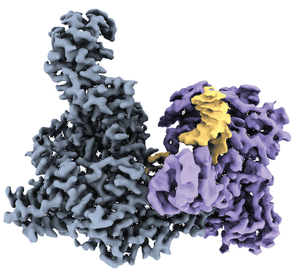 |
Vibrio cholerae DdmD-DdmE holo complex [multiple data sets in TIFF and MRC formats] | Luuk L, Jinek M [Pubmed: 38870273] [DOI: 10.1126/science.adq0534] |
1.6 TB | 2.56 Å |
| 2025-02-10 |  |
Vibrio cholerae DdmD apo complex [multiple data sets in TIFF and MRC formats] | Luuk L, Jinek M [Pubmed: 38870273] [DOI: 10.1126/science.adq0534] |
1.5 TB | 2.55 Å |
| 2025-02-10 | 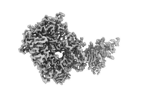 |
HSV-1 DNA polymerase-processivity factor complex in halted elongation state [multiple data sets in TIFF and MRC formats] | Gustavsson E, Grünewald K, Elias P, Hällberg BM [Pubmed: 38806233] [DOI: 10.1093/nar/gkae374] |
1.5 TB | 2.4 - 2.97 Å |
