Electron Microscopy Public Image Archive
Electron Microscopy Public Image Archive
The EMPIAR-PDBj team at Osaka University assists Asian EM researchers with the transfer of big EM image data to EMPIAR. Instead of sending the data directly to the EBI (UK) via the internet, hard drives can also be sent to Osaka University by postal mail or via a courier service. As an alternative, internet transfer to our server in Osaka is also available. If you would like to take advantage of our submission services, please contact us first by e-mail before sending the data to us.
| Release date | Imageset | Title | Authors and references | Size | Resolution |
|---|---|---|---|---|---|
| 2017-03-15 | 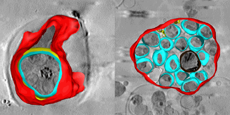 |
Soft X-ray tomography of Plasmodium falciparum infected human erythrocytes stalled in egress by the inhibitors Compound 2 and E64 [1 Soft X-ray tomograms in MRC format] | Hale VL, Saibil HR, Duke E, Fleck RA, Blackman MJ [Pubmed: 28292906] [DOI: 10.1073/pnas.1619441114] |
280.6 MB | — |
| 2023-10-13 | 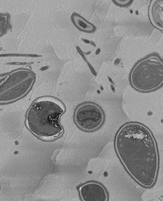 |
SBF-SEM micrographs of A. algerae microsporidia spores, 5 min germination [1215 micrographs in TIFF format] | Davydov A, Jaroenlak P, Ekiert D, Bhabha G [DOI: 10.7554/eLife.86638.1] |
226.3 GB | — |
| 2023-10-13 | 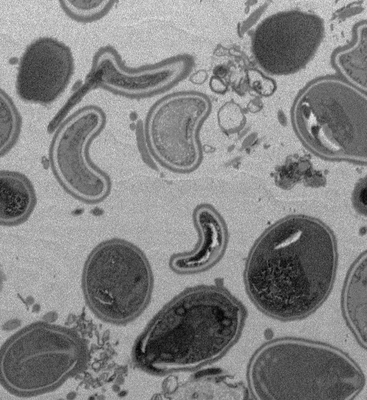 |
SBF-SEM micrographs of A. algerae microsporidia spores, 45 min germination [300 micrographs in TIFF format] | Davydov A, Jaroenlak P, Ekiert D, Bhabha G [DOI: 10.7554/eLife.86638.1] |
55.9 GB | — |
| 2021-01-08 | 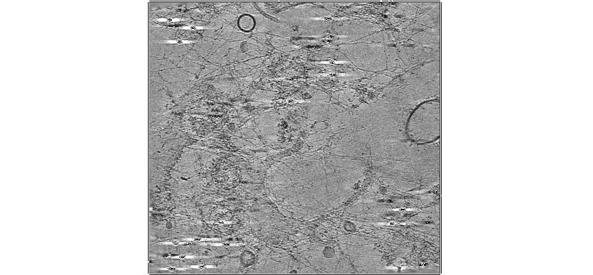 |
A lamin A/C variant causing striated muscle disease provides insights into filament organization [2 tilt series in MRC format] | Tatli M [Pubmed: 33536248] [DOI: 10.1242/jcs.256156] |
2.7 GB | — |
| 2023-10-10 | 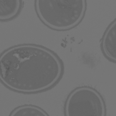 |
SBF-SEM micrographs of A. algerae spores, Ungerminated [250 micrographs in TIFF format] | Jaroenlak P, Cammer M, Davydov A, Sall J, Usmani M, Liang F, Ekiert D, Bhabha G [Pubmed: 32946515] [DOI: 10.1371/journal.ppat.1008738] |
46.6 GB | — |
| 2023-01-18 | 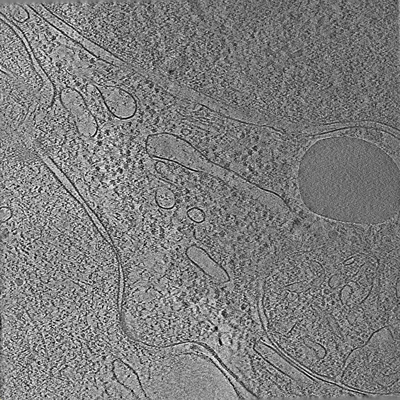 |
Tilt series of cell-cell contact of two PTK-1 cells [35 tilt series in MRC format] | Lemos M, Bezault A, Sauvanet C, Hanein D, Volkmann N [Pubmed: 36539423] [DOI: 10.1038/s41467-022-35409-9] |
1.1 GB | — |
| 2023-02-10 | 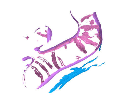 |
A surface morphometrics toolkit to quantify organellar membrane ultrastructure using cryo-electron tomography [multiple data sets in MRC format] | Barad BA, Medina M, Fuentes D, Wiseman RL, Grotjahn DA [DOI: 10.1101/2022.01.23.477440] |
126.9 GB | — |
| 2018-01-17 | 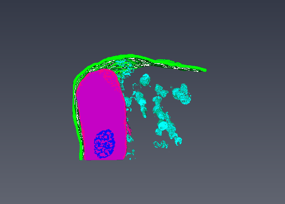 |
Serial Block Face Scanning Electron Micrscopy dataset of fetal day 64 guinea pig psoas muscle in transverse [93 micrographs in TIFF format] | Cocks ET [DOI: 10.1111/jmi.12676] |
264.4 MB | — |
| 2019-05-21 | 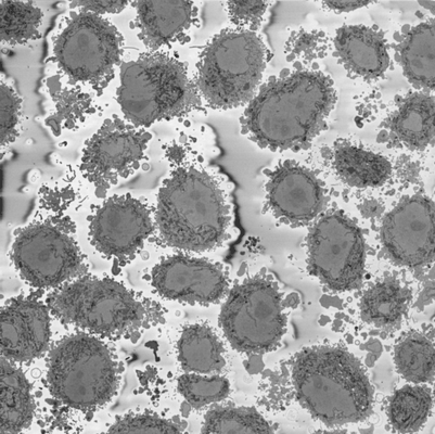 |
Serial Block Face SEM of HeLa cell pellet with 10 nm pixels and 50 nm slices (benchmark dataset) [518 micrographs in DM4 format] | Peddie CP, Jones ML, Collinson LM | 129.8 GB | — |
| 2021-02-12 | 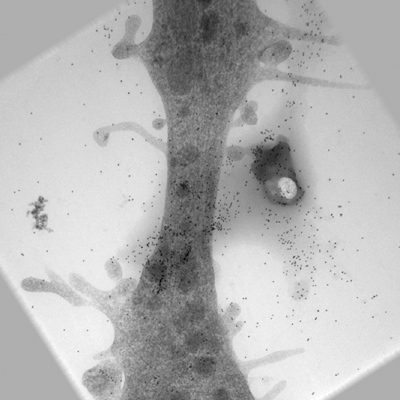 |
Ultra-high voltage electron microscope tomography tilt series of neurite section [1 tilt series in MRC format] | Nishida T, Yoshimura R, Nishi R, Imoto Y, Endo Y [Pubmed: 32133640] [DOI: 10.1111/jmi.12885] |
120.3 MB | — |
| 2018-05-30 |  |
Cryo-ET of natural chromatin from Ostreococcus tauri and Saccharyomyces cerevisiae [25 class averages in MRC format] | Cai S, Song Y, Chen C, Shi J [Pubmed: 29742050] [DOI: 10.1091/mbc.E17-07-0449] |
40.1 GB | — |
| 2021-02-12 | 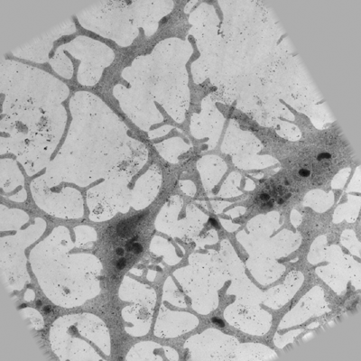 |
Ultra-high voltage electron microscope tomography tilt series of 0.7-μm-thick neurite section acquired at 6,000× magnification at an accelerating voltage of 1 MV [1 tilt series in MRC format] | Nishida T, Yoshimura R, Nishi R, Imoto Y, Endo Y [Pubmed: 32133640] [DOI: 10.1111/jmi.12885] |
120.3 MB | — |
| 2021-02-12 | 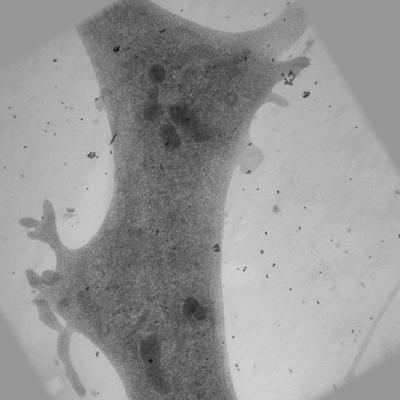 |
Ultra-high voltage electron microscope tomography tilt series of 0.7-μm-thick neurite section acquired at 15,000× magnification at an accelerating voltage of 1 MV [1 tilt series in MRC format] | Nishida T, Yoshimura R, Nishi R, Imoto Y, Endo Y [Pubmed: 32133640] [DOI: 10.1111/jmi.12885] |
120.3 MB | — |
| 2017-11-28 | 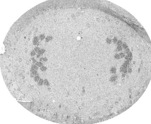 |
FIB-SEM of a dividing cell at 3.1 min after anaphase onset [1652 multi-frame micrographs composed of 1 frames each in TIFF format] | Otsuka S, Steyer AM, Schorb M, Hériché JK, Hossain MJ, Sethi S, Kueblbeck M, Schwab Y, Beck M, Ellenberg J [Pubmed: 29323269] [DOI: 10.1038/s41594-017-0001-9] |
27.0 GB | — |
| 2020-05-27 | 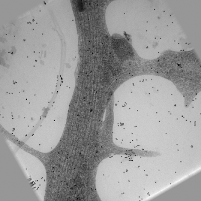 |
Ultra-high voltage electron microscope tomography tilt series of 0.7-μm-thick neurite section acquired at 20,000× magnification at an accelerating voltage of 1 MV [1 tilt series in MRC format] | Nishida T, Yoshimura R, Nishi R, Imoto Y, Endo Y [Pubmed: 32133640] [DOI: 10.1111/jmi.12885] |
120.3 MB | — |
| 2023-02-22 |  |
Light and electron microscopy continuum-resolution imaging of 3D cell cultures [19 multi-frame micrographs composed of 1 frames each in MRC format] | DImprima EDI, Garcia Montero MGM, Gawrzak, SG, Ronchi PR, Zagoriy IZ, Schwab YS, Jechlinger JS, Mahamid JM | 72.9 GB | — |
| 2017-11-30 | 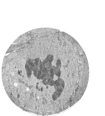 |
FIB-SEM of a dividing cell at 6.3 min after anaphase onset [3206 multi-frame micrographs composed of 1 frames each in TIFF format] | Otsuka S, Steyer AM, Schorb M, Hériché JK, Hossain MJ, Sethi S, Kueblbeck M, Schwab Y, Beck M, Ellenberg J [Pubmed: 29323269] [DOI: 10.1038/s41594-017-0001-9] |
83.2 GB | — |
| 2017-11-28 | 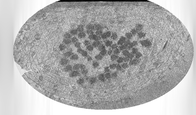 |
FIB-SEM of a dividing cell at 3.9 min after anaphase onset [2293 multi-frame micrographs composed of 1 frames each in TIFF format] | Otsuka S, Steyer AM, Schorb M, Hériché JK, Hossain MJ, Sethi S, Kueblbeck M, Schwab Y, Beck M, Ellenberg J [Pubmed: 29323269] [DOI: 10.1038/s41594-017-0001-9] |
26.8 GB | — |
| 2022-01-11 | 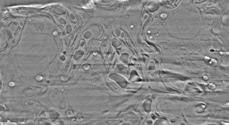 |
Cryo-FIB-SEM data on Chlamydomonas reinhardtii cells [37 micrographs in TIFF format] | Klumpe S [Pubmed: 34951584] [DOI: 10.7554/elife.70506] |
888.2 MB | — |
| 2017-11-30 | 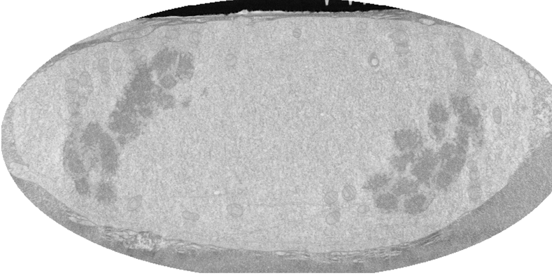 |
FIB-SEM of a dividing cell at 4.3 min after anaphase onset [1358 multi-frame micrographs composed of 1 frames each in TIFF format] | Otsuka S, Steyer AM, Schorb M, Hériché JK, Hossain MJ, Sethi S, Kueblbeck M, Schwab Y, Beck M, Ellenberg J [Pubmed: 29323269] [DOI: 10.1038/s41594-017-0001-9] |
14.0 GB | — |
| 2021-11-01 | 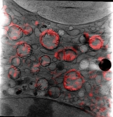 |
Cryo Soft X-ray data for tetraspeck correlation [multiple data sets in MRC format] | Groen J, Pereiro E [Pubmed: 33990802] [DOI: 10.1038/s41596-021-00522-4] |
15.1 GB | — |
| 2024-04-02 | 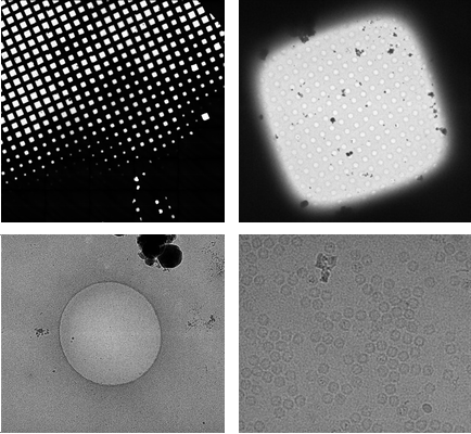 |
EMinsight: a tool to capture cryoEM microscope configuration and experimental outcomes for analysis and deposition [2605 multi-frame micrographs composed of 50 frames each in TIFF format] | Morris KL, Harrison PJ, Hatton D, Riggs S, Thiyagalingamc J | 1.2 TB | — |
| 2017-11-30 | 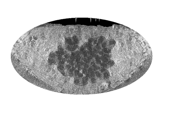 |
FIB-SEM of a dividing cell at 5.3 min after anaphase onset [1998 multi-frame micrographs composed of 1 frames each in TIFF format] | Otsuka S, Steyer AM, Schorb M, Hériché JK, Hossain MJ, Sethi S, Kueblbeck M, Schwab Y, Beck M, Ellenberg J [Pubmed: 29323269] [DOI: 10.1038/s41594-017-0001-9] |
31.2 GB | — |
| 2017-11-28 | 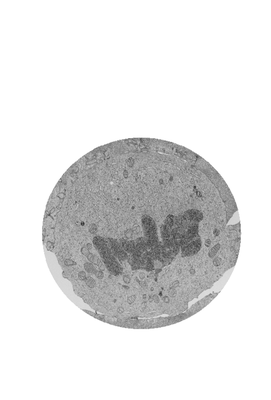 |
FIB-SEM of a dividing cell at 5.7 min after anaphase onset [2620 multi-frame micrographs composed of 1 frames each in TIFF format] | Otsuka S, Steyer AM, Schorb M, Hériché JK, Hossain MJ, Sethi S, Kueblbeck M, Schwab Y, Beck M, Ellenberg J [Pubmed: 29323269] [DOI: 10.1038/s41594-017-0001-9] |
63.0 GB | — |
| 2021-03-05 | 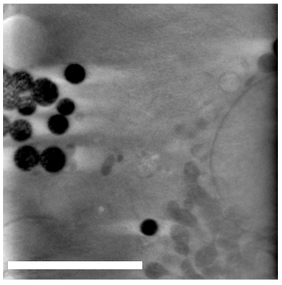 |
Dynabeads as Fiducials for SXT and SIM Correlation [700 tilt series in MRC format] | Okolo CA, Kounatidis I, Groen J, Nahas KL, Balint S, Fish T, Koronfel MA, López-Cortajarena A, Dobbie I, Pereiro E, Harkiolaki M | 1.2 GB | — |
