Electron Microscopy Public Image Archive
Electron Microscopy Public Image Archive
The EMPIAR-PDBj team at Osaka University assists Asian EM researchers with the transfer of big EM image data to EMPIAR. Instead of sending the data directly to the EBI (UK) via the internet, hard drives can also be sent to Osaka University by postal mail or via a courier service. As an alternative, internet transfer to our server in Osaka is also available. If you would like to take advantage of our submission services, please contact us first by e-mail before sending the data to us.
| Release date | Imageset | Title | Authors and references | Size | Resolution |
|---|---|---|---|---|---|
| 2022-05-20 | 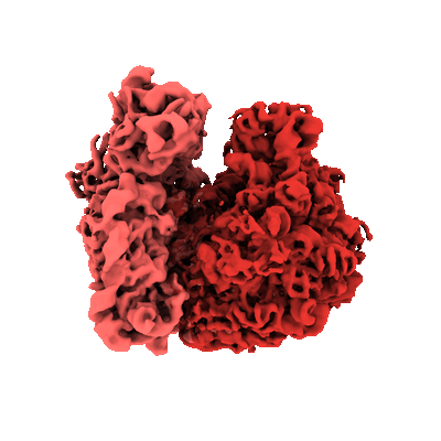 |
Parallel cryo electron tomography (PACE-tomo) of 70S ribosomes (200 kV, side-entry holder) [multiple data sets in MRC format] | Eisenstein F, Danev R [Pubmed: 36456783] [DOI: 10.1038/s41592-022-01690-1] |
222.8 GB | 5.8 - 6.5 Å |
| 2022-06-14 | 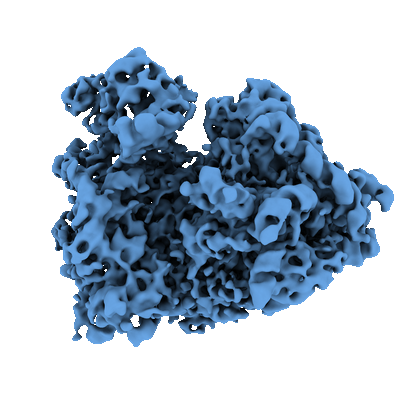 |
Parallel cryo electron tomography (PACE-tomo) of 80S ribosomes in situ [multiple data sets in MRC format] | Eisenstein F, Danev R [Pubmed: 36456783] [DOI: 10.1038/s41592-022-01690-1] |
132.3 GB | 8.2 Å |
| 2023-02-17 | 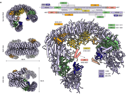 |
Cryo-EM micrographs of full-length human BIRC6 dimer [11578 multi-frame micrographs composed of 40 frames each in TIFF format] | Ehrmann JF, Grabarczyk DB, Clausen T [Pubmed: 36758105] [DOI: 10.1126/science.ade8873] |
3.0 TB | 7.0 Å |
| 2023-02-27 | 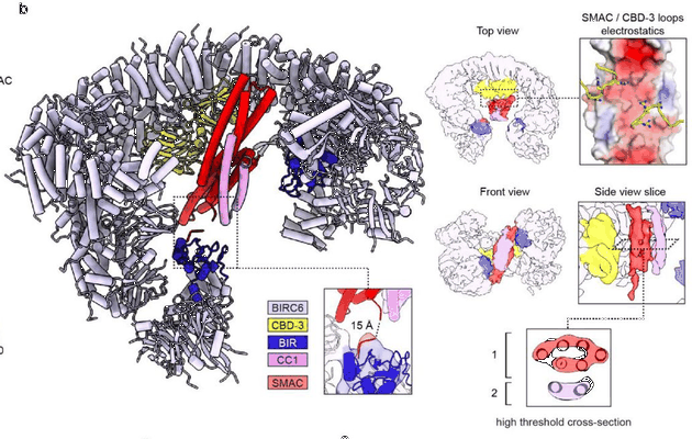 |
Cryo-EM micrographs of full-length human BIRC6 dimer with a bound DIABLO (SMAC) homodimer [multiple data sets in EER format] | Ehrmann JF, Grabarczyk DB, Clausen T [Pubmed: 36758105] [DOI: 10.1126/science.ade8873] |
24.2 TB | 7.2 Å |
| 2022-08-09 | 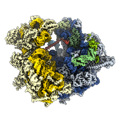 |
Single particle cryo-EM dataset of the Paranosema locustae ribosome bound to Lso2 [multiple data sets in MRC format] | Ehrenbolger K, Jespersen N, Sharma H, Sokolova YY, Tokarev YS, Vossbrinck CR, Barandun J [Pubmed: 33125369] [DOI: 10.1371/journal.pbio.3000958] |
2.0 TB | 2.9 Å |
| 2016-04-05 |  |
Subset of image stack used for 3D reconstruction [36694 micrographs in MRC format] | Egelman EH [Pubmed: 25999507] [DOI: 10.1126/science.aaa4181] |
35.9 GB | 3.8 Å |
| 2021-06-18 |  |
Single particle cryo-EM dataset of mouse heavy chain apoferritin collected on cryoARM300 with beam-image shift of 7 um [3125 multi-frame micrographs composed of 59 frames each in TIFF format] | Efremov R, Stroobants A [Pubmed: 33950012] [DOI: 10.1107/S2059798321002151] |
695.6 GB | 1.7 Å |
| 2023-05-23 | 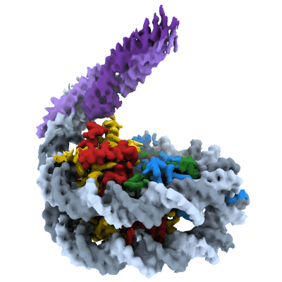 |
Cryo-EM structure of pioneer factor Cbf1 bound to the nucleosome [6339 multi-frame micrographs composed of 51 frames each in TIFF format] | Eek P, Tan S [Pubmed: 36996811] [DOI: 10.1016/j.molcel.2023.03.006] |
4.1 TB | 3.2 Å |
| 2021-11-01 | 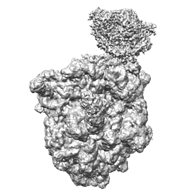 |
transcription-translation coupling complex (TEC27_NusA_NusG_CHAPSO) [1307 micrographs in MRC format] | Ebright RHE [Pubmed: 32820061] [DOI: 10.1126/science.abb5317] |
69.5 GB | 3.2 Å |
| 2021-01-15 | 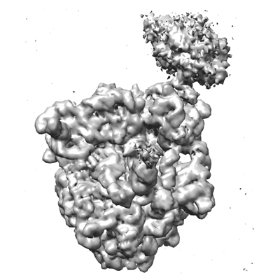 |
Transcription-translation coupling complex (TEC27_NusG_CHAPSO) [21 micrographs in MRC format] | Ebright RHE [Pubmed: 32820061] [DOI: 10.1126/science.abb5317] |
201.5 GB | 4.7 Å |
| 2023-01-30 | 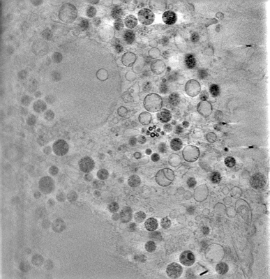 |
3D-surface reconstruction of cellular cryo-soft X-ray microscopy tomograms using semi-supervised deep learning [7 reconstructed volumes in MRC format] | Dyhr MCA, Sadeghi M, Moynova R, Knappe C, Kepsutlu Çakmak B, Werner S, Schneider G, McNally J, Noe F, Ewers H [DOI: 10.1101/2022.05.16.492055] |
23.5 GB | — |
| 2023-06-07 | 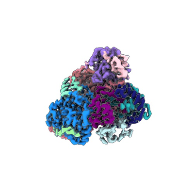 |
Cryo-electron micrographs of Hantaan virus polymerase in its apo state [2865 multi-frame micrographs composed of 50 frames each in TIFF format] | Durieux Trouilleton Q, Arragain B, Malet H [Pubmed: 37221161] [DOI: 10.1038/s41467-023-38555-w] |
660.7 GB | 3.27 Å |
| 2023-06-07 | 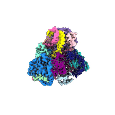 |
Cryo-electron micrographs of Hantaan virus polymerase in pre-initiation state [3261 multi-frame micrographs composed of 50 frames each in TIFF format] | Durieux Trouilleton Q, Arragain B, Malet H [Pubmed: 37221161] [DOI: 10.1038/s41467-023-38555-w] |
776.9 GB | 3.36 Å |
| 2023-06-07 | 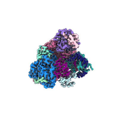 |
Cryo-electron micrographs of Hantaan virus polymerase bound to its 5' viral RNA [2547 multi-frame micrographs composed of 50 frames each in TIFF format] | Durieux Trouilleton Q, Arragain B, Malet H [Pubmed: 37221161] [DOI: 10.1038/s41467-023-38555-w] |
608.2 GB | 3.23 Å |
| 2023-06-07 | 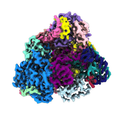 |
Cryo-electron micrographs of Hantaan virus polymerase in elongation state [3785 multi-frame micrographs composed of 50 frames each in TIFF format] | Durieux Trouilleton Q, Arragain B, Malet H [Pubmed: 37221161] [DOI: 10.1038/s41467-023-38555-w] |
927.5 GB | 3.14 Å |
| 2022-01-12 | 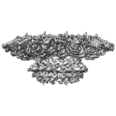 |
Motion corrected micrographs - purified Dot/Icm T4SS particles [3590 micrographs in MRC format] | Durie CL, Sheedlo MJ, Chung JM, Byrne BG, Su M, Knight T, Swanson M, Lacy DB, Ohi MD [Pubmed: 32876045] [DOI: 10.7554/eLife.59530] |
190.4 GB | 3.7 Å |
| 2023-02-28 | 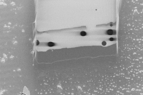 |
Cryo pFIB/SEM of PEG beads (test sample) [193 micrographs in TIFF format] | Dumoux M, Glen T, Smith JLR, Ho EML, Perdigão LMA, Pennington A, Klumpe S, Yee NBY, Farmer DA, Lai PYA, Bowles W, Kelley R, Plitzko JM, Wu L, Basham M, Clare DK, Siebert CA, Darrow MC, Naismith JH, Grange M [Pubmed: 36805107] [DOI: 10.7554/elife.83623] |
5.3 GB | — |
| 2023-02-28 | 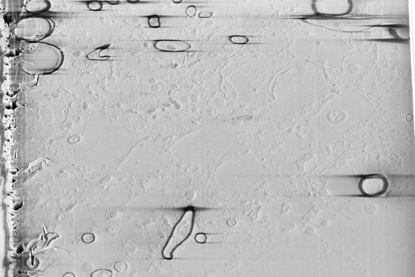 |
Cryo serial FIB SEM of mouse brain tissue [59 micrographs in TIFF format] | Dumoux M, Glen T, Smith JLR, Ho EML, Perdigão LMA, Pennington A, Klumpe S, Yee NBY, Farmer DA, Lai PYA, Bowles W, Kelley R, Plitzko JM, Wu L, Basham M, Clare DK, Siebert CA, Darrow MC, Naismith JH, Grange M [Pubmed: 36805107] [DOI: 10.7554/elife.83623] |
3.0 GB | — |
| 2023-02-17 | 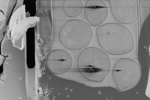 |
Cryo serial FIB/SEM of Saccharomyces cerevisiae [109 micrographs in TIFF format] | Dumoux M, Glen T, Smith JLR, Ho EML, Perdigão LMA, Pennington A, Klumpe S, Yee NBY, Farmer DA, Lai PYA, Bowles W, Kelley R, Plitzko JM, Wu L, Basham M, Clare DK, Siebert CA, Darrow MC, Naismith JH, Grange M [Pubmed: 36805107] [DOI: 10.7554/elife.83623] |
4.8 GB | — |
| 2023-02-17 | 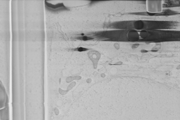 |
Cryo serial FIB/SEM of Vero cells [46 micrographs in TIFF format] | Dumoux M, Glen T, Smith JLR, Ho EML, Perdigão LMA, Pennington A, Klumpe S, Yee NBY, Farmer DA, Lai PYA, Bowles W, Kelley R, Plitzko JM, Wu L, Basham M, Clare DK, Siebert CA, Darrow MC, Naismith JH, Grange M [Pubmed: 36805107] [DOI: 10.7554/elife.83623] |
7.1 GB | — |
| 2023-02-17 | 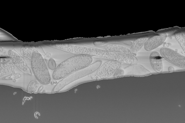 |
Cryo serial FIB/SEM of Rhodospirillum rubrum [43 micrographs in TIFF format] | Dumoux M, Glen T, Smith JLR, Ho EML, Perdigão LMA, Pennington A, Klumpe S, Yee NBY, Farmer DA, Lai PYA, Bowles W, Kelley R, Plitzko JM, Wu L, Basham M, Clare DK, Siebert CA, Darrow MC, Naismith JH, Grange M [Pubmed: 36805107] [DOI: 10.7554/elife.83623] |
5.7 GB | — |
| 2023-02-28 | 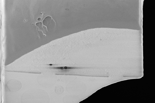 |
Cryo serial FIB/SEM of HeLa cells [46 micrographs in TIFF format] | Dumoux M, Glen T, Smith JLR, Ho EML, Perdigão LMA, Pennington A, Klumpe S, Yee NBY, Farmer DA, Lai PYA, Bowles W, Kelley R, Plitzko JM, Wu L, Basham M, Clare DK, Siebert CA, Darrow MC, Naismith JH, Grange M [Pubmed: 36805107] [DOI: 10.7554/elife.83623] |
6.7 GB | — |
| 2023-02-28 | 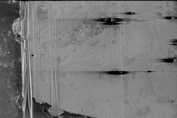 |
Cryo serial FIB/SEM of mouse heart tissue [136 micrographs in TIFF format] | Dumoux M, Glen T, Smith JLR, Ho EML, Perdigão LMA, Pennington A, Klumpe S, Yee NBY, Farmer DA, Lai PYA, Bowles W, Kelley R, Plitzko JM, Wu L, Basham M, Clare DK, Siebert CA, Darrow MC, Naismith JH, Grange M [Pubmed: 36805107] [DOI: 10.7554/elife.83623] |
18.0 GB | — |
| 2023-02-28 | 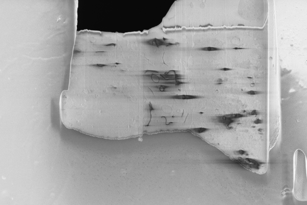 |
Cryo serial FIB/SEM of RPE-1 cells [18 micrographs in TIFF format] | Dumoux M, Glen T, Smith JLR, Ho EML, Perdigão LMA, Pennington A, Klumpe S, Yee NBY, Farmer DA, Lai PYA, Bowles W, Kelley R, Plitzko JM, Wu L, Basham M, Clare DK, Siebert CA, Darrow MC, Naismith JH, Grange M [Pubmed: 36805107] [DOI: 10.7554/elife.83623] |
826.6 MB | — |
| 2019-07-25 | 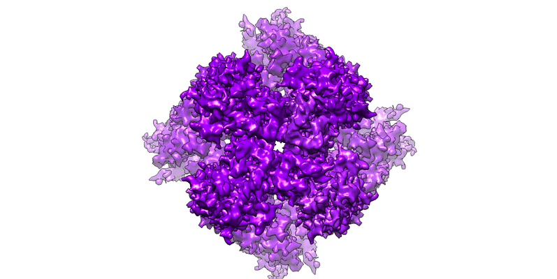 |
Open state structure of the full-length TRPV2 cation channel with a resolved pore turret domain [2447 multi-frame micrographs composed of 50 frames each in MRCS format] | Dosey TL, Wang Z, Fan G [Pubmed: 30598551] [DOI: 10.1038/s41594-018-0168-8] |
934.8 GB | 3.6 Å |
