Electron Microscopy Public Image Archive
Electron Microscopy Public Image Archive
The EMPIAR-PDBj team at Osaka University assists Asian EM researchers with the transfer of big EM image data to EMPIAR. Instead of sending the data directly to the EBI (UK) via the internet, hard drives can also be sent to Osaka University by postal mail or via a courier service. As an alternative, internet transfer to our server in Osaka is also available. If you would like to take advantage of our submission services, please contact us first by e-mail before sending the data to us.
| Release date | Imageset | Title | Authors and references | Size | Resolution |
|---|---|---|---|---|---|
| 2022-12-13 | 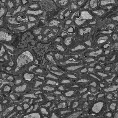 |
Focused ion beam-scanning electron microscopy links pathological myelin outfoldings to axonal changes in mice lacking Plp1 or Mag [940 micrographs in TIFF format] | Steyer AM, Möbius W [Pubmed: 36354016] [DOI: 10.1002/glia.24290] |
12.5 GB | — |
| 2022-12-13 | 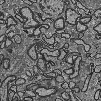 |
Focused ion beam-scanning electron microscopy links pathological myelin outfoldings to axonal changes in mice lacking Plp1 or Mag [1290 micrographs in TIFF format] | Steyer AM, Möbius W [Pubmed: 36354016] [DOI: 10.1002/glia.24290] |
22.4 GB | — |
| 2018-07-06 | 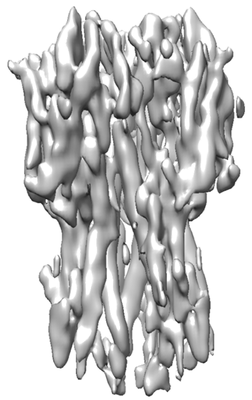 |
Single particle cryoEM of hemagglutinin with spot-to-plunge time of 500ms [multiple data sets in MRC format] | Noble AJ, Wei H, Dandey VP, Zhang Z, Potter CS, Carragher B [Pubmed: 30250056] [DOI: 10.1038/s41592-018-0139-3] |
51.1 GB | — |
| 2021-05-21 | 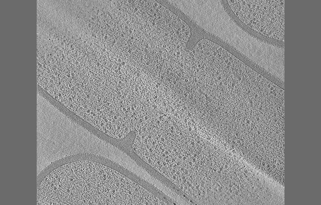 |
Tilt series of dividing vegetative and sporulating cells of Bacillus subtilis from the manuscript - Khanna et al., 2021 [multiple data sets in MRC format] | Khanna K, Lopez-Garrido J, Sugie J, Pogliano K, Villa E [Pubmed: 34018921] [DOI: 10.7554/eLife.62204] |
8.7 GB | — |
| 2022-03-14 | 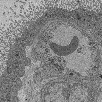 |
SBF SEM of Human term placental villi [multiple data sets in TIFF format] | Lewis RM [DOI: 10.1101/2022.01.26.477815] |
90.2 GB | — |
| 2018-08-09 |  |
Three-dimensional nanostructure of an intact microglia cell [multiple data sets in TIFF and IMOD formats] | Bolasco G, Weinhard L, Boissonnet T, Neujahr R, Gross CT [DOI: 10.3389/fnana.2018.00105] |
7.4 GB | — |
| 2020-10-14 |  |
FIB SEM images of a Zebrafish hindbrain macrophage containing 2 Toxoplasma gondii tachizoites [multiple data sets in TIFF format] | Peddie CJ, Domart MC, Collinson L [Pubmed: 32461265] [DOI: 10.1242/dmm.043091] |
1.1 TB | — |
| 2020-12-09 |  |
TEM images of a Zebrafish hindbrain cells containing Toxoplasma gondii tachizoites [multiple data sets in TIFF format] | Domart MC, Collinson L [Pubmed: 32461265] [DOI: 10.1242/dmm.043091] |
3.4 GB | — |
| 2022-03-28 |  |
Representative data from Near-native state imaging by cryo-soft-X-ray tomography reveals remodelling of cytoplasmic vesicles and mitochondria during HSV-1 infection [14 reconstructed volumes in MRC format] | Nahas KLN, Connor VC, Scherer KM, Kaminski CF, Harkiolaki M, Crump CM, Graham SC [DOI: 10.1101/2021.10.11.463900] |
9.9 GB | — |
| 2021-01-22 |  |
SBF SEM images of a Zebrafish hindbrain macrophage containing 2 Toxoplasma gondii tachizoites [multiple data sets in DM4 format] | Peddie CJ, Domart MC, Collinson L [Pubmed: 32461265] [DOI: 10.1242/dmm.043091] |
16.5 GB | — |
| 2020-12-09 |  |
SBF SEM images of a Zebrafish hindbrain macrophage containing a replicating Toxoplasma gondii tachizoite [multiple data sets in DM4 format] | Peddie CJ, Domart MC, Collinson L [Pubmed: 32461265] [DOI: 10.1242/dmm.043091] |
338.4 GB | — |
| 2020-12-09 | 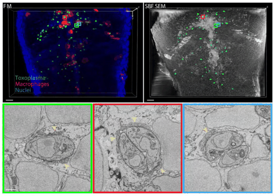 |
SBF SEM images of a Zebrafish hindbrain containing several Toxoplasma gondii tachizoites at different stages of replication [multiple data sets in DM4 and TIFF formats] | Peddie CJ, Domart MC, Collinson L [Pubmed: 32461265] [DOI: 10.1242/dmm.043091] |
175.2 GB | — |
| 2021-10-27 | 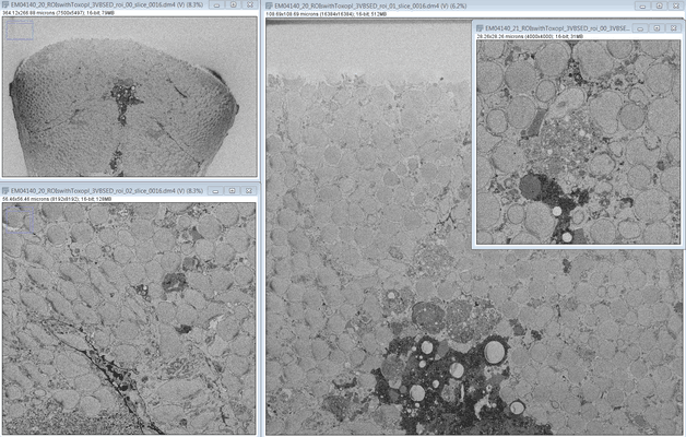 |
SBF SEM images of a Zebrafish hindbrain containing several Toxoplasma gondii tachizoites at different stages of replication [multiple data sets in DM4 format] | Peddie CJ, Domart MC, Collinson L [Pubmed: 32461265] [DOI: 10.1242/dmm.043091] |
553.9 GB | — |
| 2024-02-15 | 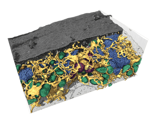 |
FIB-SEM dataset of a human bone osteosarcoma epithelial cell (U2-OS) [1168 micrographs in TIFF format] | Belevich I, Schertel A, Zaversek T, Szyrynska N, Saarnio S, Jokitalo E | 5.2 GB | — |
| 2022-03-21 |  |
CryoET of E. coli prepared with the Waffle Method [1504 multi-frame micrographs composed of 14 frames each in TIFF format] | Kelley K, Raczkowski AM, Klykov O, Jaroenlak P, Bobe D, Kopylov M, Eng ET, Bhabha G, Potter CS, Carragher B, Noble AJ [Pubmed: 35387991] [DOI: 10.1038/s41467-022-29501-3] |
94.0 GB | — |
| 2022-12-13 |  |
Focused ion beam-scanning electron microscopy links pathological myelin outfoldings to axonal changes in mice lacking Plp1 or Mag [1598 micrographs in TIFF format] | Steyer AM, Möbius W [Pubmed: 36354016] [DOI: 10.1002/glia.24290] |
31.3 GB | — |
| 2018-10-25 |  |
CryoET of bacterial RNA polymerase with several detergents [multiple data sets in MRC and RAW TEXT formats] | Chen J, Noble AJ, Kang JY, Darst SA [DOI: 10.1016/j.yjsbx.2019.100005] |
207.5 GB | — |
| 2022-03-15 | 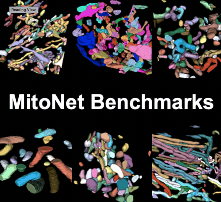 |
Seven benchmark datasets of instance segmentation of mitochondria: 6 diverse volume EM + 1 TEM (100 images) datasets [multiple data sets in TIFF format] | Narayan K, Conrad RW | 4.1 GB | — |
| 2022-12-13 |  |
Focused ion beam-scanning electron microscopy links pathological myelin outfoldings to axonal changes in mice lacking Plp1 or Mag [1553 micrographs in TIFF format] | Steyer AM, Möbius W [Pubmed: 36354016] [DOI: 10.1002/glia.24290] |
22.3 GB | — |
| 2023-11-06 |  |
Single particle cryo-EM structure of RIG-I:RNA:Riplet ternary complex [3420 multi-frame micrographs composed of 40 frames each in TIFF format] | Wang W, Pyle AM [DOI: 10.1038/s41467-023-42982-0] |
1.5 TB | — |
| 2022-12-13 |  |
Focused ion beam-scanning electron microscopy links pathological myelin outfoldings to axonal changes in mice lacking Plp1 or Mag [910 micrographs in TIFF format] | Steyer AM, Möbius W [Pubmed: 36354016] [DOI: 10.1002/glia.24290] |
13.7 GB | — |
| 2022-12-13 |  |
Focused ion beam-scanning electron microscopy links pathological myelin outfoldings to axonal changes in mice lacking Plp1 or Mag [1186 micrographs in TIFF format] | Steyer AM, Möbius W [Pubmed: 36354016] [DOI: 10.1002/glia.24290] |
28.2 GB | — |
| 2022-11-18 |  |
A high-throughput electron tomography workflow reveals over-elongated centrioles in relapsed-refractory multiple myeloma [multiple data sets in MRC format] | Köhrer S, Dittrich T, Schorb M, Weinhold N, Haberbosch I, Börmel M, Pajor G [Pubmed: 36452870] [DOI: 10.1016/j.crmeth.2022.100322] |
2.0 TB | — |
| 2023-11-13 |  |
Test subset: In situ cryo-ET dataset of Chlamydomonas reinhardtii prepared using cryo-plasmaFIB milling [18 tilt series in MRC format] | Kelley R, Zhang X, Obr M, Khavnekar S, Righetto R, Waltz F, Wietrzynski W, Michael A, Tagiltsev G, Beck F, Zhong E, Wan W, Briggs J, Plitzko J, Engel B, Kotecha A [Pubmed: 37613825] [DOI: 10.1093/micmic/ozad067.480] |
293.7 GB | — |
| 2022-11-23 |  |
Cryo-electron tomograms of RPE1 cells with comprehensive annotation of actin filaments and microtubules [multiple data sets in TIFF and MRC formats] | Cheng DWC, Goetz SK, Mahamid J [Pubmed: 36690741] [DOI: 10.1038/s41592-022-01746-2] |
32.9 GB | — |
