Tilt series of dividing vegetative and sporulating cells of Bacillus subtilis from the manuscript - Khanna et al., 2021 [multiple data sets in MRC format]
Publication:
Asymmetric localization of the cell division machinery during Bacillus subtilis sporulation
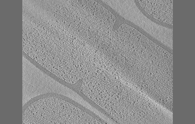
Tilt series of a dividing vegetative cell of Bacillus subtilis (Figure 3A of the manuscript Khanna et al., 2021)
Files:
Available download options:
Archives and downloads the selected files into an uncompressed zip file.
Depending on the number and size of files to be downloaded, this can take an enormous amount of time.
Also, this feature is unstable and may not work properly, and checksums of downloaded files are not verified.
We recommend using rsync, aspera, globus, etc.
Download a list of selected files.
It is possible to download files by specifying the file list with rsync command, etc.
Tomogram of a dividing vegetative cell of Bacillus subtilis (Figure 4A of the manuscript Khanna et al., 2021)
Files:
Available download options:
Archives and downloads the selected files into an uncompressed zip file.
Depending on the number and size of files to be downloaded, this can take an enormous amount of time.
Also, this feature is unstable and may not work properly, and checksums of downloaded files are not verified.
We recommend using rsync, aspera, globus, etc.
Download a list of selected files.
It is possible to download files by specifying the file list with rsync command, etc.
Tomogram of a dividing vegetative cell of Bacillus subtilis FtsZ-linker(Q-rich) strain (Figure 5A and 6A of the manuscript Khanna et al., 2021)
Files:
Available download options:
Archives and downloads the selected files into an uncompressed zip file.
Depending on the number and size of files to be downloaded, this can take an enormous amount of time.
Also, this feature is unstable and may not work properly, and checksums of downloaded files are not verified.
We recommend using rsync, aspera, globus, etc.
Download a list of selected files.
It is possible to download files by specifying the file list with rsync command, etc.
Tomogram of a dividing sporulating cell of Bacillus subtilis (Figure 7A of the manuscript Khanna et al., 2021)
Files:
Available download options:
Archives and downloads the selected files into an uncompressed zip file.
Depending on the number and size of files to be downloaded, this can take an enormous amount of time.
Also, this feature is unstable and may not work properly, and checksums of downloaded files are not verified.
We recommend using rsync, aspera, globus, etc.
Download a list of selected files.
It is possible to download files by specifying the file list with rsync command, etc.
Tomogram of a dividing sporulating cell of Bacillus subtilis (Figure 7 - figure supplement 4 of the manuscript Khanna et al., 2021)
Files:
Available download options:
Archives and downloads the selected files into an uncompressed zip file.
Depending on the number and size of files to be downloaded, this can take an enormous amount of time.
Also, this feature is unstable and may not work properly, and checksums of downloaded files are not verified.
We recommend using rsync, aspera, globus, etc.
Download a list of selected files.
It is possible to download files by specifying the file list with rsync command, etc.
Tomogram of a dividing sporulating cell of Bacillus subtilis SpoIIE null mutant (Figure 8A of the manuscript Khanna et al., 2001)
Files:
Available download options:
Archives and downloads the selected files into an uncompressed zip file.
Depending on the number and size of files to be downloaded, this can take an enormous amount of time.
Also, this feature is unstable and may not work properly, and checksums of downloaded files are not verified.
We recommend using rsync, aspera, globus, etc.
Download a list of selected files.
It is possible to download files by specifying the file list with rsync command, etc.