Electron Microscopy Public Image Archive
Electron Microscopy Public Image Archive
The EMPIAR-PDBj team at Osaka University assists Asian EM researchers with the transfer of big EM image data to EMPIAR. Instead of sending the data directly to the EBI (UK) via the internet, hard drives can also be sent to Osaka University by postal mail or via a courier service. As an alternative, internet transfer to our server in Osaka is also available. If you would like to take advantage of our submission services, please contact us first by e-mail before sending the data to us.
| Release date | Imageset | Title | Authors and references | Size | Resolution |
|---|---|---|---|---|---|
| 2014-08-07 | 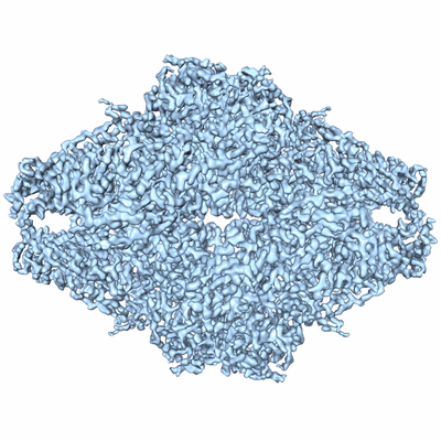 |
Structure of β-galactosidase at 3.2-Å resolution obtained by cryo-electron microscopy [multiple data sets in MRC and DM4 formats] | Bartesaghi A, Matthies D, Banerjee S, Merk A, Subramaniam S [Pubmed: 25071206] [DOI: 10.1073/pnas.1402809111] |
442.5 GB | 3.2 Å |
| 2023-04-13 | 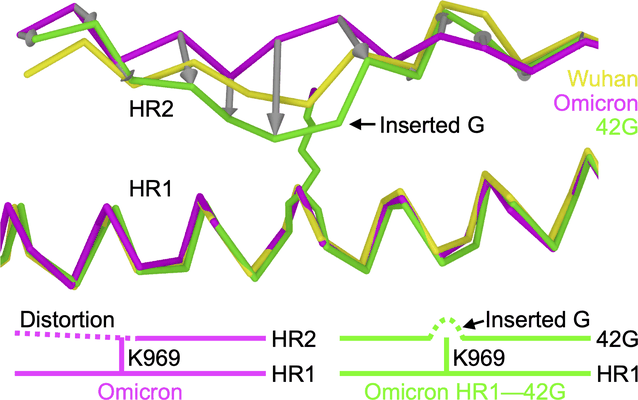 |
Structure-based design of a SARS-CoV-2 Omicron-specific inhibitor [multiple data sets in TIFF format] | Yang K, Brunger AT [Pubmed: 36940324] [DOI: 10.1073/pnas.2300360120] |
13.5 TB | 2.51 Å |
| 2022-05-17 |  |
Structures of positive allosteric modulator-bound and unbound active human calcium-sensing receptor [13082 multi-frame micrographs composed of 60 frames each in TIFF format] | Park J, Zuo H, Frangaj A, Fu Z, Yen LY, Zhang Z, Mosyak L, Slavkovich VN, Liu J, Ray KM, Cao B, Vallese F, Geng Y, Chen S, Grassucci R, Dandey VP, Tan YZ, Eng E, Lee Y, Kloss B, Liu Z, Hendrickson WA, Potter CS, Carragher B, Graziano J, Conigrave AD, Frank J, Clarke OB, Fan QR [Pubmed: 34916296] [DOI: 10.1073/pnas.2115849118] |
3.8 TB | 2.7 Å |
| 2022-09-12 |  |
Structures of the Cyanobacterial Phycobilisome in the Light-harvesting and Photoprotected States [multiple data sets in MRC and TIFF formats] | Sauer PV, Dominguez-Martin MA, Kerfeld CA [Pubmed: 36045294] [DOI: 10.1038/s41586-022-05156-4] |
16.7 TB | 2.1 - 3.5 Å |
| 2020-08-25 |  |
Structures of the human mitochondrial ribosome bound to EF-G1 reveal distinct features of mitochondrial translation elongation [stack of 6649 particles in MRC format] | Bhargava K, Datta PP, Kaushal PS, Keshavan P, Spremulli LL, Banavali NK [Pubmed: 32737313] [DOI: 10.1038/s41467-020-17715-2] |
753.5 GB | 2.96 - 3.96 Å |
| 2018-08-08 | 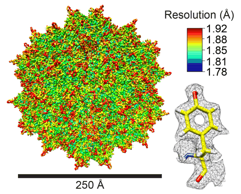 |
Sub-2 Å Single-Particle Cryo-EM Reconstruction of AAV2-L336C [multiple data sets in MRC and MRCS formats] | Tan YZ, Aiyer S, Mietzsch M, Hull JA, McKenna R, Grieger J, Samulski RJ, Baker TS, Agbandje-McKenna M, Lyumkis D [Pubmed: 30194371] [DOI: 10.1038/s41467-018-06076-6] |
5.7 TB | 1.86 Å |
| 2020-06-29 | 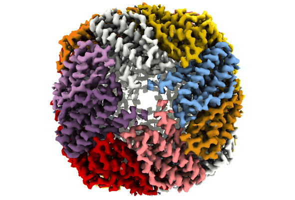 |
Sub-3 Å Apoferritin Structure Determined With Full Range of Phase Shifts Using A Single Position Of Volta Phase Plate [multiple data sets in MRC and TIFF formats] | Li K [Pubmed: 30928614] [DOI: 10.1016/j.jsb.2019.03.007] |
778.8 GB | 2.51 - 2.94 Å |
| 2015-04-24 |  |
Sub-tomogram average of a mammalian F-type ATP synthase monomer [multiple data sets in DM4 format] | Jiko C, Davies KM, Shinzawa-Itoh K, Tani K, Maeda S, Mills DJ, Tsukihara T, Fujiyoshi Y, Kuehlbrandt W, Gerle C [Pubmed: 25815585] [DOI: 10.7554/eLife.06119] |
8.9 GB | 24.0 Å |
| 2015-11-19 |  |
Sub-tomogram averaging in RELION [7 class averages in MRC format] | Bharat TA, Scheres SH [Pubmed: 27685097] [DOI: 10.1038/nprot.2016.124] |
842.8 GB | 13.0 Å |
| 2021-11-16 |  |
Subcellular architecture collodaria photosymbiosis [7 multi-frame micrographs composed of 1000 frames each in TIFF format] | Decelle JD [Pubmed: 34499794] [DOI: 10.1111/1462-2920.15766] |
10.3 GB | — |
| 2020-07-06 |  |
Subnanometer-resolution structure determination in situ by a hybrid subtomogram averaging - single particle cryoEM - workflow - on TMV [4 tilt series in MRC format] | Sanchez RM, Zhang Y, Chen W, Dietrich L, Kudryashev M [Pubmed: 32709843] [DOI: 10.1038/s41467-020-17466-0] |
37.5 GB | 5.24 Å |
| 2016-04-05 |  |
Subset of image stack used for 3D reconstruction [36694 micrographs in MRC format] | Egelman EH [Pubmed: 25999507] [DOI: 10.1126/science.aaa4181] |
35.9 GB | 3.8 Å |
| 2020-08-19 |  |
Subtomogram averaging and classification of SARS-CoV-2 Spike Proteins on intact virions [multiple data sets in TIFF and MRC formats] | Ke Z, Oton J, Cortese M, Zila V, Zivanov J, Lu JM, Peukes J, Scheres SHW, Briggs JAG [Pubmed: 32805734] [DOI: 10.1038/s41586-020-2665-2] |
372.4 GB | 7.7 - 9.9 Å |
| 2021-06-18 |  |
Subtomograms of nucleosomes extracted from cryo-tomograms of Drosophila melanogaster embryos [1 subtomograms in EM format] | Harastani M, Eltsov M, Leforestier A, Jonic S [Pubmed: 34095222] [DOI: 10.3389/fmolb.2021.663121] |
666.3 MB | — |
| 2024-02-29 |  |
Sulfolobus acidocaldarius s-layer SlaA [multiple data sets in TIFF format] | Gambelli L, McLaren MJ, Sanders K, Gaines M, Clark L, Gold VAM, Kattnig D, Sikora M, Hanus C, Isupov M, Daum B [Pubmed: 38251732] [DOI: 10.7554/eLife.84617] |
4.5 TB | 3.1 - 3.9 Å |
| 2024-02-28 |  |
Sulfolobus acidocaldarius s-layer SlaA cryoET dataset [multiple data sets in MRC format] | Gambelli L, McLaren MJ, Sanders K, Gaines M, Clark L, Gold VAM, Kattnig D, Sikora M, Hanus C, Isupov M, Daum B [Pubmed: 38251732] [DOI: 10.7554/eLife.84617] |
945.1 GB | 11.2 Å |
| 2018-05-11 |  |
T. acidophilum 20S proteasome core movies obtained using Talos Arctica operating at 200 kV equipped with a K2 – image shift used for exposure target navigation [262 multi-frame micrographs composed of 68 frames each in MRC format] | Herzik Jr MA, Wu M, Lander GC [Pubmed: 28991891] [DOI: 10.1038/nmeth.4461] |
945.5 GB | 3.3 Å |
| 2018-05-11 | 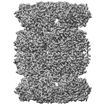 |
T. acidophilum 20S proteasome core movies obtained using Talos Arctica operating at 200 kV equipped with a K2 – stage position used for exposure target navigation [317 multi-frame micrographs composed of 68 frames each in MRC format] | Herzik Jr MA, Wu M, Lander GC [Pubmed: 28991891] [DOI: 10.1038/nmeth.4461] |
140.9 GB | 3.1 Å |
| 2015-03-06 | 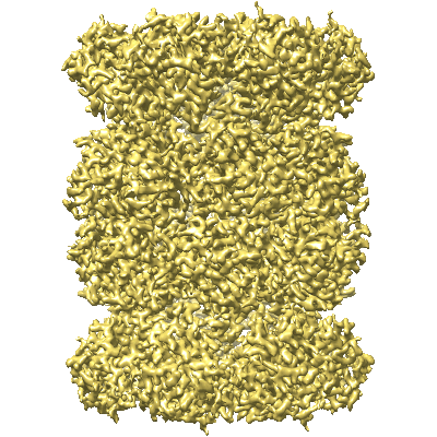 |
T20S Proteasome at 2.8 Å Resolution [multiple data sets in MRC format] | Campbell M, Veesler D, Cheng A, Potter CS, Carragher B [Pubmed: 25760083] [DOI: 10.7554/eLife.06380] |
2.0 TB | 2.8 Å |
| 2018-05-14 | 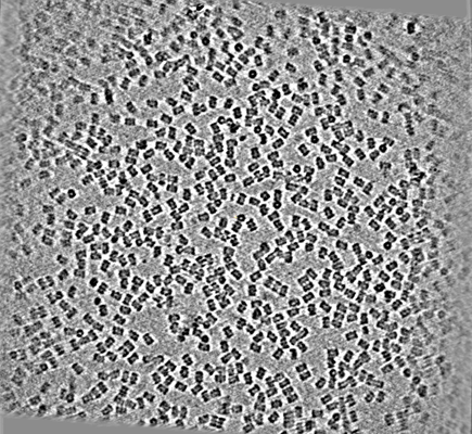 |
T20S proteasome single particle [586 micrographs in MRC format] | Noble AJ, Dandey VP, Wei H, Brasch J, Chase J, Acharya P, Tan YZ, Zhang Z, Kim LY, Scapin G, Rapp M, Eng ET, Rice MJ, Cheng A, Negro CJ, Shapiro L, Kwong PD, Jeruzalmi D, des Georges A, Potter CS, Carragher B [Pubmed: 29809143] [DOI: 10.7554/eLife.34257] |
116.1 GB | — |
| 2020-07-21 | 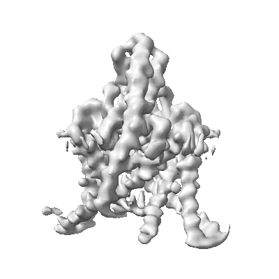 |
TASK2 in MSP1D1 lipid nanodisc at pH6.5 [3024 multi-frame micrographs composed of 50 frames each in TIFF format] | Li B, Brohawn SG [Pubmed: 32999458] [DOI: 10.1038/s41586-020-2770-2] |
2.0 TB | 3.45 Å |
| 2020-07-21 | 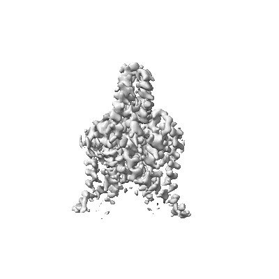 |
TASK2 in MSP1D1 lipid nanodisc at pH8.5 [3470 multi-frame micrographs composed of 50 frames each in TIFF format] | Li B, Brohawn SG [Pubmed: 32999458] [DOI: 10.1038/s41586-020-2770-2] |
2.3 TB | 3.52 Å |
| 2020-12-09 | 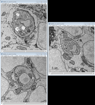 |
TEM images of a Zebrafish hindbrain cells containing Toxoplasma gondii tachizoites [multiple data sets in TIFF format] | Domart MC, Collinson L [Pubmed: 32461265] [DOI: 10.1242/dmm.043091] |
3.4 GB | — |
| 2020-08-12 |  |
TEM tomograms of Drosophila tracheal terminal cells during subcellular tube formation [multiple data sets in TIFF and MRC formats] | Mathew R, Rios-Barrera LD, Machado P, Schwab Y, Leptin M [Pubmed: 32657472] [DOI: 10.15252/embj.2020105332] |
366.7 GB | — |
| 2013-12-04 | 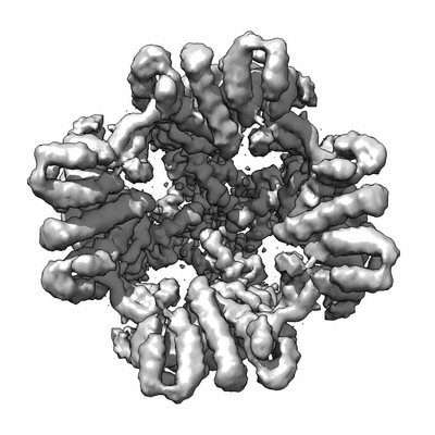 |
TRPV1 dataset taken on a K2 direct electron detector [multiple data sets in MRC format] | Liao M, Cao E, Julius D, Cheng Y [Pubmed: 24305160] [DOI: 10.1038/nature12822] |
6.3 TB | 3.275 Å |
