
Electron Microscopy Public Image Archive
Electron Microscopy Public Image Archive
Our EMPIAR-PDBj will suspend the following operations from August 10, 2024 (Saturday) to August 18, 2024 (Sunday):
Web servers and data downloads will remain available during this period.
Additionally, please be aware that EMPIAR-EBI will also be closed for annotation operations from July 25, 2024 (Thursday) to August 18, 2024 (Sunday).
Thank you.
The EMPIAR-PDBj team at Osaka University assists Asian EM researchers with the transfer of big EM image data to EMPIAR. Instead of sending the data directly to the EBI (UK) via the internet, hard drives can also be sent to Osaka University by postal mail or via a courier service. As an alternative, internet transfer to our server in Osaka is also available. If you would like to take advantage of our submission services, please contact us first by e-mail before sending the data to us.
| Release date | Imageset | Title | Authors and references | Size | Resolution |
|---|---|---|---|---|---|
| 2023-02-17 | 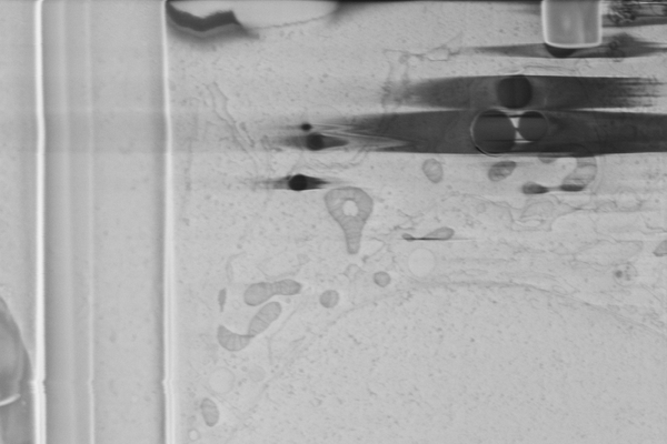 |
Cryo serial FIB/SEM of Vero cells [46 micrographs in TIFF format] | Dumoux M, Glen T, Smith JLR, Ho EML, Perdigão LMA, Pennington A, Klumpe S, Yee NBY, Farmer DA, Lai PYA, Bowles W, Kelley R, Plitzko JM, Wu L, Basham M, Clare DK, Siebert CA, Darrow MC, Naismith JH, Grange M [Pubmed: 36805107] [DOI: 10.7554/elife.83623] |
7.1 GB | — |
| 2016-06-28 | 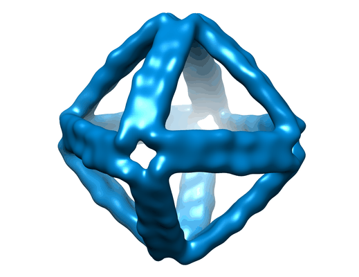 |
Designer nanoscale DNA assemblies programmed from the top down [100 micrographs in MRC format] | Veneziano R, Ratanalert S, Zhang K, Zhang F, Yan H, Chiu W, Bathe M [Pubmed: 27229143] [DOI: 10.1126/science.aaf4388] |
7.3 GB | 25.0 Å |
| 2018-08-09 | 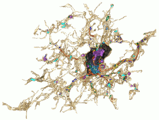 |
Three-dimensional nanostructure of an intact microglia cell [multiple data sets in TIFF and IMOD formats] | Bolasco G, Weinhard L, Boissonnet T, Neujahr R, Gross CT [DOI: 10.3389/fnana.2018.00105] |
7.4 GB | — |
| 2021-10-18 | 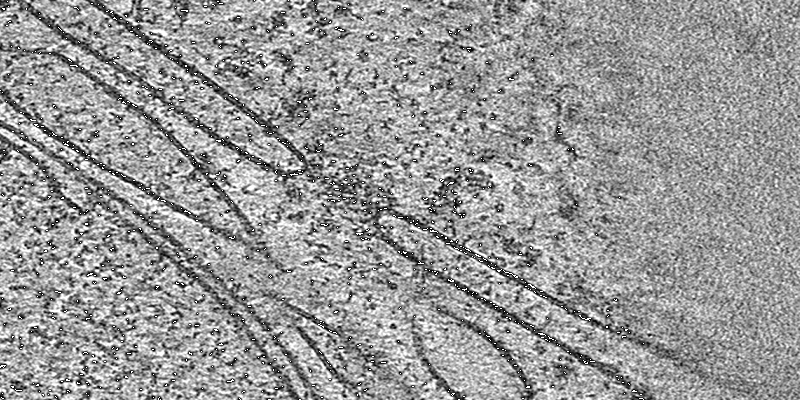 |
Cryo electron tomography of FIB-milled lamella of human DLD-1 cells [3 tilt series in MRC format] | Schuller AP, Wojtynek M, Mankus D, Tatli M, Kronenberg-Tenga R, Regmi SG, Dip PV, Lytton-Jean AKR, Brignole EJ, Dasso M, Weis K, Medalia O, Schwartz TU [Pubmed: 34646014] [DOI: 10.1038/s41586-021-03985-3] |
8.0 GB | 33.0 - 39.0 Å |
| 2021-10-18 | 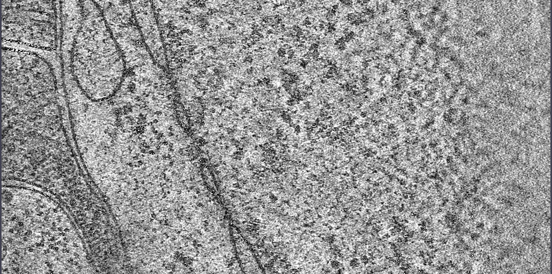 |
Cryo electron tomography of FIB-milled lamella of human DLD-1 cells [3 tilt series in MRC format] | Schuller AP, Wojtynek M, Mankus D, Tatli M, Kronenberg-Tenga R, Regmi SG, Dip PV, Lytton-Jean AKR, Brignole EJ, Dasso M, Weis K, Medalia O, Schwartz TU [Pubmed: 34646014] [DOI: 10.1038/s41586-021-03985-3] |
8.0 GB | 33.0 - 39.0 Å |
| 2015-11-04 | 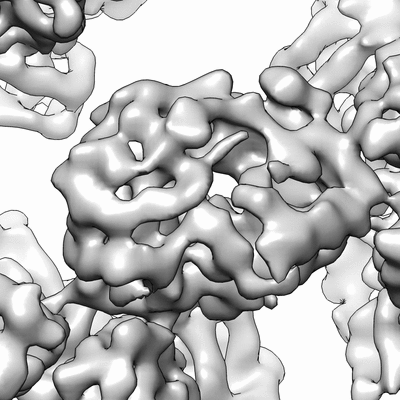 |
In vitro assembled bacteriophage phi6 polymerase complex [stack of 798 particles in MRC format] | Ilca SL, Kotecha A, Sun X, Poranen MP, Stuart DI, Huiskonen JT [Pubmed: 26534841] [DOI: 10.1038/ncomms9843] |
8.1 GB | 7.9 Å |
| 2023-05-12 | 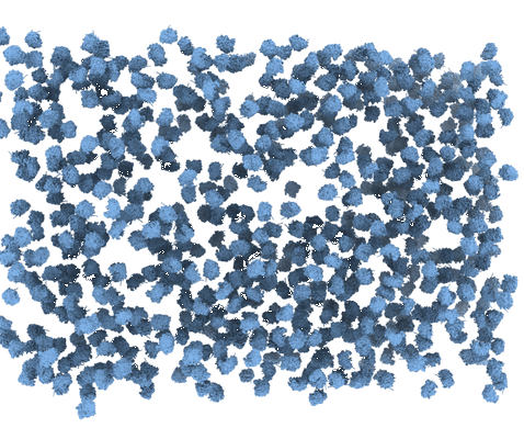 |
Quantification of gallium cryo-FIB milling damage in biological lamella [multiple data sets in MRC format] | Lucas BA, Grigorieff N [Pubmed: 37216561] [DOI: 10.1073/pnas.2301852120] |
8.1 GB | — |
| 2022-10-17 | 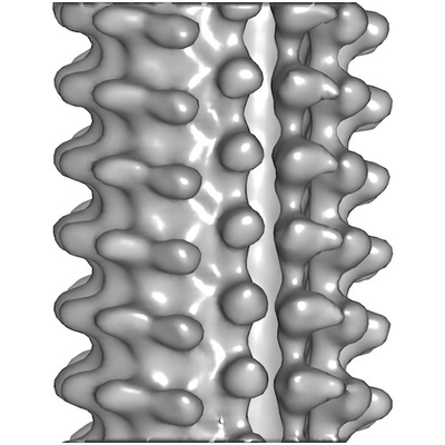 |
Cryo-electron tomography of microtubules assembled from purified porcine brain tubulin in the presence of GTP [4 reconstructed volumes in MRC format] | Guyomar C, Bousquet C, Ku S, Heumann J, Guilloux G, Gaillard N, Heichette C, Duchesne L, Steinmetz MO, Gibeaux R, Chrétien D [Pubmed: 36503602] [DOI: 10.7554/eLife.83021] |
8.4 GB | 25.0 Å |
| 2024-01-16 | 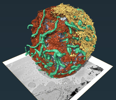 |
REEP3 and REEP4 determine the tubular morphology of the endoplasmic reticulum during mitosis [multiple data sets in DM4 and TIFF formats] | Belevich I, Jokitalo E [Pubmed: 30995177] [DOI: 10.1091/mbc.e18-11-0698] |
8.7 GB | — |
| 2021-05-21 | 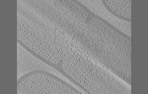 |
Tilt series of dividing vegetative and sporulating cells of Bacillus subtilis from the manuscript - Khanna et al., 2021 [multiple data sets in MRC format] | Khanna K, Lopez-Garrido J, Sugie J, Pogliano K, Villa E [Pubmed: 34018921] [DOI: 10.7554/eLife.62204] |
8.7 GB | — |
| 2024-07-16 | 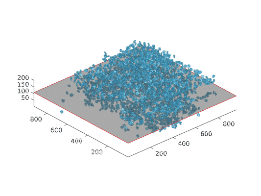 |
Cryo electron tomography of chromatin condensed by PRC1-CBX8 [multiple data sets in MRC format] | Uckelmann M, Levina V, Taveneau C, de Marco A, Davidovich C | 8.7 GB | — |
| 2024-06-04 | 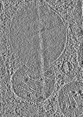 |
Cryo-electron tomogram of mitochondria in cryo-FIB milled mouse embryonic fibroblasts with OPA1 overexpression [258 multi-frame micrographs composed of 4 frames each in TIFF format] | Fry MY, Navarro PP, Ananda VY, Ge Y, McDonald JL, Hakim P, Luce BE, Lugo CM, Chao LH [Pubmed: 38225406] [DOI: 10.1038/s44318-024-00027-2] |
8.8 GB | — |
| 2015-04-24 | 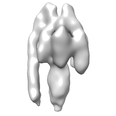 |
Sub-tomogram average of a mammalian F-type ATP synthase monomer [multiple data sets in DM4 format] | Jiko C, Davies KM, Shinzawa-Itoh K, Tani K, Maeda S, Mills DJ, Tsukihara T, Fujiyoshi Y, Kuehlbrandt W, Gerle C [Pubmed: 25815585] [DOI: 10.7554/eLife.06119] |
8.9 GB | 24.0 Å |
| 2017-12-18 | 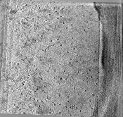 |
CryoET of Mtb 20S proteasome single particle [multiple data sets in MRC format] | Noble AJ, Dandey VP, Wei H, Brasch J, Chase J, Acharya P, Tan YZ, Zhang Z, Kim LY, Scapin G, Rapp M, Eng ET, Rice MJ, Cheng A, Negro CJ, Shapiro L, Kwong PD, Jeruzalmi D, des Georges A, Potter CS, Carragher B [Pubmed: 29809143] [DOI: 10.7554/eLife.34257] |
9.1 GB | — |
| 2021-06-09 | 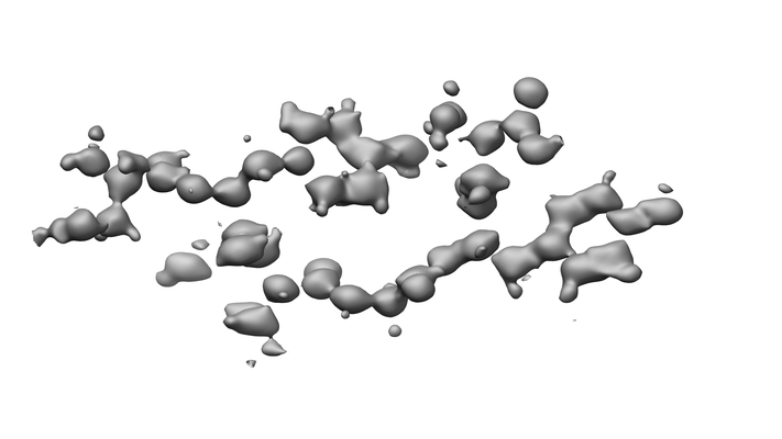 |
120kV MicroED structure of FUS (37-42) SYSGYS solved from merged datasets at 0.60 A [8 diffraction images in SMV format] | Zhou H, Luo F, Luo Z, Li D, Liu C, Li X [Pubmed: 31334636] [DOI: 10.1021/acs.analchem.9b01162] |
9.2 GB | 0.6 Å |
| 2021-06-09 | 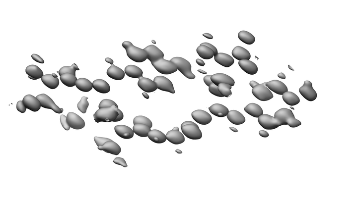 |
200kV MicroED structure of FUS (37-42) SYSGYS solved from merged datasets at 0.65 A [8 diffraction images in SMV format] | Zhou H, Luo F, Luo Z, Li D, Liu C, Li X [Pubmed: 31334636] [DOI: 10.1021/acs.analchem.9b01162] |
9.5 GB | 0.65 Å |
| 2022-10-07 | 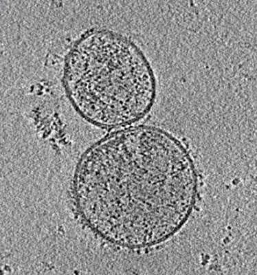 |
Tilt series of SARS-CoV-2 spike-bearing virus-like particles (VLPs) interacting with hACE2-bearing extracellular vesicles (tEVs), showing various intermediate states of the SARS-CoV-2 spike protein [6 tilt series in MRC format] | Marcink TC, Porotto M, des Georges A, Moscona A [Pubmed: 35984891] [DOI: 10.1126/sciadv.abo3153] |
9.6 GB | — |
| 2021-06-18 | 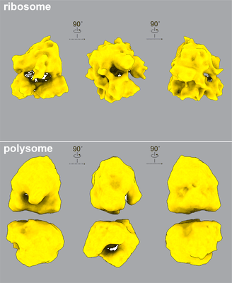 |
cryo-FIB and cryo-ET study of ribosome and polysome structures in E. coli [3 tilt series in MRC format] | Xiang YJ, Chang YJ, Liu J, Jacobs-Wagner C [Pubmed: 34186018] [DOI: 10.1016/j.cell.2021.05.037] |
9.7 GB | 20.0 - 27.0 Å |
| 2024-06-05 | 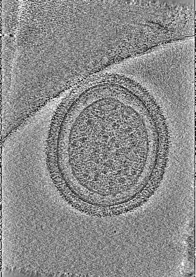 |
Native structure of a human GBP defense complex for cell-autonomous immunity to infection [280 tilt series in TIFF format] | MacMicking JDM, Zhu SZ [Pubmed: 38422126] [DOI: 10.1126/science.abm9903] |
9.7 GB | 17.0 Å |
| 2022-03-28 | 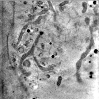 |
Representative data from Near-native state imaging by cryo-soft-X-ray tomography reveals remodelling of cytoplasmic vesicles and mitochondria during HSV-1 infection [14 reconstructed volumes in MRC format] | Nahas KLN, Connor VC, Scherer KM, Kaminski CF, Harkiolaki M, Crump CM, Graham SC [DOI: 10.1101/2021.10.11.463900] |
9.9 GB | — |
| 2021-11-26 | 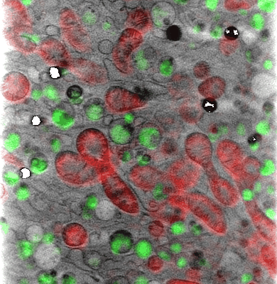 |
Reconstructed cryo soft X-ray tomography dataset of a treated NIH-3T3 cell with corresponding correlated cryo-3D-SIM channels [300 reconstructed volumes in TIFF format] | Groen J, Pereiro E [Pubmed: 34909150] [DOI: 10.1039/d1sc04183e] |
10.1 GB | — |
| 2018-07-27 | 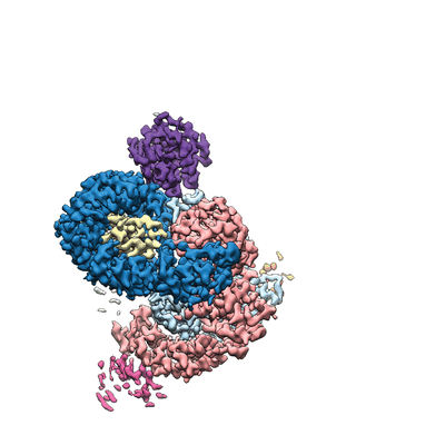 |
Single particle cryo-EM dataset of the flexible and variable oligomeric state complex AP-1:Arf1:tetherin-HIV-Nef [stack of 53841 particles in MRCS format] | Morris KL, Buffalo CZ, Hurley JH [Pubmed: 30053425] [DOI: 10.1016/j.cell.2018.07.004] |
10.1 GB | 3.73 - 4.27 Å |
| 2021-11-16 | 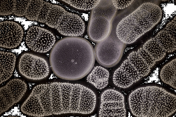 |
Subcellular architecture collodaria photosymbiosis [7 multi-frame micrographs composed of 1000 frames each in TIFF format] | Decelle JD [Pubmed: 34499794] [DOI: 10.1111/1462-2920.15766] |
10.3 GB | — |
| 2022-05-03 | 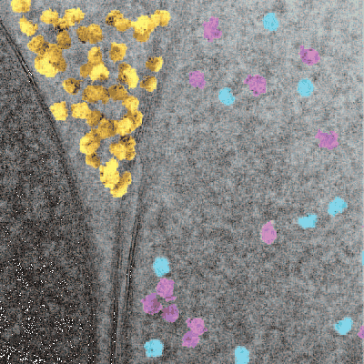 |
In situ single particle classification reveals distinct 60S maturation intermediates in cells [multiple data sets in MRC format] | Lucas BA, Zhang K, Loerch S, Grigorieff N [DOI: 10.1101/2022.04.10.487797] |
10.5 GB | — |
| 2014-06-19 | 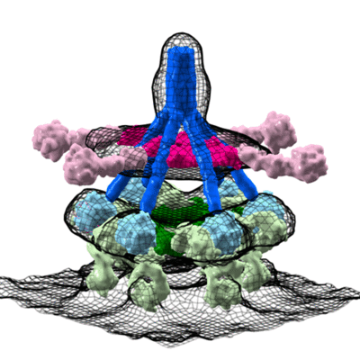 |
Cryo-electron tomography average of an C1-IgG complex [27 class averages in MRC format] | Diebolder CA, Beurskens FJ, de Jong RN, Koning RI, Strumane K, Lindorfer MA, Voorhorst M, Ugurlar D, Rosati S, Heck AJR, van de Winkel JGJ, Wilson IA, Koster AJ, Taylor RP, Ollmann-Saphire E, Burton DR, Schuurman J, Gros P, Parren PWHI [Pubmed: 24626930] [DOI: 10.1126/science.1248943] |
10.8 GB | 66.0 Å |
