Electron Microscopy Public Image Archive
Electron Microscopy Public Image Archive
The EMPIAR-PDBj team at Osaka University assists Asian EM researchers with the transfer of big EM image data to EMPIAR. Instead of sending the data directly to the EBI (UK) via the internet, hard drives can also be sent to Osaka University by postal mail or via a courier service. As an alternative, internet transfer to our server in Osaka is also available. If you would like to take advantage of our submission services, please contact us first by e-mail before sending the data to us.
| Release date | Imageset | Title | Authors and references | Size | Resolution |
|---|---|---|---|---|---|
| 2022-02-24 |  |
Affinity-purified PoxtA in complex with 70S ribosomes from Enterococcus faecalis [3640 multi-frame micrographs composed of 40 frames each in TIFF format] | Crowe-McAuliffe CT, Murina V, Hauryliuk V, Wilson DN [Pubmed: 35387982] [DOI: 10.1038/s41467-022-29274-9] |
321.8 GB | 2.4 Å |
| 2018-08-15 |  |
The first reconstruction of beta-galactosidase solved by cryoARM200 [1338 multi-frame micrographs composed of 49 frames each in TIFF format] | Kato T, Terehara N, Namba K | 321.4 GB | 2.6 Å |
| 2023-09-22 |  |
Cryo-EM particle stacks of the small dataset of ∆KsgA 30S plus KsgA sample [stack of 588015 particles in MRCS format] | Sun J, Kinman LF, Jahagirdar D, Ortega J, Davis JH [Pubmed: 37653244] [DOI: 10.1038/s41594-023-01078-5] |
320.1 GB | 2.8 Å |
| 2022-08-19 |  |
Single particle cryo-EM of the human CST•Polα/Primase (POLA1 FL) complex in a recruitment state [multiple data sets in MRC format] | Cai SW [Pubmed: 35578024] [DOI: 10.1038/s41594-022-00766-y] |
317.1 GB | 16.0 Å |
| 2024-04-17 | 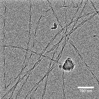 |
CryoEM movies of IAPP-S20G timecourse (6 week, FT11) [2350 multi-frame micrographs composed of 30 frames each in TIFF format] | Wilkinson M, Xu Y, Thacker D, Taylor AIP, Fisher DG, Gallardo RU, Radford SE, Ranson NA [Pubmed: 38134875] [DOI: 10.1016/j.cell.2023.11.025] |
316.2 GB | 3.1 - 3.4 Å |
| 2022-12-09 | 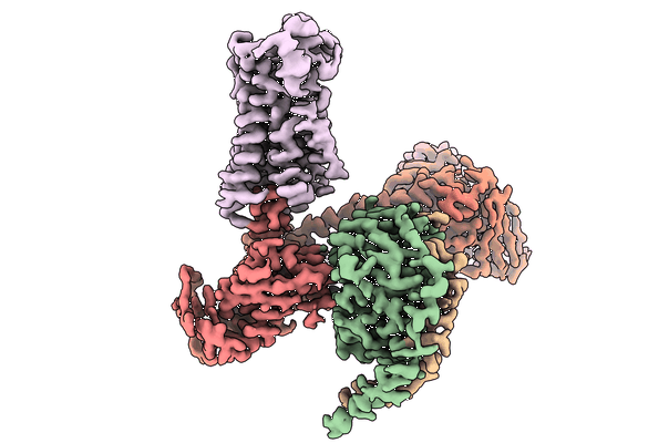 |
CryoEM structure of hM3Dq-miniGq in complex with CNO [3577 micrographs in MRC format] | Fay J, Roth B, Zhang S [Pubmed: 36450989] [DOI: 10.1038/s41586-022-05489-0] |
314.1 GB | 2.79 Å |
| 2021-01-22 | 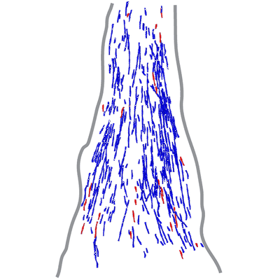 |
Actin filament structure from focal adhesions of mouse embryonic fibroblasts [7 tilt series in MRC format] | Martins B, Sorrentino S, Chung WL, Tatli M, Medalia O, Eibauer M [Pubmed: 33476550] [DOI: 10.1016/j.str.2020.12.014] |
314.1 GB | 17.0 Å |
| 2022-09-09 | 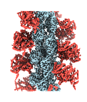 |
cryo-EM structure of the ADP state wild type myosin-15-F-actin complex [1624 multi-frame micrographs composed of 24 frames each in TIFF format] | Gong R, Bird JE, Alushin GM [Pubmed: 35857845] [DOI: 10.1126/sciadv.abl4733] |
313.4 GB | 3.63 - 4.15 Å |
| 2023-10-03 | 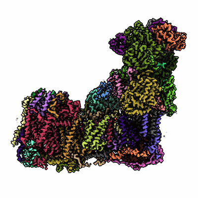 |
Single particle cryo-EM dataset of mitochondrial complex I from Mus musculus inhibited by IACS-2858 - 1 [1957 multi-frame micrographs composed of 1 frames each in MRC format] | Chung I, Hirst J [Pubmed: 33990335] [DOI: 10.1126/sciadv.abg4000] |
312.6 GB | 3.04 Å |
| 2020-06-30 | 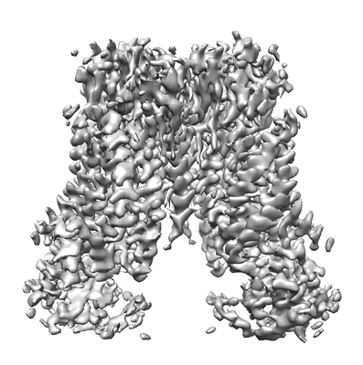 |
Cryo-EM structure of TMEM16F in digitonin with calcium bound [stack of 2505 particles in MRC format] | Feng S, Dang S, Han TW, Ye W, Jin P, Cheng T, Li J, Jan YN, Jan LY, Cheng Y [Pubmed: 31291589] [DOI: 10.1016/j.celrep.2019.06.023] |
308.8 GB | 3.5 Å |
| 2024-04-17 | 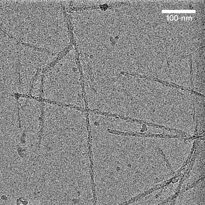 |
CryoEM movies of IAPP-S20G timecourse (3 week repeat, FT14) [2317 multi-frame micrographs composed of 40 frames each in TIFF format] | Wilkinson M, Xu Y, Thacker D, Taylor AIP, Fisher DG, Gallardo RU, Radford SE, Ranson NA [Pubmed: 38134875] [DOI: 10.1016/j.cell.2023.11.025] |
308.5 GB | 3.0 Å |
| 2021-10-19 | 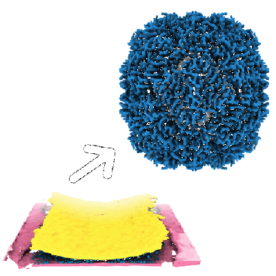 |
Apoferritin, TMV and T20S proteasome in nanofluidic channels [multiple data sets in TIFF format] | Huber ST, Sarajlic E, Huijink R, Weis F, Evers WH, Jakobi AJ [Pubmed: 35060902] [DOI: 10.7554/eLife.72629] |
308.3 GB | 3.0 - 5.4 Å |
| 2021-11-30 |  |
E. coli MsbA in DDM in complex with G247 [3485 micrographs in MRC format] | Thélot FA, Liao M [Pubmed: 34554829] [DOI: 10.1126/science.abi9009] |
307.0 GB | 3.55 Å |
| 2020-09-02 |  |
Native Pyruvate Dehydrogenase Complex from Neurospora crassa [4887 micrographs in MRC format] | Forsberg BO, Aibara S, Howard RJ, Mortezaei N, Lindahl E [Pubmed: 32938938] [DOI: 10.1038/s41467-020-18401-z] |
305.5 GB | 4.1 - 4.4 Å |
| 2022-11-17 |  |
Defocus and Volta potential phase plate cryo-electron tomography of S. pombe cryo-FIB lamellae with comprehensive annotations of structures and macromolecules [multiple data sets in TIFF and MRC formats] | Goetz SK, Mahamid J [Pubmed: 36690741] [DOI: 10.1038/s41592-022-01746-2] |
305.5 GB | 9.3 - 34.0 Å |
| 2022-01-18 |  |
High-resolution mapping of metal ions reveals principles of surface layer assembly in Caulobacter crescentus cells [multiple data sets in TIFF and MRC formats] | von Kugelgen A, Bharat TAM [Pubmed: 34800371] [DOI: 10.1016/j.str.2021.10.012] |
304.4 GB | 4.37 Å |
| 2022-05-25 |  |
SARS-CoV-2 spike protein S:D614G + S:A222V variant [4841 micrographs in MRC format] | Ginex T, Marco-Marín C, Wieczór M, Mata CP, Krieger J, Ruiz-Rodriguez P, López-Redondo ML, Francés-Gómez C, Melero R, Sánchez-Sorzano CÓ, Martínez M, Gougeard N, Forcada-Nadal A, Zamora-Caballero S, Gozalbo-Rovira R, Sanz-Frasquet C, Arranz R, Bravo J, Rubio V, Marina A, Geller R, Comas I, Gil C, Coscolla M, Orozco M, Llácer JL, Carazo JM [Pubmed: 35816514] [DOI: 10.1371/journal.ppat.1010631] |
303.8 GB | 3.4 Å |
| 2022-08-12 |  |
In situ cryo-electron tomography of the C. reinhardtii ciliary transition zone [multiple data sets in MRC and EM formats] | van den Hoek H, Klena N, Jordan MA, Alvarez Viar G, Righetto RD, Schaffer M, Erdmann PS, Wan W, Geimer S, Plitzko JM, Baumeister W, Pigino G, Hamel V, Guichard P, Engel BD [Pubmed: 35901159] [DOI: 10.1126/science.abm6704] |
302.0 GB | — |
| 2017-12-18 |  |
CryoET of apoferritin single particle [multiple data sets in MRC format] | Noble AJ, Dandey VP, Wei H, Brasch J, Chase J, Acharya P, Tan YZ, Zhang Z, Kim LY, Scapin G, Rapp M, Eng ET, Rice MJ, Cheng A, Negro CJ, Shapiro L, Kwong PD, Jeruzalmi D, des Georges A, Potter CS, Carragher B [Pubmed: 29809143] [DOI: 10.7554/eLife.34257] |
301.1 GB | — |
| 2021-03-05 |  |
Structure of the human U4/U6.U5 tri-snRNP [4812 micrographs in MRC format] | Stark H [Pubmed: 26912367] [DOI: 10.1126/science.aad2085] |
300.9 GB | 7.0 Å |
| 2021-08-27 |  |
AL amyloid fibril from a lambda 3 light chain [1964 multi-frame micrographs composed of 40 frames each in TIFF format] | Radamaker L, Schmidt M, Fändrich M [Pubmed: 33558536] [DOI: 10.1038/s41467-021-21126-2] |
297.8 GB | 3.2 - 3.4 Å |
| 2023-01-03 |  |
Movies of apoferritin collected at different dose rates on the Direct Electron Apollo direct detector - 15 eps [972 multi-frame micrographs composed of 76 frames each in TIFF format] | Peng R, Fu X, Mendez JH, Randolph PS, Bammes BE, Stagg SM [Pubmed: 36578473] [DOI: 10.1016/j.yjsbx.2022.100080] |
297.2 GB | — |
| 2024-04-12 |  |
Cryo-electron tomography data of Emiliania huxleyi virus 201 for subtomogram averaging [283 tilt series in TIFF format] | Homola M, Büttner CR, Füzik T, Nováček J, Chaillet M, Förster F, Plevka P [DOI: 10.1101/2023.06.30.547180] |
294.8 GB | 13.0 Å |
| 2018-12-13 |  |
Beta-2-microglobulin fibrils with multiple polymorphs formed at pH 2 [5549 micrographs in MRC format] | Iadanza MG [Pubmed: 30375379] [DOI: 10.1038/s41467-018-06761-6] |
294.3 GB | 3.975 Å |
| 2023-11-13 |  |
Test subset: In situ cryo-ET dataset of Chlamydomonas reinhardtii prepared using cryo-plasmaFIB milling [18 tilt series in MRC format] | Kelley R, Zhang X, Obr M, Khavnekar S, Righetto R, Waltz F, Wietrzynski W, Michael A, Tagiltsev G, Beck F, Zhong E, Wan W, Briggs J, Plitzko J, Engel B, Kotecha A [Pubmed: 37613825] [DOI: 10.1093/micmic/ozad067.480] |
293.7 GB | — |
