Electron Microscopy Public Image Archive
Electron Microscopy Public Image Archive
The EMPIAR-PDBj team at Osaka University assists Asian EM researchers with the transfer of big EM image data to EMPIAR. Instead of sending the data directly to the EBI (UK) via the internet, hard drives can also be sent to Osaka University by postal mail or via a courier service. As an alternative, internet transfer to our server in Osaka is also available. If you would like to take advantage of our submission services, please contact us first by e-mail before sending the data to us.
| Release date | Imageset | Title | Authors and references | Size | Resolution |
|---|---|---|---|---|---|
| 2022-07-18 |  |
Structure of SARS-CoV-2 M protein in lipid nanodiscs [7588 multi-frame micrographs composed of 1251 frames each in EER format] | Dolan KA, Dutta M, Kern DM, Kotecha A, Voth GA, Brohawn SG [Pubmed: 36264056] [DOI: 10.7554/eLife.81702] |
4.0 TB | 3.52 Å |
| 2022-07-12 |  |
Single particle Data of the activated B. subtilis ClpC arranged as a tetramer of hexamers [4002 multi-frame micrographs composed of 40 frames each in MRC format] | Morrale FE, Meinhart A, Haselbach D, Clausen T [Pubmed: 35662409] [DOI: 10.1016/j.cell.2022.05.009] |
3.1 TB | 3.7 - 10.0 Å |
| 2024-01-23 |  |
Cryo electron microscopy micrographs of high molecular weight fractions from yeast native cell extracts [multiple data sets in MRC format] | Schmidt L, Tueting C, Kyrilis F, Hamdi F, Semchonok DA, Kastritis PL [DOI: 10.1101/2022.07.15.498668] |
4.7 TB | 3.78 - 8.1 Å |
| 2022-11-11 |  |
Tilt series of SARS-CoV-2 virions, Wuhan/Alpha/Beta/Delta variants [multiple data sets in MRC format] | Calder LJ, Calcraft T, Hussain S, Harvey R, Rosenthal PB [Pubmed: 36357779] [DOI: 10.1038/s42003-022-04183-1] |
120.5 GB | 27.75 - 44.4 Å |
| 2022-07-12 | 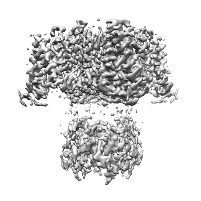 |
Human Kv1.3 [multiple data sets in TIFF format] | Meyerson JR, Selvakumar P [Pubmed: 35788586] [DOI: 10.1038/s41467-022-31285-5] |
1.4 TB | 2.89 Å |
| 2022-07-08 | 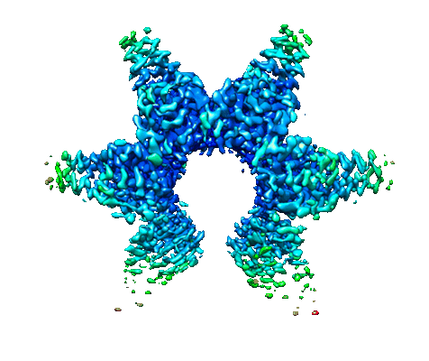 |
Minimal protein-only RNase P structure reveals insights into tRNA precursor recognition and catalysis [2370 multi-frame micrographs composed of 48 frames each in TIFF format] | Teramoto T, Koyasu T, Adachi N, Kawasaki M, Moriya T, Numata T, Senda T, Kakuta Y [Pubmed: 34339732] [DOI: 10.1016/j.jbc.2021.101028] |
2.0 TB | 3.62 Å |
| 2022-08-08 | 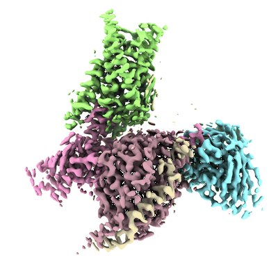 |
Cryo-EM structure of MrgD-Gi complex with beta-alanine [17087 multi-frame micrographs composed of 66 frames each in TIFF format] | Kawamoto A [Pubmed: 35840655] [DOI: 10.1038/s42003-022-03668-3] |
4.1 TB | 3.1 - 3.2 Å |
| 2022-07-27 | 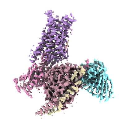 |
Cryo-EM structure of apo-state MrgD-Gi complex [14985 multi-frame micrographs composed of 66 frames each in TIFF format] | Kawamoto A [Pubmed: 35840655] [DOI: 10.1038/s42003-022-03668-3] |
3.5 TB | 2.9 - 3.1 Å |
| 2022-07-01 |  |
Single-particle cryo-EM dataset of the Vairimorpha necatrix ribosome [3284 multi-frame micrographs composed of 32 frames each in TIFF format] | Barandun J, Hunziker M, Vossbrinck CR, Klinge S [Pubmed: 31332387] [DOI: 10.1038/s41564-019-0514-6] |
1.0 TB | 3.26 - 3.7 Å |
| 2022-08-09 |  |
Single particle cryo-EM dataset of the Paranosema locustae ribosome bound to Lso2 [multiple data sets in MRC format] | Ehrenbolger K, Jespersen N, Sharma H, Sokolova YY, Tokarev YS, Vossbrinck CR, Barandun J [Pubmed: 33125369] [DOI: 10.1371/journal.pbio.3000958] |
2.0 TB | 2.9 Å |
| 2022-11-11 |  |
Cryo-EM structure of full-length human immunoglobulin M [43331 micrographs in MRC format] | Chen Q, Rosenthal PB, Tolar P [Pubmed: 36274064] [DOI: 10.1038/s41467-022-34090-2] |
2.6 TB | 4.4 Å |
| 2022-08-12 |  |
In situ cryo-electron tomography of the C. reinhardtii ciliary transition zone [multiple data sets in MRC and EM formats] | van den Hoek H, Klena N, Jordan MA, Alvarez Viar G, Righetto RD, Schaffer M, Erdmann PS, Wan W, Geimer S, Plitzko JM, Baumeister W, Pigino G, Hamel V, Guichard P, Engel BD [Pubmed: 35901159] [DOI: 10.1126/science.abm6704] |
302.0 GB | — |
| 2022-07-12 |  |
Non-conventional octameric structure of C-phycocyanin [2036 multi-frame micrographs composed of 49 frames each in TIFF format] | Minato T, Teramoto T, Adachi N, Hung NK, Yamada K, Kawasaki M, Akutsu M, Moriya T, Senda T, Ogo S, Kakuta Y, Yoon KS [Pubmed: 34716405] [DOI: 10.1038/s42003-021-02767-x] |
1.8 TB | 3.06 - 3.71 Å |
| 2022-07-26 |  |
Human Kv1.3 with A0194009G09 nanobodies [multiple data sets in TIFF format] | Meyerson JR, Selvakumar P [Pubmed: 35788586] [DOI: 10.1038/s41467-022-31285-5] |
3.3 TB | 3.25 Å |
| 2022-07-26 |  |
Human Kv1.3 with a Fab-ShK fusion [5079 multi-frame micrographs composed of 48 frames each in TIFF format] | Meyerson JR, Selvakumar P [Pubmed: 35788586] [DOI: 10.1038/s41467-022-31285-5] |
2.0 TB | 3.39 Å |
| 2023-02-14 |  |
Nudaurelia capensis omega virus maturation intermediate captured at pH6.25 (insect cell expressed VLPs) [multiple data sets in MRC format] | Castells-Graells R, Hesketh EL, Johnson JE, Ranson NA, Lawson DM, Lomonossoff GP | 8.6 TB | 4.8 Å |
| 2023-02-13 |  |
Nudaurelia capensis omega virus maturation intermediates captured at pH5.6 (insect cell expressed VLPs) [multiple data sets in MRC format] | Castells-Graells R, Hesketh EL, Johnson JE, Ranson NA, Lawson DM, Lomonossoff GP | 13.7 TB | 4.8 Å |
| 2022-09-20 |  |
Human amino acid transporter EAAT2 [multiple data sets in TIFF format] | Kato T, Kusakizako T, Yamashita K, Nishizawa T, Nureki O [Pubmed: 35953475] [DOI: 10.1038/s41467-022-32442-6] |
1.5 TB | 3.49 - 3.58 Å |
| 2022-10-31 |  |
Cryo-EM reconstruction of P.falciparum kinesin-8B motor domain in no nucleotide state bound to tubulin dimer [4075 micrographs in MRC format] | Liu T, Shilliday F, Cook AD, Moores CA [Pubmed: 36384964] [DOI: 10.1038/s41467-022-34710-x] |
216.2 GB | 3.3 - 4.3 Å |
| 2022-09-20 |  |
Cryo-electron tomography of Cryo-FIB milled dividing E. coli ftsN SPOR domain deletion strain. [60 multi-frame micrographs composed of 4 frames each in MRC format] | Navarro PP, Vettiger A, Ananda VY, Llopis PM, Allolio C, Bernhardt TG, Chao LH [Pubmed: 36097171] [DOI: 10.1038/s41564-022-01210-z] |
189.1 GB | — |
| 2022-09-20 | 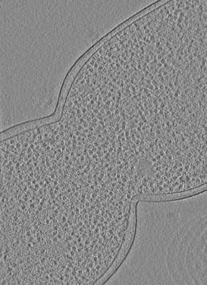 |
Cryo-electron tomography of Cryo-FIB milled dividing E. coli ftsL* strain. [60 multi-frame micrographs composed of 4 frames each in MRC format] | Navarro PP, Vettiger A, Ananda VY, Llopis PM, Allolio C, Bernhardt TG, Chao LH [Pubmed: 36097171] [DOI: 10.1038/s41564-022-01210-z] |
67.2 GB | — |
| 2022-09-20 | 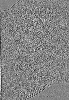 |
Cryo-electron tomography of Cryo-FIB milled dividing E. coli envC and/or nlpD deletion strain. [60 multi-frame micrographs composed of 4 frames each in TIFF format] | Navarro PP, Vettiger A, Ananda VY, Llopis PM, Allolio C, Bernhardt TG, Chao LH [Pubmed: 36097171] [DOI: 10.1038/s41564-022-01210-z] |
73.0 GB | — |
| 2022-09-20 | 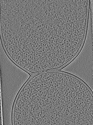 |
Cryo-electron tomography of Cryo-FIB milled dividing E. coli. [60 multi-frame micrographs composed of 4 frames each in TIFF format] | Navarro PP, Vettiger A, Ananda VY, Llopis PM, Allolio C, Bernhardt TG, Chao LH [Pubmed: 36097171] [DOI: 10.1038/s41564-022-01210-z] |
81.5 GB | — |
| 2022-07-18 | 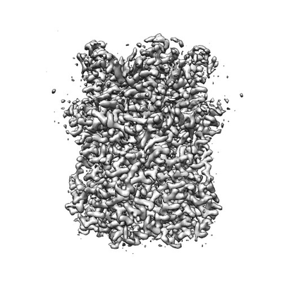 |
Thylakoid-located voltage-dependent chloride channel VCCN1 [multiple data sets in TIFF format] | Hagino T, Kato T, Kasuya G, Kobayashi K, Kusakizako T, Hamamoto S, Sobajima T, Fujiwara Y, Yamashita K, Kawasaki H, Maturana AD, Nishizawa T, Nureki O [Pubmed: 35523970] [DOI: 10.1038/s41467-022-30292-w] |
2.2 TB | 2.7 - 3.0 Å |
| 2022-07-26 |  |
Micrographs of PI3-kinase SH3 amyloid fibrils [multiple data sets in MRC format] | Röder C, Schröder GF [Pubmed: 31434882] [DOI: 10.1038/s41467-019-11320-8] |
32.0 GB | 3.4 Å |
