Electron Microscopy Public Image Archive
Electron Microscopy Public Image Archive
The EMPIAR-PDBj team at Osaka University assists Asian EM researchers with the transfer of big EM image data to EMPIAR. Instead of sending the data directly to the EBI (UK) via the internet, hard drives can also be sent to Osaka University by postal mail or via a courier service. As an alternative, internet transfer to our server in Osaka is also available. If you would like to take advantage of our submission services, please contact us first by e-mail before sending the data to us.
| Release date | Imageset | Title | Authors and references | Size | Resolution |
|---|---|---|---|---|---|
| 2023-03-27 | 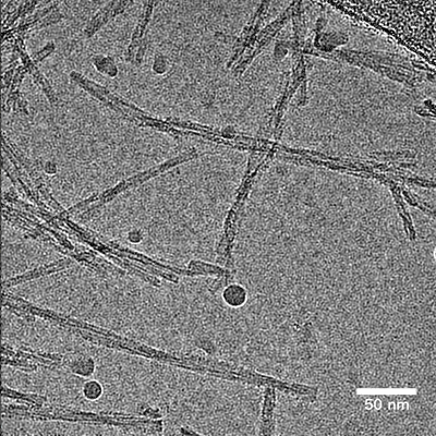 |
Cryo electron microscopy of beta-2-microglobulin amyloid fibrils for the variant D76N (in vitro, pH 6.2) [3849 multi-frame micrographs composed of 36 frames each in TIFF format] | Wilkinson M, Gallardo RU, Martinez RM, Guthertz N, So M, Aubrey LD, Radford SE, Ranson NA [Pubmed: 36864041] [DOI: 10.1038/s41467-023-36791-8] |
575.5 GB | 3.0 - 4.1 Å |
| 2020-12-02 | 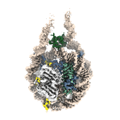 |
Single-particle cryo-EM micrographs of a chromatosome containing chimeric linker histone gH1.10-ncH1.4 [1426 multi-frame micrographs composed of 40 frames each in TIFF format] | Zhou BR [Pubmed: 33238161] [DOI: 10.1016/j.molcel.2020.10.038] |
571.7 GB | 3.03 Å |
| 2019-07-29 | 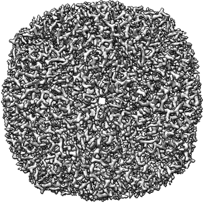 |
Movies of horse spleen apoferritin on multifunctional ultrastable graphene supports for electron cryomicroscopy [480 multi-frame micrographs composed of 38 frames each in MRCS format] | Naydenova K, Peet MJ, Russo CJ [Pubmed: 31127045] [DOI: 10.1073/pnas.1904766116] |
570.0 GB | 2.1 Å |
| 2023-07-31 | 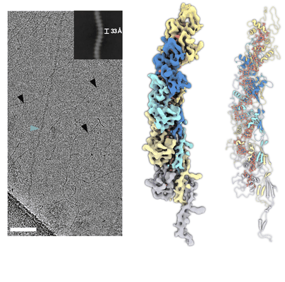 |
Cryo-EM micrographs of AvECN cytochrome nanowires [2572 multi-frame micrographs composed of 40 frames each in TIFF format] | Baquero DP, Cvirkaite-Krupovic V, Egelman EH, Krupovic M, Wang F [Pubmed: 37290436] [DOI: 10.1016/j.cell.2023.05.012] |
555.1 GB | 3.9 Å |
| 2021-10-27 | 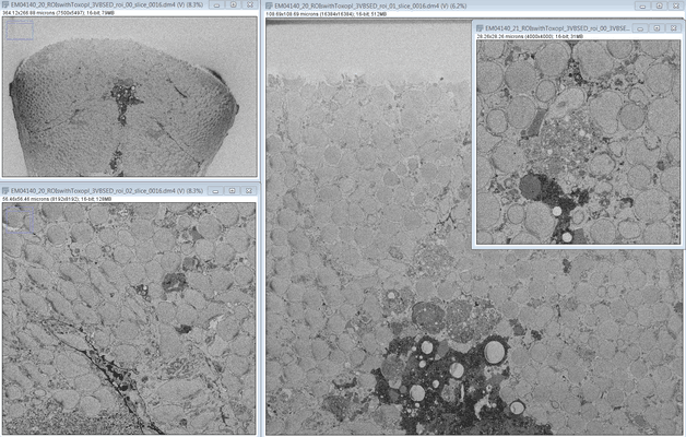 |
SBF SEM images of a Zebrafish hindbrain containing several Toxoplasma gondii tachizoites at different stages of replication [multiple data sets in DM4 format] | Peddie CJ, Domart MC, Collinson L [Pubmed: 32461265] [DOI: 10.1242/dmm.043091] |
553.9 GB | — |
| 2024-03-18 |  |
Seeded Aβ(1-40) amyloid fibril [3018 multi-frame micrographs composed of 40 frames each in TIFF format] | Pfeiffer PB, Schmidt M, Fändrich M [Pubmed: 38158175] [DOI: 10.1016/j.jmb.2023.168422] |
550.2 GB | 2.97 Å |
| 2020-04-24 |  |
Mouse heavy-chain apoferritin movies obtained using a Talos Arctica (200 kV) equipped with a K2 [1679 multi-frame micrographs composed of 90 frames each in TIFF format] | Wu M, Lander GC, Herzik MA [Pubmed: 32647824] [DOI: 10.1016/j.yjsbx.2020.100020] |
549.5 GB | 1.75 Å |
| 2020-06-30 |  |
Structural basis for strand-transfer inhibitor binding to HIV intasomes [multiple data sets in MRC format] | Passos DO, Li M, Jóźwik IK, Zhao XZ, Santos-Martins D, Yang R, Smith SJ, Jeon Y, Forli S, Hughes SH, Burke TR, Craigie R, Lyumkis D [Pubmed: 32001521] [DOI: 10.1126/science.aay8015] |
544.6 GB | 2.8 Å |
| 2020-04-24 |  |
Rabbit muscle aldolase movies obtained using a Talos Arctica (200 kV) equipped with a K2 [3316 multi-frame micrographs composed of 44 frames each in TIFF format] | Wu M, Lander GC, Herzik MA [Pubmed: 32647824] [DOI: 10.1016/j.yjsbx.2020.100020] |
543.3 GB | 2.13 Å |
| 2020-11-24 |  |
Motion-corrected micrographs and extracted particle images of the 70S ribosome from the human pathogen Acinetobacter baumannii in complex with tigecycline [multiple data sets in MRC and MRCS formats] | Nicholson D, Edwards TA, O'Neill AJ, Ranson NA [Pubmed: 32857965] [DOI: 10.1016/j.str.2020.08.004] |
538.5 GB | 2.5 - 3.0 Å |
| 2020-05-27 |  |
Atomic resolution structure of apoferritin [3370 multi-frame micrographs composed of 434 frames each in EER format] | Nakane T, Kotecha A, Sente A, McMullan G, Masiulis S, Brown PMGE, Grigoras IT, Malinauskaite L, Malinauskas T, Miehling J, Uchański T, Yu L, Karia D, Pechnikova EV, de Jong E, Keizer J, Bischoff M, McCormack J, Tiemeijer P, Hardwick SW, Chirgadze DY, Murshudov G, Aricescu AR, Scheres SHW [Pubmed: 33087931] [DOI: 10.1038/s41586-020-2829-0] |
538.1 GB | 1.22 Å |
| 2023-10-17 |  |
3D reconstructions of parasite development and the intracellular niche of the microsporidian pathogen E. intestinalis [multiple data sets in DM4 format] | Antao NVA, Lam CKL, Davydov AD, Riggi MR, Sall JS, Petzold CP, Liang FL, Iwasa JI, Ekiert DCE, Bhabha GB [Pubmed: 37425741] [DOI: 10.1101/2023.07.02.547383] |
537.9 GB | — |
| 2020-12-04 | 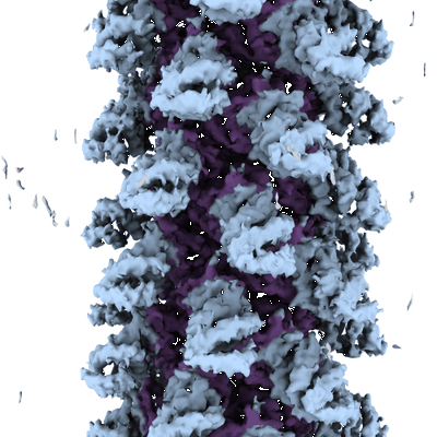 |
NLRP1-CT filament [1994 multi-frame micrographs composed of 50 frames each in TIFF format] | Robert Hollingsworth L, David L, Li Y, Wu H [Pubmed: 33420033] [DOI: 10.1038/s41467-020-20320-y] |
534.7 GB | 3.72 Å |
| 2021-11-02 |  |
Cryo electron microscopy of ex-vivo murine SAA amyloid fibrils [1429 multi-frame micrographs composed of 40 frames each in TIFF format] | Schmidt MS, Fändrich MF [Pubmed: 30846696] [DOI: 10.1038/s41467-019-09033-z] |
533.9 GB | 3.0 Å |
| 2022-01-25 |  |
Single particle cryo-EM dataset of sarkosyl-insoluble fraction from the frontal cortex of an individual with Alzheimer’s disease of amyloid-β 42 filaments [1762 multi-frame micrographs composed of 40 frames each in TIFF format] | Yang Y, Arseni D, Zhang WJ, Huang M, Lovestam SKA, Schweighauser M, Kotecha A, Murzin AG, Peak-Chew S, Macdonald J, Lavenir I, Garringer HJ, Gelpi E, Newell KL, Kovacs GG, Vidal R, Ghetti B, Falcon B, Scheres SHW, Goedert M [Pubmed: 35025654] [DOI: 10.1126/science.abm7285] |
531.9 GB | 2.5 Å |
| 2022-04-25 |  |
Cryo electron microscopy of in vitro recombinant SAA1.1 amyloid fibrils [multiple data sets in TIFF and JPEG formats] | Schmidt MS [Pubmed: 33579941] [DOI: 10.1038/s41467-021-21129-z] |
525.4 GB | 2.73 - 2.95 Å |
| 2023-08-20 |  |
AP2 bound to Y-cargo motif of Tgn38 in the presence of heparin [5958 micrographs in MRC format] | Partlow EA, Cannon KS, Hollopeter G, Baker RW [Pubmed: 35347313] [DOI: 10.1038/s41594-022-00749-z] |
523.1 GB | 4.7 Å |
| 2022-09-09 |  |
Cryo-EM structure of the rigor state Jordan myosin-15-F-actin complex [2641 multi-frame micrographs composed of 24 frames each in TIFF format] | Gong R, Bird JE, Alushin GM [Pubmed: 35857845] [DOI: 10.1126/sciadv.abl4733] |
519.5 GB | 3.76 - 4.18 Å |
| 2023-10-23 |  |
Cryo-EM structure of DIDS-bound human Anion Exchanger 1 [5910 micrographs in MRC format] | Capper MJ, Yang S, Stone AC, Vatansever S, Zilberg G, Mathiharan YK, Habib R, Hutchinson K, Zhao Y, Schlessinger A, Mezei M, Osman R, Zhang B, Wacker D [Pubmed: 37679563] [DOI: 10.1038/s41594-023-01085-6] |
518.9 GB | 2.95 Å |
| 2021-02-05 |  |
Affinity-purified RqcH-ribosome-associated quality control complexes from Bacillus subtilis [7178 multi-frame micrographs composed of 20 frames each in TIFF format] | Crowe-McAuliffe CT, Takada H, Murina V, Atkinson GC, Wilson DN, Hauryliuk V [Pubmed: 33259810] [DOI: 10.1016/j.molcel.2020.11.002] |
514.6 GB | 2.6 - 3.5 Å |
| 2015-09-01 | 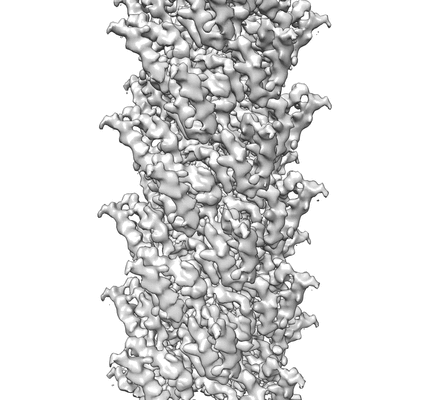 |
New movie data for MAVS CARD C1 filaments [512 multi-frame micrographs composed of 16 frames each in MRC format] | Chew PL, Ng TS, Lok SM, Xu H, He X, Zheng H, Huang LJ, Hou F, Yu Z, de la Cruz MJ, Borkowski B, Zhang X, Chen ZJ, Jiang QX [Pubmed: 26314863] [DOI: 10.7554/eLife.07546] |
512.1 GB | 4.2 Å |
| 2023-05-22 | 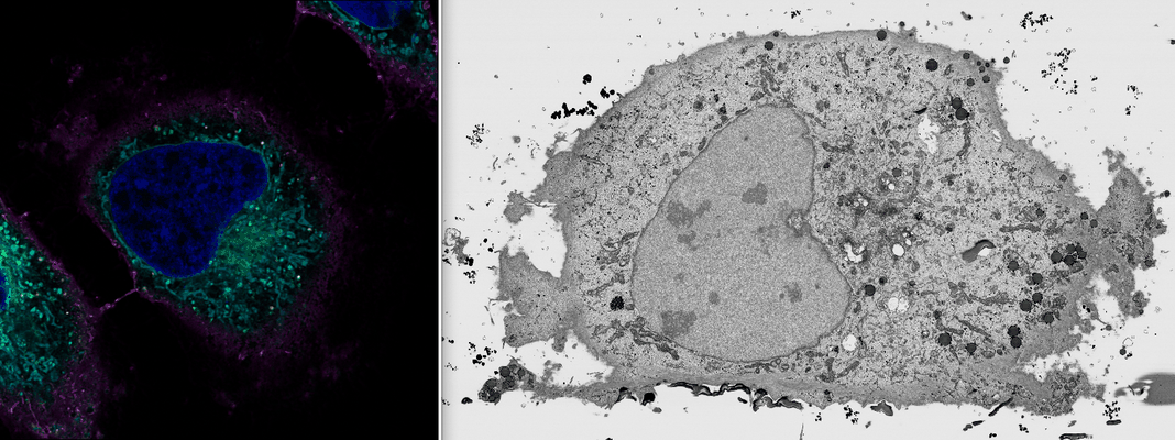 |
Benchmark FIB SEM data (#2) of HeLa cells previously imaged by Zeiss LSM900 Airyscan microscopy [multiple data sets in TIFF format] | Peddie CJ, Domart MC, Collinson LM [DOI: 10.1101/2023.05.11.540445] |
511.9 GB | — |
| 2024-03-18 |  |
Ex vivo murine AApoAII amyloid fibril [3083 multi-frame micrographs composed of 40 frames each in TIFF format] | Andreotti S, Schmidt M, Fändrich M [Pubmed: 38199491] [DOI: 10.1016/j.jmb.2024.168441] |
510.6 GB | 2.4 - 2.6 Å |
| 2022-09-09 | 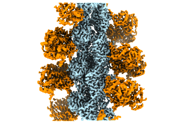 |
cryo-EM structure of the rigor state wild type myosin-15-F-actin complex [1485 multi-frame micrographs composed of 40 frames each in TIFF format] | Gong R, Bird JE, Alushin GM [Pubmed: 35857845] [DOI: 10.1126/sciadv.abl4733] |
506.7 GB | 2.83 - 3.17 Å |
| 2020-10-16 |  |
E. coli 50S ribosome bound to compound 21 [1064 multi-frame micrographs composed of 80 frames each in TIFF format] | Pellegrino J, Lee DJ, Fraser JS, Seiple IB [Pubmed: 32968273] [DOI: 10.1038/s41586-020-2761-3] |
503.5 GB | 2.9 Å |
