Electron Microscopy Public Image Archive
Electron Microscopy Public Image Archive
The EMPIAR-PDBj team at Osaka University assists Asian EM researchers with the transfer of big EM image data to EMPIAR. Instead of sending the data directly to the EBI (UK) via the internet, hard drives can also be sent to Osaka University by postal mail or via a courier service. As an alternative, internet transfer to our server in Osaka is also available. If you would like to take advantage of our submission services, please contact us first by e-mail before sending the data to us.
| Release date | Imageset | Title | Authors and references | Size | Resolution |
|---|---|---|---|---|---|
| 2019-05-09 | 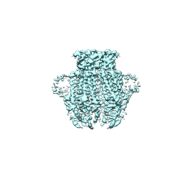 |
apo-LRRC8A in MSP2N2 nanodiscs [1779 multi-frame micrographs composed of 50 frames each in MRCS format] | Kern DM [Pubmed: 30775971] [DOI: 10.7554/eLife.42636] |
739.5 GB | 4.18 Å |
| 2023-11-13 | 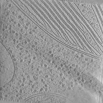 |
Test subset: In situ cryo-ET dataset of Chlamydomonas reinhardtii prepared using cryo-plasmaFIB milling [18 tilt series in MRC format] | Kelley R, Zhang X, Obr M, Khavnekar S, Righetto R, Waltz F, Wietrzynski W, Michael A, Tagiltsev G, Beck F, Zhong E, Wan W, Briggs J, Plitzko J, Engel B, Kotecha A [Pubmed: 37613825] [DOI: 10.1093/micmic/ozad067.480] |
293.7 GB | — |
| 2024-04-09 |  |
In situ cryo-ET dataset of Chlamydomonas reinhardtii prepared using cryo-plasmaFIB milling [multiple data sets in EER and MRC formats] | Kelley R, Khavnekar S, Zhang X, Obr M, Chakraborty S, Koh AF, Heebner J, Righetto R, Waltz F, McCafferty C, Van den Hoek H, Wietrzynski W, Van Der Stappen P, Michael A, Van Dorst S, Tagiltsev G, Beck F, Zhong E, Wan W, Briggs J, Plitzko J, Engel B, Kotecha A [Pubmed: 37613825] [DOI: 10.1093/micmic/ozad067.480] |
27.7 TB | — |
| 2022-10-28 | 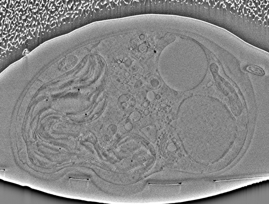 |
Chlamydomonas Cryo-Slice and View on Thermo Scientific Helios 5 Hydra PFIB [477 micrographs in TIFF format] | Kelley R, Khavnekar S, Wietrzynski W, Plitzko J, Kotecha A | 4.4 GB | — |
| 2022-03-21 | 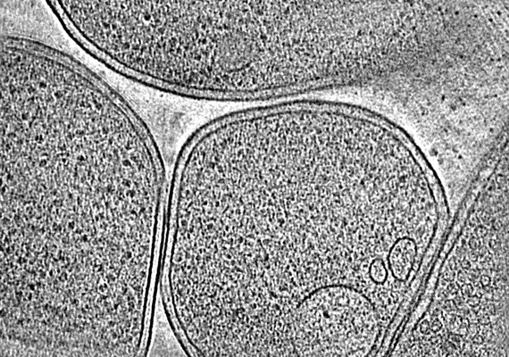 |
CryoET of E. coli prepared with the Waffle Method [1504 multi-frame micrographs composed of 14 frames each in TIFF format] | Kelley K, Raczkowski AM, Klykov O, Jaroenlak P, Bobe D, Kopylov M, Eng ET, Bhabha G, Potter CS, Carragher B, Noble AJ [Pubmed: 35387991] [DOI: 10.1038/s41467-022-29501-3] |
94.0 GB | — |
| 2020-10-09 |  |
Single particle cryo electron microscopy of aldolase (rabbit, muscle) using beam-tilt on Talos Arctica [1625 micrographs in MRC format] | Kearns S, Cianfrocco MA [Pubmed: 33209328] [DOI: 10.1107/S2052252520013482] |
87.6 GB | 2.8 - 4.9 Å |
| 2020-08-19 |  |
Cryo-EM structure of SARS-CoV-2 Spike Proteins on intact virions [7982 multi-frame micrographs composed of 48 frames each in TIFF format] | Ke Z, Qu K, Cortese M, Zila V, Nakane T, Xiong X, Scheres SHW, Briggs JAG [Pubmed: 32805734] [DOI: 10.1038/s41586-020-2665-2] |
2.1 TB | 3.5 - 4.1 Å |
| 2020-08-19 |  |
Subtomogram averaging and classification of SARS-CoV-2 Spike Proteins on intact virions [multiple data sets in TIFF and MRC formats] | Ke Z, Oton J, Cortese M, Zila V, Zivanov J, Lu JM, Peukes J, Scheres SHW, Briggs JAG [Pubmed: 32805734] [DOI: 10.1038/s41586-020-2665-2] |
372.4 GB | 7.7 - 9.9 Å |
| 2022-11-14 |  |
Cryo-EM structures of the translocational binary toxin complex CDTa-bound CDTb-pore [11284 multi-frame micrographs composed of 84 frames each in TIFF format] | Kawamoto A, Yamada T, Yoshida T, Sato Y, Kato T, Tsuge H [Pubmed: 36253419] [DOI: 10.1038/s41467-022-33888-4] |
3.3 TB | 2.56 - 2.64 Å |
| 2022-08-08 |  |
Cryo-EM structure of MrgD-Gi complex with beta-alanine [17087 multi-frame micrographs composed of 66 frames each in TIFF format] | Kawamoto A [Pubmed: 35840655] [DOI: 10.1038/s42003-022-03668-3] |
4.1 TB | 3.1 - 3.2 Å |
| 2022-07-27 |  |
Cryo-EM structure of apo-state MrgD-Gi complex [14985 multi-frame micrographs composed of 66 frames each in TIFF format] | Kawamoto A [Pubmed: 35840655] [DOI: 10.1038/s42003-022-03668-3] |
3.5 TB | 2.9 - 3.1 Å |
| 2024-02-13 |  |
Cryo electron microscopy of Virus-like Particle based on PVY coat protein [502 multi-frame micrographs composed of 40 frames each in TIFF format] | Kavcic L, Kezar A [Pubmed: 38233506] [DOI: 10.1038/s42004-024-01100-x] |
379.4 GB | 2.99 - 3.34 Å |
| 2024-03-26 |  |
Cryo electron microscopy of Virus-like Particle based on PVY coat protein with dC40 deletion [6215 multi-frame micrographs composed of 40 frames each in TIFF format] | Kavcic L, Kezar A [Pubmed: 38233506] [DOI: 10.1038/s42004-024-01100-x] |
5.8 TB | 3.09 - 3.5 Å |
| 2024-02-13 |  |
Cryo electron microscopy of Virus-like Particle based on PVY coat protein with dC79 deletion [491 multi-frame micrographs composed of 41 frames each in TIFF format] | Kavcic L, Kezar A [Pubmed: 38233506] [DOI: 10.1038/s42004-024-01100-x] |
377.4 GB | 3.2 Å |
| 2024-03-26 |  |
Cryo electron microscopy of assemblies based on truncated PVY coat protein [2862 multi-frame micrographs composed of 44 frames each in TIFF format] | Kavcic L, Kezar A [Pubmed: 38233506] [DOI: 10.1038/s42004-024-01100-x] |
2.3 TB | 2.93 - 3.62 Å |
| 2024-02-29 |  |
Cryo electron microscopy of assemblies based on truncated PVY coat protein with K176C mutation [9480 multi-frame micrographs composed of 32 frames each in TIFF format] | Kavcic L, Kezar A [Pubmed: 38233506] [DOI: 10.1038/s42004-024-01100-x] |
957.1 GB | 2.99 - 3.16 Å |
| 2024-02-13 |  |
Cryo electron microscopy of Virus-like Particle based on PVY coat protein with T43C and D136C mutation [461 multi-frame micrographs composed of 38 frames each in TIFF format] | Kavcic L, Kezar A [Pubmed: 38233506] [DOI: 10.1038/s42004-024-01100-x] |
332.6 GB | 2.41 Å |
| 2022-06-13 |  |
Structural and functional analyses of the tridomain-nonribosomal peptide synthetase FmoA3 for 4-methyloxazoline ring formation [1886 multi-frame micrographs composed of 39 frames each in TIFF format] | Katsuyama Y, Sone K, Harada A, Kawai S, Urano N, Adachi N, Moriya T, Kawasaki M, Shin-Ya K, Senda T, Ohnishi Y [Pubmed: 33783097] [DOI: 10.1002/anie.202102760] |
1.4 TB | 3.55 Å |
| 2018-08-15 |  |
The first reconstruction of beta-galactosidase solved by cryoARM200 [1338 multi-frame micrographs composed of 49 frames each in TIFF format] | Kato T, Terehara N, Namba K | 321.4 GB | 2.6 Å |
| 2019-02-19 |  |
The 1.54 Å structure of Apoferritin by CRYOARM300 with cold-FEG [972 multi-frame micrographs composed of 50 frames each in TIFF format] | Kato T, Makino F, Nakane T, Terahara N, Kaneko T, Shimizu Y, Motoki S, Ishikawa I, Yonekura K, Namba K [DOI: 10.1017/S1431927619005725] |
145.9 GB | 1.54 Å |
| 2022-09-20 |  |
Human amino acid transporter EAAT2 [multiple data sets in TIFF format] | Kato T, Kusakizako T, Yamashita K, Nishizawa T, Nureki O [Pubmed: 35953475] [DOI: 10.1038/s41467-022-32442-6] |
1.5 TB | 3.49 - 3.58 Å |
| 2024-02-06 | 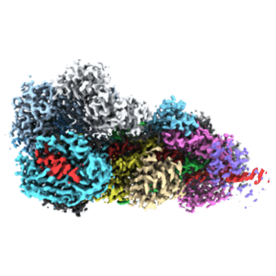 |
RNA-triggered protein cleavage and cell growth arrest by the type III-E CRISPR nuclease-protease [multiple data sets in TIFF format] | Kato K, Okazaki S, Ishikawa J, Isayama Y, Nishizawa T, Nishimasu H [Pubmed: 36423304] [DOI: 10.1126/science.add7347] |
2.6 TB | 2.49 - 2.84 Å |
| 2024-02-01 | 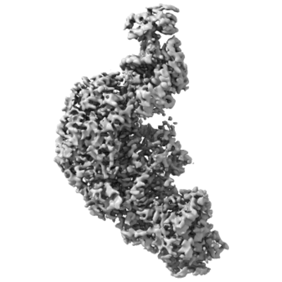 |
Structure and engineering of the type III-E CRISPR-Cas7-11 effector complex [2781 multi-frame micrographs composed of 64 frames each in TIFF format] | Kato K, Okazaki S, Isayama Y, Nishizawa T, Nishimasu H [Pubmed: 35643083] [DOI: 10.1016/j.cell.2022.05.003] |
853.6 GB | 2.45 Å |
| 2020-12-04 | 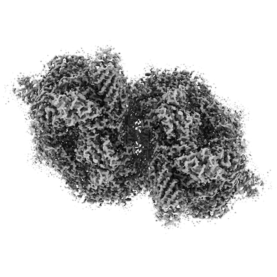 |
Cryo-EM Structure of PSII at 1.95 angstrom resolution [2160 multi-frame micrographs composed of 50 frames each in TIFF format] | Kato K, Miyazaki N, Hamaguchi T, Nakajima Y, Akita F, Yonekura K, Shen J [Pubmed: 33753866] [DOI: 10.1038/s42003-021-01919-3] |
427.6 GB | 1.95 Å |
| 2018-08-23 | 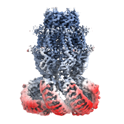 |
CryoEM structure of human LRRC8A [5805 multi-frame micrographs composed of 40 frames each in TIFF format] | Kasuya G, Nakane T, Yokoyama T, Shirouzu M, Ishitani R, Nureki O [Pubmed: 30127360] [DOI: 10.1038/s41594-018-0109-6] |
2.9 TB | 4.25 Å |
