Electron Microscopy Public Image Archive
Electron Microscopy Public Image Archive
The EMPIAR-PDBj team at Osaka University assists Asian EM researchers with the transfer of big EM image data to EMPIAR. Instead of sending the data directly to the EBI (UK) via the internet, hard drives can also be sent to Osaka University by postal mail or via a courier service. As an alternative, internet transfer to our server in Osaka is also available. If you would like to take advantage of our submission services, please contact us first by e-mail before sending the data to us.
| Release date | Imageset | Title | Authors and references | Size | Resolution |
|---|---|---|---|---|---|
| 2022-05-27 | 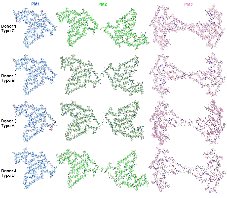 |
Cryo electron-microscopy of TMEM106B fibrils extracted from four FTLD-TDP patient brains [multiple data sets in TIFF format] | Jiang YX, Cao Q, Sawaya MR, Abskharon R, Ge P, DeTure M, Dickson DW, Fu JY, Ogorzalek Loo RR, Loo JA, Eisenberg DS [Pubmed: 35344984] [DOI: 10.1038/s41586-022-04670-9] |
3.4 TB | 2.9 - 3.7 Å |
| 2023-03-03 |  |
Cryo-iDPC-STEM structure of TMV - convergence semi-angle 4.0 mrad [multiple data sets in TIFF format] | Lazić I, Wirix M, Leidl ML, Sachse C [Pubmed: 36064775] [DOI: 10.1038/s41592-022-01586-0] |
1.2 GB | 3.5 Å |
| 2022-08-08 |  |
Architecture of the human erythrocyte ankyrin-1 complex [multiple data sets in TIFF, MRC and MRCS formats] | Vallese F, Clarke OB [Pubmed: 35835865] [DOI: 10.1038/s41594-022-00792-w] |
5.2 TB | 2.4 Å |
| 2022-07-29 |  |
Cryo-EM structure of the GOLD-domain seven-transmembrane protein TMEM87A [7060 multi-frame micrographs composed of 50 frames each in TIFF format] | Hoel CM, Zhang L, Brohawn SG [Pubmed: 36373655] [DOI: 10.7554/eLife.81704] |
4.7 TB | 4.74 Å |
| 2022-05-27 |  |
Human Xkr8-Basigin complex bound to Fab fragment [1998 multi-frame micrographs composed of 48 frames each in TIFF format] | Sakuragi T, Tsutsumi A, Kikkawa M, Nagata S [Pubmed: 34625749] [DOI: 10.1038/s41594-021-00665-8] |
451.5 GB | 3.8 Å |
| 2022-07-08 | 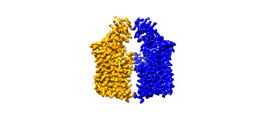 |
Cryo-EM structure of human KCC1 bound with inhibitor VU0463271 [5833 multi-frame micrographs composed of 40 frames each in TIFF format] | Cao E [Pubmed: 35759661] [DOI: 10.1073/pnas.2109083119] |
3.9 TB | 3.5 Å |
| 2022-05-31 | 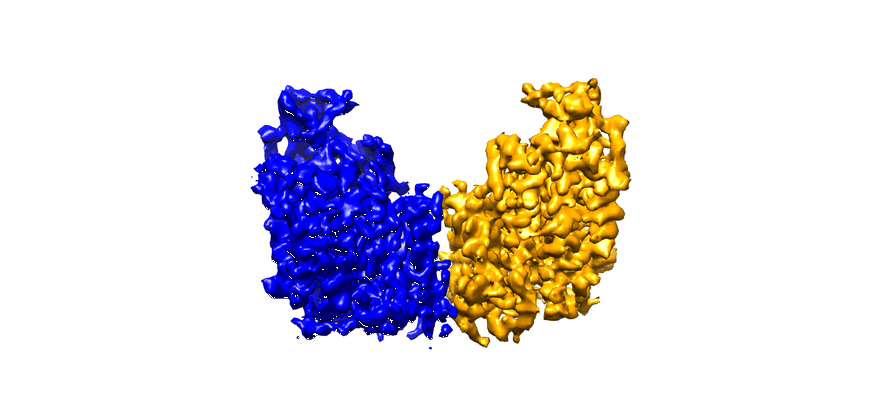 |
Cryo-EM structure of human NKCC1 K289NA492E bound with bumetanide [4273 multi-frame micrographs composed of 40 frames each in TIFF format] | Zhao YZ [Pubmed: 35585053] [DOI: 10.1038/s41467-022-30407-3] |
2.1 TB | 3.6 Å |
| 2022-05-27 | 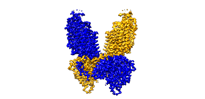 |
Cryo-EM structure of human NKCC1 K289NA492EL671C bound with bumetanide [3891 multi-frame micrographs composed of 40 frames each in TIFF format] | Zhao YZ [Pubmed: 35585053] [DOI: 10.1038/s41467-022-30407-3] |
1.8 TB | 2.9 Å |
| 2022-05-27 | 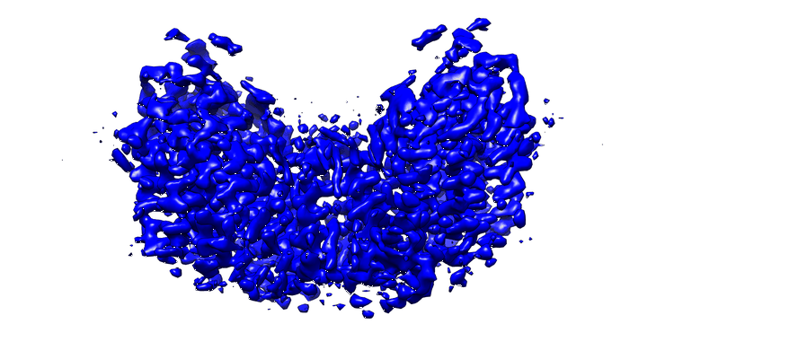 |
Cryo-EM structure of human NKCC1 K289NA492E bound with Furosemide [3405 multi-frame micrographs composed of 40 frames each in TIFF format] | Zhao Y [Pubmed: 35585053] [DOI: 10.1038/s41467-022-30407-3] |
1.6 TB | 3.4 Å |
| 2022-06-07 | 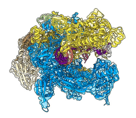 |
Cryo-EM structure of Bacillus subtilis RNA Polymerase elongation complex [4486 multi-frame micrographs composed of 40 frames each in MRC format] | Newing T, Oakley A, Miller M, Dawson C, Brown S, Bouwer J, Tolun G, Lewis P [Pubmed: 33339820] [DOI: 10.1038/s41467-020-20157-5] |
2.3 TB | 3.36 Å |
| 2022-06-07 | 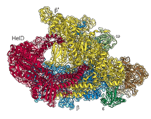 |
Cryo-EM structure of Bacillus subtilis RNA Polymerase in complex with HelD [3656 multi-frame micrographs composed of 60 frames each in MRC format] | Newing T, Oakley A, Miller M, Dawson C, Brown S, Bouwer J, Tolun G, Lewis P [Pubmed: 33339820] [DOI: 10.1038/s41467-020-20157-5] |
666.2 GB | 3.36 Å |
| 2022-09-23 |  |
Structure of pre-60S particle bound to DRG1(AFG2) [multiple data sets in TIFF format] | Prattes M, Grishkovskaya I, Hodirnau VV, Bergler H, Haselbach D [Pubmed: 36097293] [DOI: 10.1038/s41594-022-00832-5] |
3.8 TB | 3.2 - 3.8 Å |
| 2022-06-07 |  |
Cryo-EM Structure of the Hyperpolarization-Activated Potassium Channel KAT1 [1503 multi-frame micrographs composed of 40 frames each in MRC format] | Clark MD, Contreras GF, Shen R, Perozo E [Pubmed: 32461693] [DOI: 10.1038/s41586-020-2335-4] |
459.8 GB | 3.5 - 3.8 Å |
| 2022-06-01 |  |
Crosshair, semi-automated targeting for electron microscopy with a motorised ultramicrotome [multiple data sets in TIFF and PNG formats] | Meechan K, Guan W, Riedinger A, Stankova V, Yoshimura A, Pipitone R, Milberger A, Schaar H, Romero-Brey I, Templin R, Peddie C J, Schieber N L, Jones M L, Collinson L, Schwab Y | 151.0 GB | — |
| 2022-06-13 |  |
Structure of bile acid transporter NTCP [multiple data sets in TIFF format] | Asami J, Shimizu T, Ohto U [Pubmed: 35580629] [DOI: 10.1038/s41586-022-04845-4] |
8.6 TB | 3.11 - 3.55 Å |
| 2022-06-07 |  |
Cryo-EM structure of the gastric proton pump complexed with revaprazan [multiple data sets in TIFF format] | Abe K, Tanaka S, Morita M, Yamagishi T [Pubmed: 35604136] [DOI: 10.1021/acs.jmedchem.2c00338] |
2.1 TB | 2.76 Å |
| 2022-07-12 |  |
In situ cryo-electron tomography of T. kivui cells [multiple data sets in MRC and TIFF formats] | Dietrich HM, Righetto RD, Kumar A, Wietrzynski W, Trischler R, Schuller SK, Wagner J, Schwarz FM, Engel BD, Müller V, Schuller JM [Pubmed: 35859174] [DOI: 10.1038/s41586-022-04971-z] |
373.4 GB | 17.0 Å |
| 2022-06-13 | 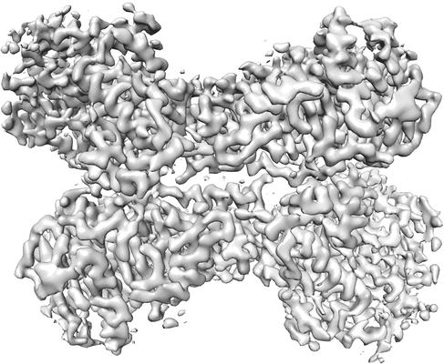 |
Structural and functional analyses of the tridomain-nonribosomal peptide synthetase FmoA3 for 4-methyloxazoline ring formation [1886 multi-frame micrographs composed of 39 frames each in TIFF format] | Katsuyama Y, Sone K, Harada A, Kawai S, Urano N, Adachi N, Moriya T, Kawasaki M, Shin-Ya K, Senda T, Ohnishi Y [Pubmed: 33783097] [DOI: 10.1002/anie.202102760] |
1.4 TB | 3.55 Å |
| 2023-02-13 | 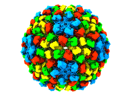 |
Nudaurelia capensis omega virus maturation intermediate captured at pH5.9 (insect cell expressed VLPs) [multiple data sets in MRC format] | Castells-Graells R, Hesketh EL, Johnson JE, Ranson NA, Lawson DM, Lomonossoff GP | 8.3 TB | 3.92 Å |
| 2022-06-27 |  |
DNA polymerase D temporarily connects primase to the CMG-like helicase before interacting with proliferating cell nuclear antigen [2378 multi-frame micrographs composed of 39 frames each in TIFF format] | Oki K, Yamagami T, Nagata M, Mayanagi K, Shirai T, Adachi N, Numata T, Ishino S, Ishino Y [Pubmed: 33849056] [DOI: 10.1093/nar/gkab243] |
1.7 TB | 7.1 Å |
| 2022-07-22 |  |
Structure of the human RAD17-RFC clamp loader and 9-1-1 checkpoint clamp bound to a dsDNA-ssDNA junction [8271 multi-frame micrographs composed of 882 frames each in EER format] | Day M, Oliver AW, Pearl LH [Pubmed: 35819203] [DOI: 10.1093/nar/gkac588] |
4.4 TB | 3.59 Å |
| 2023-02-01 |  |
Cryo-EM data and 2DTM results of entire sections of differentiated ER-HoxB8 cells [multiple data sets in TIFF and MRC formats] | Elferich JE, Schiroli GS, Scadden DS, Grigorieff NG [Pubmed: 36382886] [DOI: 10.7554/elife.80980] |
1.3 TB | — |
| 2023-01-03 | 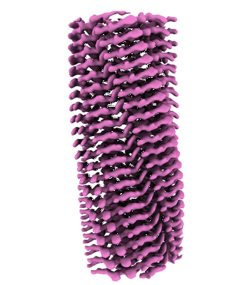 |
Cryo-EM structure of hnRNPDL amyloid fibrils [multiple data sets in TIFF format] | Chaves-Sanjuan A, Garcia-Pardo J, Bartolome-Nafria A, Gil-Garcia M, Visentin C, Bolognesi M, Ricagno S, Ventura S [Pubmed: 36646699] [DOI: 10.1038/s41467-023-35854-0] |
880.1 GB | 2.5 Å |
| 2023-02-14 | 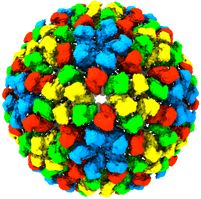 |
Nudaurelia capensis omega virus procapsid at pH7.6 (insect cell expressed VLPs) [multiple data sets in MRC format] | Castells-Graells R, Hesketh EL, Johnson JE, Ranson NA, Lawson DM, Lomonossoff GP | 8.5 TB | 4.88 Å |
| 2023-03-15 | 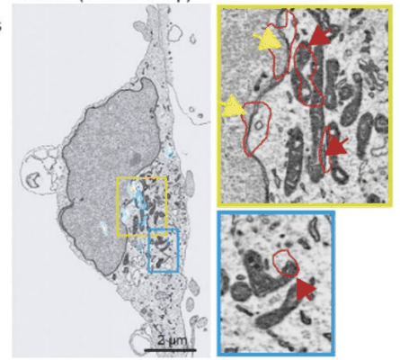 |
Performing Correlative Light and Electron Microscopy to reveal the structural organization and location of alpha-synuclein aggregation hotspots inside the neuron. [multiple data sets in DM4 and TIFF formats] | Choi ML, Chappard A, Singh BP, Maclachlan C, Rodrigues M, Fedotova E, Berezhnov AV, De S, Peddie C, Athauda D, Viridi GS, Zhang W, Evans JR, Wernick A, Zanjani ZS, Angelova PR, Esteras N, Vinikurov A, Morris K, Jeacock K, Tosatto L, Little D, Gissen P, Collinson L, Clarke DJ, Kunath T, Klenerman D, Abramov AY, Horrocks MH, Gandhi S [DOI: 10.1101/2022.06.07.494932] |
88.4 GB | — |
