Electron Microscopy Public Image Archive
Electron Microscopy Public Image Archive
The EMPIAR-PDBj team at Osaka University assists Asian EM researchers with the transfer of big EM image data to EMPIAR. Instead of sending the data directly to the EBI (UK) via the internet, hard drives can also be sent to Osaka University by postal mail or via a courier service. As an alternative, internet transfer to our server in Osaka is also available. If you would like to take advantage of our submission services, please contact us first by e-mail before sending the data to us.
| Release date | Imageset | Title | Authors and references | Size | Resolution |
|---|---|---|---|---|---|
| 2022-05-10 |  |
CEM-MitoLab: a dataset of ~22K cellular EM 2D images with label maps of ~135K mitochondrial instances, for deep learning [43720 micrographs in TIFF format] | Narayan K, Conrad RW | 2.8 GB | — |
| 2022-05-11 |  |
Cryo-EM SPA dataset of Megadalton-range protein communities from a Chaetomium thermophilum native cell extract [2808 multi-frame micrographs composed of 13 frames each in MRC format] | Skalidis IS, Kyrilis FLK, Tüting CT, Müller JM, Sorokina MS, Hamdi FH, Sadian YS, Chojnowski GC, Kastritis PLK [Pubmed: 34836937] [DOI: 10.1038/s41467-021-27287-4] |
1.1 TB | 3.84 - 4.52 Å |
| 2022-05-11 |  |
Tilt series and tomograms of cells expressing different non structural proteins (NSPs) of SARS-CoV-2 [multiple data sets in MRC format] | Polshchuk RS, Polishchuk E, De Matteis MA [Pubmed: 35551511] [DOI: 10.1038/s41586-022-04835-6] |
107.1 GB | — |
| 2022-05-17 |  |
Structures of positive allosteric modulator-bound and unbound active human calcium-sensing receptor [13082 multi-frame micrographs composed of 60 frames each in TIFF format] | Park J, Zuo H, Frangaj A, Fu Z, Yen LY, Zhang Z, Mosyak L, Slavkovich VN, Liu J, Ray KM, Cao B, Vallese F, Geng Y, Chen S, Grassucci R, Dandey VP, Tan YZ, Eng E, Lee Y, Kloss B, Liu Z, Hendrickson WA, Potter CS, Carragher B, Graziano J, Conigrave AD, Frank J, Clarke OB, Fan QR [Pubmed: 34916296] [DOI: 10.1073/pnas.2115849118] |
3.8 TB | 2.7 Å |
| 2022-05-17 |  |
Cryo-EM structure of the human ATP13A2 [multiple data sets in TIFF format] | Tomita A, Daiho T, Kusakizako T, Yamashita K, Ogasawara S, Murata T, Nishizawa T, Nureki O [Pubmed: 34798056] [DOI: 10.1016/j.molcel.2021.11.001] |
3.8 TB | 3.54 - 3.92 Å |
| 2022-05-17 |  |
Conformational rearrangements upon start codon recognition in human 48S translation initiation complex [multiple data sets in MRC and MRCS formats] | Yi SH, Petrychenko V, Schliep JE, Goyal A, Linden A, Chari A, Urlaub H, Stark H, Rodnina MV, Adio S, Fischer N [Pubmed: 35489072] [DOI: 10.1093/nar/gkac283] |
1.1 TB | 3.7 - 4.7 Å |
| 2022-05-20 | 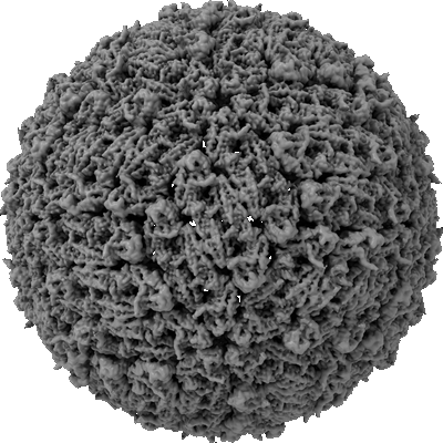 |
Single-particle reconstruction of Tick-borne encephalitis virus (Final reconstruction) [multiple data sets in TIFF format] | Pulkkinen LIA, Barrass SV, Domanska A, Överby AK, Anastasina M, Butcher SJ [Pubmed: 35458522] [DOI: 10.3390/v14040792] |
16.4 TB | 3.3 Å |
| 2022-05-20 | 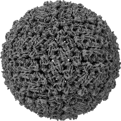 |
Single-particle reconstruction of Tick-borne encephalitis virus (Preparation 1) [14135 multi-frame micrographs composed of 30 frames each in MRC format] | Pulkkinen LIA, Barrass SV, Domanska A, Överby AK, Anastasina M, Butcher SJ [Pubmed: 35458522] [DOI: 10.3390/v14040792] |
6.2 TB | 3.5 Å |
| 2022-05-20 | 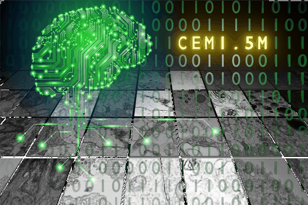 |
CEM1.5M : a cellular EM dataset containing ~1.5 x 10⁶ unlabeled 2D image patches curated for deep learning [1592753 micrographs in TIFF format] | Narayan K | 57.6 GB | — |
| 2022-05-20 | 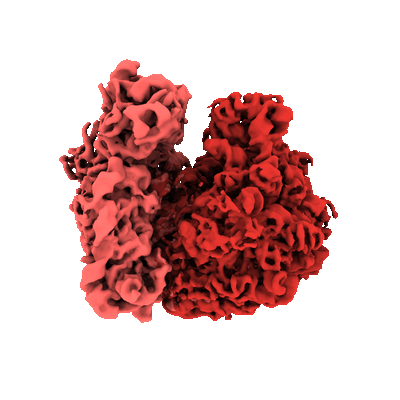 |
Parallel cryo electron tomography (PACE-tomo) of 70S ribosomes (200 kV, side-entry holder) [multiple data sets in MRC format] | Eisenstein F, Danev R [Pubmed: 36456783] [DOI: 10.1038/s41592-022-01690-1] |
222.8 GB | 5.8 - 6.5 Å |
| 2022-05-24 |  |
Cryo-EM data used for the determination of LACV-L structure in transcription early-elongation state [2510 multi-frame micrographs composed of 60 frames each in TIFF format] | Arragain B, Durieux Trouilleton Q, Baudin F, Provaznik J, Azevedo N, Cusack S, Schoehn G, Malet H [Pubmed: 35173159] [DOI: 10.1038/s41467-022-28428-z] |
721.4 GB | 3.3 Å |
| 2022-05-24 |  |
The complex of dephosphorylated human cystic fibrosis transmembrane conductance regulator (CFTR) and Lumacaftor (VX-809) [3191 multi-frame micrographs composed of 50 frames each in TIFF format] | Fiedorczuk K, Chen J [Pubmed: 34995514] [DOI: 10.1016/j.cell.2021.12.009] |
1.4 TB | 3.9 Å |
| 2022-05-24 |  |
The complex of phosphorylated human cystic fibrosis transmembrane conductance regulator (CFTR) with ATP/Mg and Tezacaftor (VX-661) [4257 multi-frame micrographs composed of 50 frames each in TIFF format] | Fiedorczuk K, Chen J [Pubmed: 34995514] [DOI: 10.1016/j.cell.2021.12.009] |
1.8 TB | 3.8 Å |
| 2022-05-25 |  |
SARS-CoV-2 spike protein S:D614G + S:A222V variant [4841 micrographs in MRC format] | Ginex T, Marco-Marín C, Wieczór M, Mata CP, Krieger J, Ruiz-Rodriguez P, López-Redondo ML, Francés-Gómez C, Melero R, Sánchez-Sorzano CÓ, Martínez M, Gougeard N, Forcada-Nadal A, Zamora-Caballero S, Gozalbo-Rovira R, Sanz-Frasquet C, Arranz R, Bravo J, Rubio V, Marina A, Geller R, Comas I, Gil C, Coscolla M, Orozco M, Llácer JL, Carazo JM [Pubmed: 35816514] [DOI: 10.1371/journal.ppat.1010631] |
303.8 GB | 3.4 Å |
| 2022-05-25 |  |
A drug and ATP binding site in type 1 ryanodine receptor [6890 multi-frame micrographs composed of 50 frames each in TIFF format] | Melville Z, Dridi H, Yuan Q, Reiken S, Wronska A, Liu Y, Clarke OB, Marks AR [Pubmed: 35580609] [DOI: 10.1016/j.str.2022.04.010] |
1.5 TB | 2.45 Å |
| 2022-05-26 |  |
Cryo TEM single particle dataset of purified ChAdOx1/ADZ-1222 [2875 multi-frame micrographs composed of 40 frames each in TIFF format] | Boyd RJ, Baker AB [Pubmed: 34851659] [DOI: 10.1126/sciadv.abl8213] |
1.0 TB | 3.07 Å |
| 2022-05-27 |  |
Cryo electron-microscopy of TMEM106B fibrils extracted from four FTLD-TDP patient brains [multiple data sets in TIFF format] | Jiang YX, Cao Q, Sawaya MR, Abskharon R, Ge P, DeTure M, Dickson DW, Fu JY, Ogorzalek Loo RR, Loo JA, Eisenberg DS [Pubmed: 35344984] [DOI: 10.1038/s41586-022-04670-9] |
3.4 TB | 2.9 - 3.7 Å |
| 2022-05-27 |  |
Human Xkr8-Basigin complex bound to Fab fragment [1998 multi-frame micrographs composed of 48 frames each in TIFF format] | Sakuragi T, Tsutsumi A, Kikkawa M, Nagata S [Pubmed: 34625749] [DOI: 10.1038/s41594-021-00665-8] |
451.5 GB | 3.8 Å |
| 2022-05-27 |  |
Cryo-EM structure of human NKCC1 K289NA492EL671C bound with bumetanide [3891 multi-frame micrographs composed of 40 frames each in TIFF format] | Zhao YZ [Pubmed: 35585053] [DOI: 10.1038/s41467-022-30407-3] |
1.8 TB | 2.9 Å |
| 2022-05-27 |  |
Cryo-EM structure of human NKCC1 K289NA492E bound with Furosemide [3405 multi-frame micrographs composed of 40 frames each in TIFF format] | Zhao Y [Pubmed: 35585053] [DOI: 10.1038/s41467-022-30407-3] |
1.6 TB | 3.4 Å |
| 2022-05-31 |  |
Cryo-EM structure of human NKCC1 K289NA492E bound with bumetanide [4273 multi-frame micrographs composed of 40 frames each in TIFF format] | Zhao YZ [Pubmed: 35585053] [DOI: 10.1038/s41467-022-30407-3] |
2.1 TB | 3.6 Å |
| 2022-05-31 | 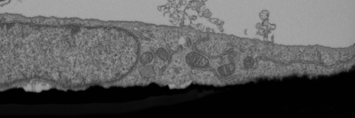 |
FIB-SEM of U2OS cell at leading edge of a wound assay, LPA stimulated, polarized [1798 multi-frame micrographs composed of 1 frames each in TIFF format] | Costa J, Pinto A, Machado P | 4.1 GB | — |
| 2022-05-31 | 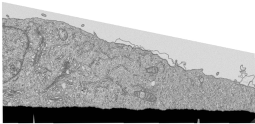 |
FIB-SEM of U2OS cell at leading edge of a wound assay, non-LPA stimulated, non-polarized [998 multi-frame micrographs composed of 1 frames each in TIFF format] | Costa J, Pinto A, Machado P | 5.7 GB | — |
| 2022-06-01 | 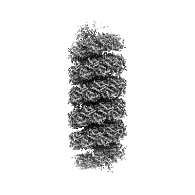 |
Micrographs of viruses ATV and AFV6 [791 multi-frame micrographs composed of 40 frames each in TIFF format] | Wang F, Cvirkaite-Krupovic V, Krupovic M, Egelman EH [Pubmed: 35325592] [DOI: 10.1016/j.cell.2022.02.019] |
218.0 GB | 3.9 Å |
| 2022-06-01 |  |
Crosshair, semi-automated targeting for electron microscopy with a motorised ultramicrotome [multiple data sets in TIFF and PNG formats] | Meechan K, Guan W, Riedinger A, Stankova V, Yoshimura A, Pipitone R, Milberger A, Schaar H, Romero-Brey I, Templin R, Peddie C J, Schieber N L, Jones M L, Collinson L, Schwab Y | 151.0 GB | — |
