Electron Microscopy Public Image Archive
Electron Microscopy Public Image Archive
Our EMPIAR-PDBj will suspend the following operations from August 10, 2024 (Saturday) to August 18, 2024 (Sunday):
Web servers and data downloads will remain available during this period.
Additionally, please be aware that EMPIAR-EBI will also be closed for annotation operations from July 25, 2024 (Thursday) to August 18, 2024 (Sunday).
Thank you.
大阪大学的EMPIAR-PDBj团队为亚洲EM研究人员向EMPIAR传送大型EM图像数据提供服务。 除了通过互联网将数据直接传送到EBI(UK),研究人员还可以通过邮政或快递服务将数据硬盘发送到大阪大学,或者通过互联网传送到设置于大阪大学的服务器,然后由我们代为传送至数据登录网站。 如果您想使用此项服务,请先通过 电子邮件 与我们联系。
| Release date | Imageset | Title | Authors and references | Size | Resolution |
|---|---|---|---|---|---|
| 2022-09-20 |  |
Cryo-electron tomography of Cryo-FIB milled dividing E. coli ftsN SPOR domain deletion strain. [60 multi-frame micrographs composed of 4 frames each in MRC format] | Navarro PP, Vettiger A, Ananda VY, Llopis PM, Allolio C, Bernhardt TG, Chao LH [Pubmed: 36097171] [DOI: 10.1038/s41564-022-01210-z] |
189.1 GB | — |
| 2023-08-18 |  |
Cryo electron tomography of human choriocarcinoma cells [9 multi-frame micrographs composed of 10 frames each in MRC format] | Tun WM, Darrow MC, Basham M [DOI: 10.1017/S2633903X23000107] |
115.3 GB | — |
| 2019-11-18 |  |
cryo-ET of cryo-FIB milled yeast cell in which scs2/22 ist2 are deleted [multiple data sets in MRC format] | Hoffmann PC, Bharat TAM, Wozny MR, Boulanger J, Miller EA, Kukulski W [Pubmed: 31743663] [DOI: 10.1016/j.devcel.2019.09.019] |
12.8 GB | — |
| 2022-09-20 |  |
Cryo-electron tomography of Cryo-FIB milled dividing E. coli ftsL* strain. [60 multi-frame micrographs composed of 4 frames each in MRC format] | Navarro PP, Vettiger A, Ananda VY, Llopis PM, Allolio C, Bernhardt TG, Chao LH [Pubmed: 36097171] [DOI: 10.1038/s41564-022-01210-z] |
67.2 GB | — |
| 2024-07-16 |  |
Cryo electron tomography of chromatin condensed by PRC1-CBX8 [multiple data sets in MRC format] | Uckelmann M, Levina V, Taveneau C, de Marco A, Davidovich C | 8.7 GB | — |
| 2019-11-18 |  |
cryo-ET of cryo-FIB milled yeast cell in which scs2/22 ist2 are deleted with high intracellular calcium [121 tilt series in MRC format] | Hoffmann PC, Bharat TAM, Wozny MR, Boulanger J, Miller EA, Kukulski W [Pubmed: 31743663] [DOI: 10.1016/j.devcel.2019.09.019] |
14.3 GB | — |
| 2022-09-20 |  |
Cryo-electron tomography of Cryo-FIB milled dividing E. coli envC and/or nlpD deletion strain. [60 multi-frame micrographs composed of 4 frames each in TIFF format] | Navarro PP, Vettiger A, Ananda VY, Llopis PM, Allolio C, Bernhardt TG, Chao LH [Pubmed: 36097171] [DOI: 10.1038/s41564-022-01210-z] |
73.0 GB | — |
| 2019-11-18 |  |
cryo-ET of cryo-FIB milled yeast cell, in which Tcb3-GFP is overexpressed and scs2/22 ist2 tcb1/2 are deleted [117 tilt series in MRC format] | Hoffmann PC, Bharat TAM, Wozny MR, Boulanger J, Miller EA, Kukulski W [Pubmed: 31743663] [DOI: 10.1016/j.devcel.2019.09.019] |
3.1 GB | — |
| 2022-09-20 | 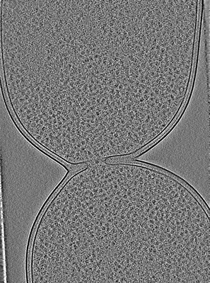 |
Cryo-electron tomography of Cryo-FIB milled dividing E. coli. [60 multi-frame micrographs composed of 4 frames each in TIFF format] | Navarro PP, Vettiger A, Ananda VY, Llopis PM, Allolio C, Bernhardt TG, Chao LH [Pubmed: 36097171] [DOI: 10.1038/s41564-022-01210-z] |
81.5 GB | — |
| 2019-10-07 | 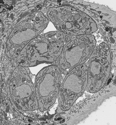 |
Processed FIB SEM images of a parasitophorous vacuole containing Toxoplasma gondii ∆CAP parasites. [1 multi-frame micrographs composed of 1 frames each in MRC format] | Hunt A, Russell MRG, Wagener J, Kent R, Carmeille R, Peddie CJ, Collinson L, Heaslip A, Ward GE, Treeck M [Pubmed: 31577230] [DOI: 10.7554/elife.50598] |
898.1 MB | — |
| 2016-07-06 | 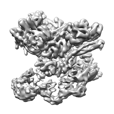 |
Unsupervised single-particle deep classification via statistical manifold learning [multiple data sets in MRC format] | Wu J, Ma YB, Congdon C, Brett B, Chen S, Xu Y, Ouyang Q, Mao Y | 28.2 GB | — |
| 2019-10-07 | 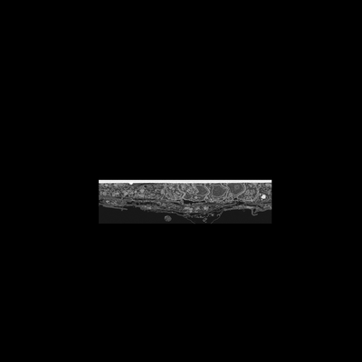 |
Raw FIB SEM images of a parasitophorous vacuole containing Toxoplasma gondii ∆CAP parasites, complemented with CAP. [4529 micrographs in TIFF format] | Hunt A, Russell MRG, Wagener J, Kent R, Carmeille R, Peddie CJ, Collinson L, Heaslip A, Ward GE, Treeck M [Pubmed: 31577230] [DOI: 10.7554/eLife.50598] |
605.2 GB | — |
| 2016-11-18 | 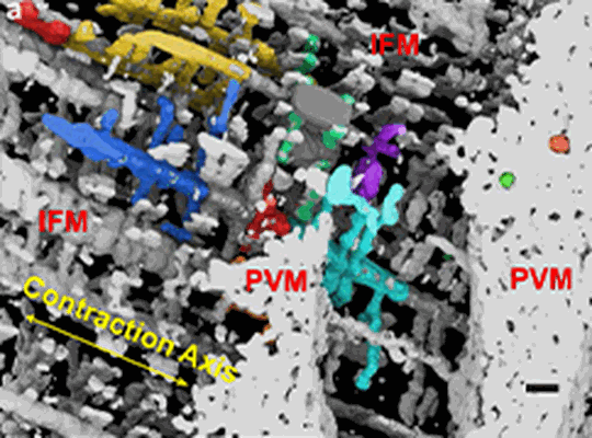 |
Focused Ion Beam-Scanning Electron Microscopy of mitochondrial reticulum in murine skeletal muscle [291 reconstructed volumes in MRC format] | Glancy B, Hartnell LM, Malide D, Yu ZX, Combs CA, Connelly PS, Subramaniam S, Balaban RS [Pubmed: 26223627] [DOI: 10.1038/nature14614] |
1.5 GB | — |
| 2019-10-07 |  |
Raw FIB SEM images of a parasitophorous vacuole containing Toxoplasma gondii ∆CAP parasites. [4490 micrographs in TIFF format] | Hunt A, Russell MRG, Wagener J, Kent R, Carmeille R, Peddie CJ, Collinson L, Heaslip A, Ward GE, Treeck M [Pubmed: 31577230] [DOI: 10.7554/eLife.50598] |
273.8 GB | — |
| 2019-10-07 | 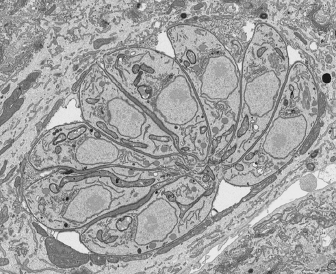 |
Processed FIB SEM images of a parasitophorous vacuole containing Toxoplasma gondii ∆CAP parasites, complemented with CAP. [1 multi-frame micrographs composed of 1 frames each in MRC format] | Hunt A, Russell MRG, Wagener J, Kent R, Carmeille R, Peddie CJ, Collinson L, Heaslip A, Ward GE, Treeck M [Pubmed: 31577230] [DOI: 10.7554/elife.50598] |
583.8 MB | — |
| 2021-11-26 |  |
Reconstructed cryo soft X-ray tomography dataset of a treated NIH-3T3 cell with corresponding correlated cryo-3D-SIM channels [300 reconstructed volumes in TIFF format] | Groen J, Pereiro E [Pubmed: 34909150] [DOI: 10.1039/d1sc04183e] |
10.1 GB | — |
| 2021-05-28 |  |
CLEM/FIB-SEM Imaging of T Cells after the Formation of Signaling Microclusters at the Immunological Synapse [160 micrographs in TIFF format] | Narayan K | 26.6 MB | — |
| 2020-02-28 |  |
Three-Dimensional Reconstructions of Mouse Circumvallate Taste Buds Using Serial Blockface Scanning Electron Microscopy: I. Cell Types and the Apical Region of the Taste Bud [1194 multi-frame micrographs composed of 1 frames each in TIFF format] | Yang R, Dzowo YK, Wilson CE, Russell RL, Kidd GJ, Salcedo E, Lasher RS, Kinnamon JC, Finger TE [Pubmed: 31587284] [DOI: 10.1002/cne.24779] |
184.9 GB | — |
| 2022-01-12 |  |
Cryo-FIB-SEM volume in a Sum159 human cell line [20 micrographs in TIFF format] | Klumpe S, Fung HKH, Goetz SK, Zagoriy I, Hampoelz B, Zhang X, Erdmann PS, Baumbach J, Müller CW, Beck M, Plitzko JM, Mahamid J [Pubmed: 34951584] [DOI: 10.7554/elife.70506] |
120.1 MB | — |
| 2020-12-21 | 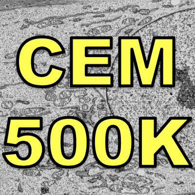 |
CEM500K - A large-scale heterogeneous unlabeled cellular electron microscopy image dataset for deep learning. [496544 micrographs in TIFF format] | Conrad RW, Narayan K [DOI: 10.1101/2020.12.11.421792] |
16.6 GB | — |
| 2021-01-08 | 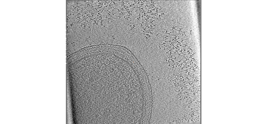 |
Nanoscale view of the Clostridium thermocellum cellulosome during cellulose degradation reveals an ecological strategy leading to phenotypic heterogeneity [37 tilt series in MRC format] | Tatli M, Moraïs S, Tovar-Herrera OE, Bomble Y, Bayer EA, Medalia O, Mizrahi I | 1004.3 MB | — |
| 2017-03-15 | 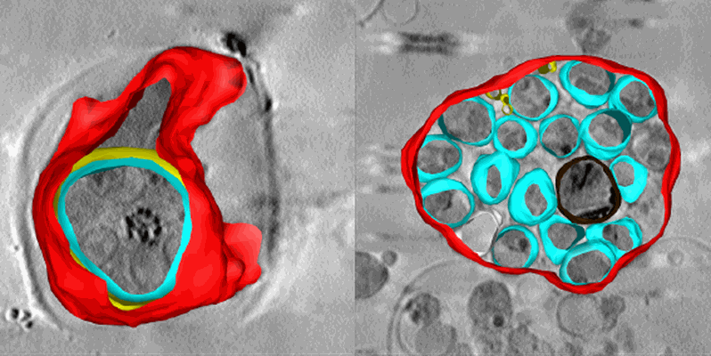 |
Soft X-ray tomography of Plasmodium falciparum infected human erythrocytes stalled in egress by the inhibitors Compound 2 and E64 [1 Soft X-ray tomograms in MRC format] | Hale VL, Saibil HR, Duke E, Fleck RA, Blackman MJ [Pubmed: 28292906] [DOI: 10.1073/pnas.1619441114] |
280.6 MB | — |
| 2023-10-13 | 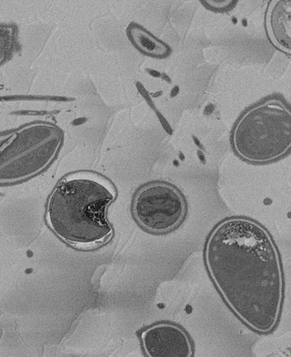 |
SBF-SEM micrographs of A. algerae microsporidia spores, 5 min germination [1215 micrographs in TIFF format] | Davydov A, Jaroenlak P, Ekiert D, Bhabha G [DOI: 10.7554/eLife.86638.1] |
226.3 GB | — |
| 2023-10-13 | 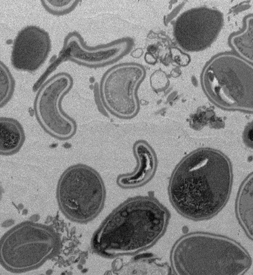 |
SBF-SEM micrographs of A. algerae microsporidia spores, 45 min germination [300 micrographs in TIFF format] | Davydov A, Jaroenlak P, Ekiert D, Bhabha G [DOI: 10.7554/eLife.86638.1] |
55.9 GB | — |
| 2021-01-08 |  |
A lamin A/C variant causing striated muscle disease provides insights into filament organization [2 tilt series in MRC format] | Tatli M [Pubmed: 33536248] [DOI: 10.1242/jcs.256156] |
2.7 GB | — |
