Electron Microscopy Public Image Archive
Electron Microscopy Public Image Archive
大阪大学的EMPIAR-PDBj团队为亚洲EM研究人员向EMPIAR传送大型EM图像数据提供服务。 除了通过互联网将数据直接传送到EBI(UK),研究人员还可以通过邮政或快递服务将数据硬盘发送到大阪大学,或者通过互联网传送到设置于大阪大学的服务器,然后由我们代为传送至数据登录网站。 如果您想使用此项服务,请先通过 电子邮件 与我们联系。
| Release date | Imageset | Title | Authors and references | Size | Resolution |
|---|---|---|---|---|---|
| 2020-10-14 | 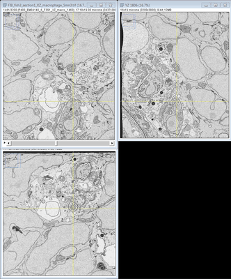 |
FIB SEM images of a Zebrafish hindbrain macrophage containing 2 Toxoplasma gondii tachizoites [multiple data sets in TIFF format] | Peddie CJ, Domart MC, Collinson L [Pubmed: 32461265] [DOI: 10.1242/dmm.043091] |
1.1 TB | — |
| 2020-10-12 | 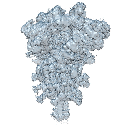 |
Cryo electron microscopy of SARS-CoV-2 spike in prefusion state [3207 multi-frame micrographs composed of 30 frames each in MRC format] | Carazo JM [Pubmed: 33063791] [DOI: 10.1107/S2052252520012725] |
2.1 TB | 3.0 - 3.3 Å |
| 2020-10-09 |  |
Structure of two nucleosomes bridged by human PARP2 [multiple data sets in MRCS format] | Gaullier G, Morgan GP, Luger K [Pubmed: 33141820] [DOI: 10.1371/journal.pone.0240932] |
677.3 GB | 10.5 Å |
| 2020-10-09 | 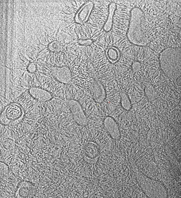 |
Human delta protocadherin 1 full ectodomains on membranes, tomogram 2 [multiple data sets in TIFF, JPEG and MRC formats] | Harrison OJ, Brasch J, Katsamba PS, Ahlsen G, Noble AJ, Dan H, Sampogna R, Potter CS, Carragher B, Honig B, Shapiro L [Pubmed: 32101743] [DOI: 10.1016/j.celrep.2020.02.003] |
230.3 GB | — |
| 2020-10-09 | 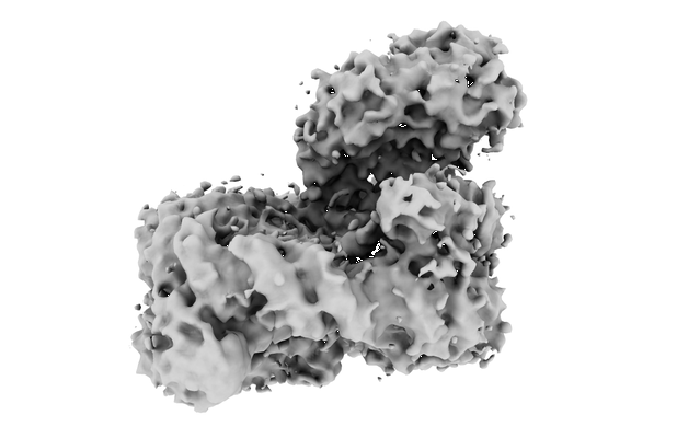 |
mouse cGAS with nucleosomes from 293T [2979 multi-frame micrographs composed of 40 frames each in MRC format] | Zhao B, Xu P, Rowlett CM, Jing T, Shinde O, Lei Y, West AP, Liu WR, Li P [Pubmed: 32911481] [DOI: 10.1038/s41586-020-2749-z] |
1.5 TB | 4.36 Å |
| 2020-10-09 | 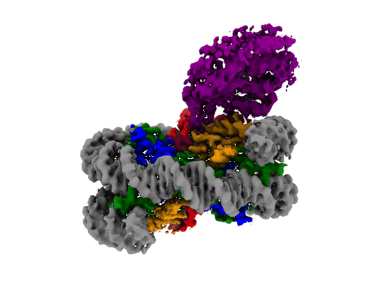 |
mouse cGAS with reconstituted nucleosome [5353 multi-frame micrographs composed of 40 frames each in MRC format] | Zhao B, Xu P [Pubmed: 32911481] [DOI: 10.1038/s41586-020-2749-z] |
767.6 GB | 2.98 Å |
| 2020-10-09 |  |
Single particle cryo electron microscopy of aldolase (rabbit, muscle) using beam-tilt on Talos Arctica [1625 micrographs in MRC format] | Kearns S, Cianfrocco MA [Pubmed: 33209328] [DOI: 10.1107/S2052252520013482] |
87.6 GB | 2.8 - 4.9 Å |
| 2020-10-02 | 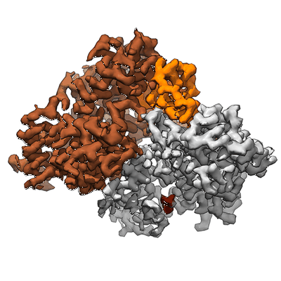 |
Single-particle cryo-EM of the human CDK-activating kinase in complex with ATP-gamma-S [multiple data sets in TIFF format] | Greber BJ, Perez-Bertoldi JM, Lim K, Iavarone AT, Toso DB, Nogales E [Pubmed: 32855301] [DOI: 10.1073/pnas.2009627117] |
3.5 TB | 2.8 Å |
| 2020-10-02 |  |
Connexin-46/50 in a dynamic lipid environment resolved by CryoEM at 1.9 Å [2087 multi-frame micrographs composed of 150 frames each in TIFF format] | Flores JA, Haddad BG, Dolan KA, Myers JB, Yoshioka CC, Copperman J, Zuckerman DM, Reichow SL [Pubmed: 32859914] [DOI: 10.1038/s41467-020-18120-5] |
4.1 TB | 1.94 - 2.5 Å |
| 2020-09-30 |  |
Structure of the Bacterial Ribosome at 2 Å Resolution [multiple data sets in TIFF format] | Watson ZL, Ward FR, Méheust R, Ad O, Schepartz A, Banfield JF, Cate JH [Pubmed: 32924932] [DOI: 10.7554/eLife.60482] |
2.1 TB | 1.98 Å |
| 2020-09-28 |  |
Single-particle cryo-EM of the human CDK-activating kinase in complex with THZ1 [4453 multi-frame micrographs composed of 69 frames each in TIFF format] | Greber BJ, Perez-Bertoldi JM, Lim K, Iavarone AT, Toso DB, Nogales E [Pubmed: 32855301] [DOI: 10.1073/pnas.2009627117] |
2.4 TB | 3.3 Å |
| 2020-09-28 |  |
Cryo electron microscopy of SARS-CoV-2 stabilized spike in prefusion state [3511 multi-frame micrographs composed of 40 frames each in TIFF format] | Carazo JM [Pubmed: 33063791] [DOI: 10.1107/S2052252520012725] |
865.0 GB | 2.9 Å |
| 2020-09-25 |  |
A "drug sweeping" state of the TriABC triclosan efflux pump from Pseudomonas aeruginosa [multiple data sets in MRCS format] | Fabre L, Abigail LT, Ntreh AT, Amira A, Yazidi A, Inga IV, Weeks JW, Leus IV, Jon JW, Sudipta S, Ruickoldt J, Jakob J, Rouiller I, Zgurskaya HI, Isabelle I, Sygusch J, Helen HI, Jurgen J [Pubmed: 32966762] [DOI: 10.1016/j.str.2020.09.001] |
238.7 GB | 4.3 - 20.0 Å |
| 2020-09-11 |  |
Cryo-EM structures of remodeler-nucleosome intermediates suggest allosteric control through the nucleosome [719 multi-frame micrographs composed of 30 frames each in MRCS format] | Armache J-P, Gamarra N, Johnson SL, Leonard JD, Wu S, Narlikar G, Cheng Y [Pubmed: 31210637] [DOI: 10.7554/eLife.46057] |
1.4 TB | 3.39 Å |
| 2020-09-11 |  |
Spatial Intra- and Intercellular Alignment of Respiratory Cilia and its Relation to Function [16 multi-frame micrographs composed of 100 frames each in MRC format] | Schneiter M, Halm S, Odriozola A, Mogel H, Rička J, Stoffel MH, Zuber B, Frenz M, Tschanz SA [DOI: 10.1101/735332] |
67.5 GB | — |
| 2020-09-11 |  |
Human Parainfluenza Virus Fusion Complex Glycoproteins Imaged In Action On Authentic Viral Surfaces [2 tilt series in MRC format] | Marcink TC, Wang T, des Georges A, Porotto M, Moscona A [Pubmed: 32956394] [DOI: 10.1371/journal.ppat.1008883] |
11.6 GB | 17.18 Å |
| 2020-09-03 | 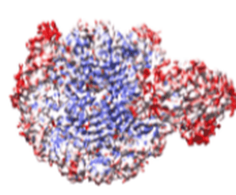 |
ISWI-NCP complex in the ADPBeF-bound state [stack of 166165 particles in MRCS format] | Yan L, Wu H, Li X, Gao N, Chen Z [Pubmed: 30872815] [DOI: 10.1038/s41594-019-0199-9] |
41.7 GB | 3.37 Å |
| 2020-09-02 | 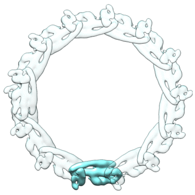 |
CryoEM reconstruction of ESCRT-III filament composed of IST1 NTD R16E K27E double mutant [stack of 4556 particles in MRCS format] | Nguyen HC, Talledge N, McCullough J, Sharma A, Moss FR, Iwasa JH, Vershinin MD, Sundquist WI, Frost A [Pubmed: 32251413] [DOI: 10.1038/s41594-020-0404-x] |
1.7 GB | 7.2 Å |
| 2020-09-02 | 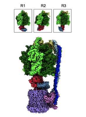 |
Structural basis of redox modulation on chloroplast ATP synthase (reduced form) [2063 micrographs in MRC format] | Yang JH, Williams D, Kandiah E, Fromme P, Chiu PL [Pubmed: 32879423] [DOI: 10.1038/s42003-020-01221-8] |
109.9 GB | 3.05 - 4.34 Å |
| 2020-09-02 | 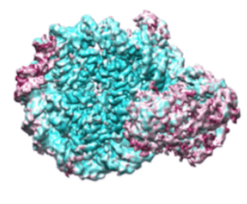 |
ISWI-NCP complex in the ADP-bound state [stack of 168430 particles in MRCS format] | Yan L, Wu H, Li X, Gao N, Chen Z [Pubmed: 32123390] [DOI: 10.1038/s41594-020-0388-6] |
36.2 GB | 2.9 Å |
| 2020-09-02 |  |
Native Pyruvate Dehydrogenase Complex from Neurospora crassa [4887 micrographs in MRC format] | Forsberg BO, Aibara S, Howard RJ, Mortezaei N, Lindahl E [Pubmed: 32938938] [DOI: 10.1038/s41467-020-18401-z] |
305.5 GB | 4.1 - 4.4 Å |
| 2020-08-28 |  |
Cryo-EM structures of four polymorphic TDP-43 amyloid cores [multiple data sets in MRC format] | Cao Q, Boyer DR, Sawaya MR, Ge P, Eisenberg DS [Pubmed: 31235914] [DOI: 10.1038/s41594-019-0248-4] |
4.5 TB | 3.3 - 3.8 Å |
| 2020-08-25 |  |
FAK structure from single particle analysis of 2D crystals [multiple data sets in MRC, TIFF and MRCS formats] | Acebron I, Righetto RD, Biyani N, Chami M, Boskovic J, Stahlberg H, Lietha D [Pubmed: 32779739] [DOI: 10.15252/embj.2020104743] |
1.8 TB | 5.96 - 6.32 Å |
| 2020-08-25 |  |
Structures of the human mitochondrial ribosome bound to EF-G1 reveal distinct features of mitochondrial translation elongation [stack of 6649 particles in MRC format] | Bhargava K, Datta PP, Kaushal PS, Keshavan P, Spremulli LL, Banavali NK [Pubmed: 32737313] [DOI: 10.1038/s41467-020-17715-2] |
753.5 GB | 2.96 - 3.96 Å |
| 2020-08-19 | 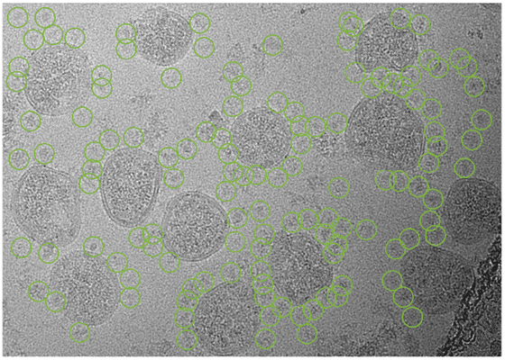 |
Cryo-EM structure of SARS-CoV-2 Spike Proteins on intact virions [7982 multi-frame micrographs composed of 48 frames each in TIFF format] | Ke Z, Qu K, Cortese M, Zila V, Nakane T, Xiong X, Scheres SHW, Briggs JAG [Pubmed: 32805734] [DOI: 10.1038/s41586-020-2665-2] |
2.1 TB | 3.5 - 4.1 Å |
