Electron Microscopy Public Image Archive
Electron Microscopy Public Image Archive
大阪大学的EMPIAR-PDBj团队为亚洲EM研究人员向EMPIAR传送大型EM图像数据提供服务。 除了通过互联网将数据直接传送到EBI(UK),研究人员还可以通过邮政或快递服务将数据硬盘发送到大阪大学,或者通过互联网传送到设置于大阪大学的服务器,然后由我们代为传送至数据登录网站。 如果您想使用此项服务,请先通过 电子邮件 与我们联系。
| Release date | Imageset | Title | Authors and references | Size | Resolution |
|---|---|---|---|---|---|
| 2022-05-27 | 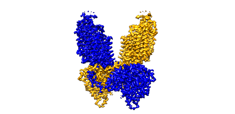 |
Cryo-EM structure of human NKCC1 K289NA492EL671C bound with bumetanide [3891 multi-frame micrographs composed of 40 frames each in TIFF format] | Zhao YZ [Pubmed: 35585053] [DOI: 10.1038/s41467-022-30407-3] |
1.8 TB | 2.9 Å |
| 2022-05-27 | 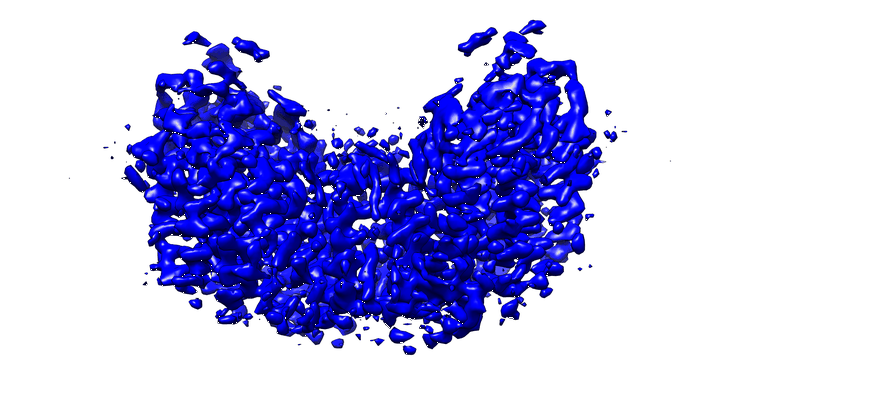 |
Cryo-EM structure of human NKCC1 K289NA492E bound with Furosemide [3405 multi-frame micrographs composed of 40 frames each in TIFF format] | Zhao Y [Pubmed: 35585053] [DOI: 10.1038/s41467-022-30407-3] |
1.6 TB | 3.4 Å |
| 2022-05-26 |  |
Cryo TEM single particle dataset of purified ChAdOx1/ADZ-1222 [2875 multi-frame micrographs composed of 40 frames each in TIFF format] | Boyd RJ, Baker AB [Pubmed: 34851659] [DOI: 10.1126/sciadv.abl8213] |
1.0 TB | 3.07 Å |
| 2022-05-25 |  |
SARS-CoV-2 spike protein S:D614G + S:A222V variant [4841 micrographs in MRC format] | Ginex T, Marco-Marín C, Wieczór M, Mata CP, Krieger J, Ruiz-Rodriguez P, López-Redondo ML, Francés-Gómez C, Melero R, Sánchez-Sorzano CÓ, Martínez M, Gougeard N, Forcada-Nadal A, Zamora-Caballero S, Gozalbo-Rovira R, Sanz-Frasquet C, Arranz R, Bravo J, Rubio V, Marina A, Geller R, Comas I, Gil C, Coscolla M, Orozco M, Llácer JL, Carazo JM [Pubmed: 35816514] [DOI: 10.1371/journal.ppat.1010631] |
303.8 GB | 3.4 Å |
| 2022-05-25 |  |
A drug and ATP binding site in type 1 ryanodine receptor [6890 multi-frame micrographs composed of 50 frames each in TIFF format] | Melville Z, Dridi H, Yuan Q, Reiken S, Wronska A, Liu Y, Clarke OB, Marks AR [Pubmed: 35580609] [DOI: 10.1016/j.str.2022.04.010] |
1.5 TB | 2.45 Å |
| 2022-05-24 | 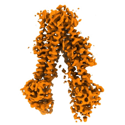 |
The complex of dephosphorylated human cystic fibrosis transmembrane conductance regulator (CFTR) and Lumacaftor (VX-809) [3191 multi-frame micrographs composed of 50 frames each in TIFF format] | Fiedorczuk K, Chen J [Pubmed: 34995514] [DOI: 10.1016/j.cell.2021.12.009] |
1.4 TB | 3.9 Å |
| 2022-05-24 | 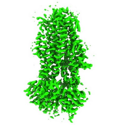 |
The complex of phosphorylated human cystic fibrosis transmembrane conductance regulator (CFTR) with ATP/Mg and Tezacaftor (VX-661) [4257 multi-frame micrographs composed of 50 frames each in TIFF format] | Fiedorczuk K, Chen J [Pubmed: 34995514] [DOI: 10.1016/j.cell.2021.12.009] |
1.8 TB | 3.8 Å |
| 2022-05-24 | 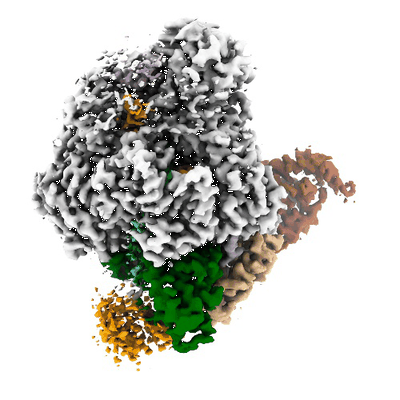 |
Cryo-EM data used for the determination of LACV-L structure in transcription early-elongation state [2510 multi-frame micrographs composed of 60 frames each in TIFF format] | Arragain B, Durieux Trouilleton Q, Baudin F, Provaznik J, Azevedo N, Cusack S, Schoehn G, Malet H [Pubmed: 35173159] [DOI: 10.1038/s41467-022-28428-z] |
721.4 GB | 3.3 Å |
| 2022-05-20 | 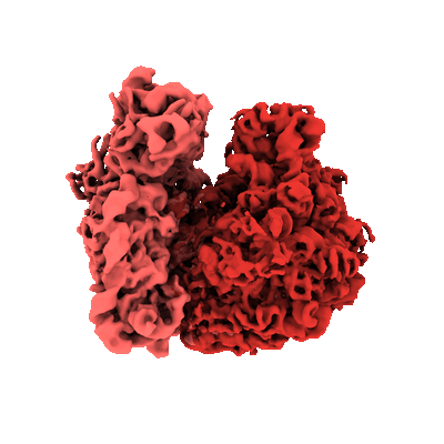 |
Parallel cryo electron tomography (PACE-tomo) of 70S ribosomes (200 kV, side-entry holder) [multiple data sets in MRC format] | Eisenstein F, Danev R [Pubmed: 36456783] [DOI: 10.1038/s41592-022-01690-1] |
222.8 GB | 5.8 - 6.5 Å |
| 2022-05-20 | 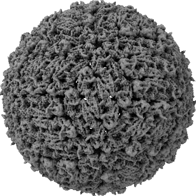 |
Single-particle reconstruction of Tick-borne encephalitis virus (Final reconstruction) [multiple data sets in TIFF format] | Pulkkinen LIA, Barrass SV, Domanska A, Överby AK, Anastasina M, Butcher SJ [Pubmed: 35458522] [DOI: 10.3390/v14040792] |
16.4 TB | 3.3 Å |
| 2022-05-20 | 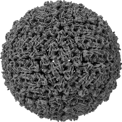 |
Single-particle reconstruction of Tick-borne encephalitis virus (Preparation 1) [14135 multi-frame micrographs composed of 30 frames each in MRC format] | Pulkkinen LIA, Barrass SV, Domanska A, Överby AK, Anastasina M, Butcher SJ [Pubmed: 35458522] [DOI: 10.3390/v14040792] |
6.2 TB | 3.5 Å |
| 2022-05-20 | 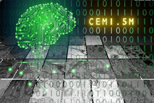 |
CEM1.5M : a cellular EM dataset containing ~1.5 x 10⁶ unlabeled 2D image patches curated for deep learning [1592753 micrographs in TIFF format] | Narayan K | 57.6 GB | — |
| 2022-05-17 |  |
Structures of positive allosteric modulator-bound and unbound active human calcium-sensing receptor [13082 multi-frame micrographs composed of 60 frames each in TIFF format] | Park J, Zuo H, Frangaj A, Fu Z, Yen LY, Zhang Z, Mosyak L, Slavkovich VN, Liu J, Ray KM, Cao B, Vallese F, Geng Y, Chen S, Grassucci R, Dandey VP, Tan YZ, Eng E, Lee Y, Kloss B, Liu Z, Hendrickson WA, Potter CS, Carragher B, Graziano J, Conigrave AD, Frank J, Clarke OB, Fan QR [Pubmed: 34916296] [DOI: 10.1073/pnas.2115849118] |
3.8 TB | 2.7 Å |
| 2022-05-17 |  |
Cryo-EM structure of the human ATP13A2 [multiple data sets in TIFF format] | Tomita A, Daiho T, Kusakizako T, Yamashita K, Ogasawara S, Murata T, Nishizawa T, Nureki O [Pubmed: 34798056] [DOI: 10.1016/j.molcel.2021.11.001] |
3.8 TB | 3.54 - 3.92 Å |
| 2022-05-17 |  |
Conformational rearrangements upon start codon recognition in human 48S translation initiation complex [multiple data sets in MRC and MRCS formats] | Yi SH, Petrychenko V, Schliep JE, Goyal A, Linden A, Chari A, Urlaub H, Stark H, Rodnina MV, Adio S, Fischer N [Pubmed: 35489072] [DOI: 10.1093/nar/gkac283] |
1.1 TB | 3.7 - 4.7 Å |
| 2022-05-11 |  |
Cryo-EM SPA dataset of Megadalton-range protein communities from a Chaetomium thermophilum native cell extract [2808 multi-frame micrographs composed of 13 frames each in MRC format] | Skalidis IS, Kyrilis FLK, Tüting CT, Müller JM, Sorokina MS, Hamdi FH, Sadian YS, Chojnowski GC, Kastritis PLK [Pubmed: 34836937] [DOI: 10.1038/s41467-021-27287-4] |
1.1 TB | 3.84 - 4.52 Å |
| 2022-05-11 |  |
Tilt series and tomograms of cells expressing different non structural proteins (NSPs) of SARS-CoV-2 [multiple data sets in MRC format] | Polshchuk RS, Polishchuk E, De Matteis MA [Pubmed: 35551511] [DOI: 10.1038/s41586-022-04835-6] |
107.1 GB | — |
| 2022-05-10 |  |
CEM-MitoLab: a dataset of ~22K cellular EM 2D images with label maps of ~135K mitochondrial instances, for deep learning [43720 micrographs in TIFF format] | Narayan K, Conrad RW | 2.8 GB | — |
| 2022-05-03 |  |
In situ single particle classification reveals distinct 60S maturation intermediates in cells [multiple data sets in MRC format] | Lucas BA, Zhang K, Loerch S, Grigorieff N [DOI: 10.1101/2022.04.10.487797] |
10.5 GB | — |
| 2022-05-03 | 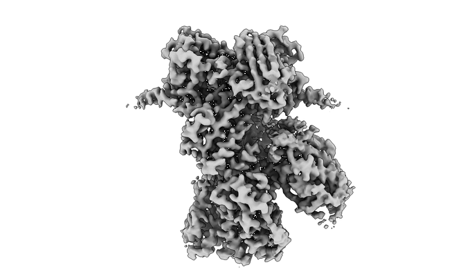 |
Cryo-EM Structures of Glucocorticoid Receptor-Hsp90-p23 [the GR Maturation Complex], Hsp90-p23, and MBP-Hsp90-p23 [multiple data sets in MRC format] | Noddings CM, Wang RY, Agard DA [Pubmed: 34937936] [DOI: 10.1038/s41586-021-04236-1] |
494.1 GB | 2.56 - 3.63 Å |
| 2022-04-29 | 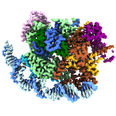 |
Structure of transcription factor UAF in complex with TBP and 35S rRNA promoter DNA [multiple data sets in TIFF format] | Baudin F, Murciano B, Fung HKH, Fromm SA, Mattei S, Mahamid J, Müller CW [Pubmed: 35442737] [DOI: 10.1126/sciadv.abn5725] |
3.1 TB | 2.8 Å |
| 2022-04-29 | 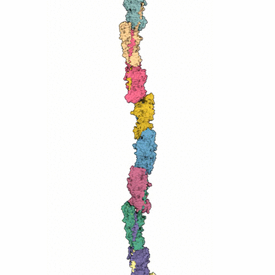 |
Raw data for "Structure of polymerized type V pilin reveals assembly mechanism involving protease-mediated strand exchange" [1153 micrographs in MRC format] | Shibata S, Shoji M, Okada K, Matsunami H, Matthews M, Imada K, Nakayama K, Wolf M [Pubmed: 32284566] [DOI: 10.1038/s41564-020-0705-1] |
72.1 GB | 3.6 Å |
| 2022-04-29 |  |
Cryogenic electron microscopy structure of full length human meta vinculin [3005 multi-frame micrographs composed of 40 frames each in TIFF format] | Izard T, Rangarajan ES [Pubmed: 33440717] [DOI: 10.3390/ijms22020645] |
744.8 GB | 4.15 - 4.5 Å |
| 2022-04-29 |  |
Cryo electron microscopy of wild-type hyaluronan synthase with UDP [3062 multi-frame micrographs composed of 40 frames each in TIFF format] | Maloney FP, Kuklewicz J, Zimmer J [Pubmed: 35355017] [DOI: 10.1038/s41586-022-04534-2] |
818.1 GB | 3.1 Å |
| 2022-04-26 |  |
Parallel cryo electron tomography (PACE-tomo) of 70S ribosomes [multiple data sets in TIFF and MRC formats] | Eisenstein F, Danev R [Pubmed: 36456783] [DOI: 10.1038/s41592-022-01690-1] |
282.6 GB | 3.1 Å |
