Electron Microscopy Public Image Archive
Electron Microscopy Public Image Archive
大阪大学的EMPIAR-PDBj团队为亚洲EM研究人员向EMPIAR传送大型EM图像数据提供服务。 除了通过互联网将数据直接传送到EBI(UK),研究人员还可以通过邮政或快递服务将数据硬盘发送到大阪大学,或者通过互联网传送到设置于大阪大学的服务器,然后由我们代为传送至数据登录网站。 如果您想使用此项服务,请先通过 电子邮件 与我们联系。
| Release date | Imageset | Title | Authors and references | Size | Resolution |
|---|---|---|---|---|---|
| 2022-11-29 | 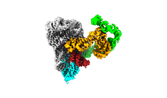 |
CryoEM micrographs of a group II intron retroelement (apoRNP) [8005 multi-frame micrographs composed of 40 frames each in TIFF format] | Chung KC, Xu LX, Pyle AMP [Pubmed: 36356138] [DOI: 10.1126/science.abq2844] |
3.1 TB | 3.1 Å |
| 2022-11-28 | 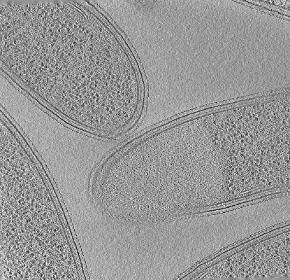 |
Cryo-electron tomography of FIB-milled Caulobacter crescentus expressing WT PopZ [multiple data sets in MRC and TIFF formats] | Lasker K, Lam V, Villa E [Pubmed: 36163138] [DOI: 10.1038/s41467-022-33221-z] |
7.1 GB | — |
| 2022-11-28 | 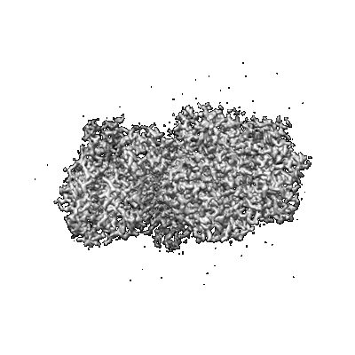 |
Cryo-EM Structure of Formate Dehydrogenase 1 from Methylorubrum extorquens AM1 [7650 multi-frame micrographs composed of 40 frames each in TIFF format] | Yoshikawa T, Makino F, Miyata T, Suzuki Y, Tanaka H, Namba K, Kano K, Sowa K, Kitazumi Y, Shirai O [Pubmed: 35535582] [DOI: 10.1039/d2cc01541b] |
1.4 TB | 2.19 Å |
| 2022-11-25 | 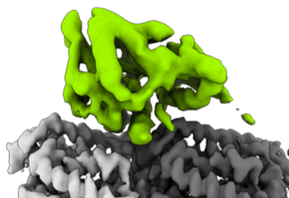 |
Cryo-EM dataset of P.berghei kinesin-8B motor domain in AMPPNP state bound to tubulin dimer [1026 micrographs in MRC format] | Liu T, Shilliday F, Cook AD, Moores CA [Pubmed: 36384964] [DOI: 10.1038/s41467-022-34710-x] |
54.4 GB | 3.3 Å |
| 2022-11-23 |  |
Cryo-electron tomograms of RPE1 cells with comprehensive annotation of actin filaments and microtubules [multiple data sets in TIFF and MRC formats] | Cheng DWC, Goetz SK, Mahamid J [Pubmed: 36690741] [DOI: 10.1038/s41592-022-01746-2] |
32.9 GB | — |
| 2022-11-21 |  |
Micrographs of Rigid-Rod Aromatics BP-NBD [5610 multi-frame micrographs composed of 40 frames each in TIFF format] | Yi M, Wang F, Tan W, Hsieh JT, Egelman EH, Xu B [Pubmed: 35849554] [DOI: 10.1021/jacs.2c05491] |
1.2 TB | 2.4 Å |
| 2022-11-21 |  |
Tau Paired Helical Filament from Alzheimer's Disease incubated with EGCG for 3 hours [6406 multi-frame micrographs composed of 30 frames each in MRCS format] | Seidler PM, Murray KA, Boyer DR, Ge P, Sawaya MR, Eisenberg DS [Pubmed: 36114178] [DOI: 10.1038/s41467-022-32951-4] |
1.7 TB | 3.8 Å |
| 2022-11-21 |  |
Tau Paired Helical Filament from Alzheimer's Disease incubated 1 hr. with EGCG [3890 multi-frame micrographs composed of 48 frames each in TIFF format] | Seidler PM, Murray KA, Boyer DR, Ge P, Sawaya MR, Eisenberg DS [Pubmed: 36114178] [DOI: 10.1038/s41467-022-32951-4] |
3.7 TB | 3.3 Å |
| 2022-11-21 | 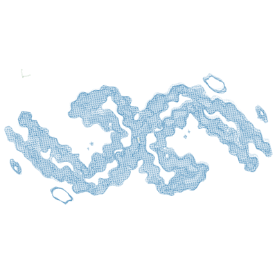 |
Tau Paired Helical Filament from Alzheimer's Disease not incubated with EGCG [2651 multi-frame micrographs composed of 48 frames each in TIFF format] | Seidler PM, Murray KA, Boyer DR, Ge P, Sawaya MR, Eisenberg DS [Pubmed: 36114178] [DOI: 10.1038/s41467-022-32951-4] |
2.5 TB | 3.4 Å |
| 2022-11-18 | 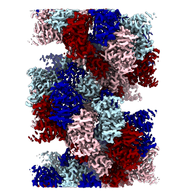 |
Cryo-EM reconstruction of TNKS2 SAM-PARP filament [multiple data sets in MRCS, MRC and TIFF formats] | Mariotti L, Inian O, Desfosses A, Beuron F, Morris E.P, Guettler S [Pubmed: 36418402] [DOI: 10.1038/s41586-022-05449-8] |
11.8 TB | 2.98 Å |
| 2022-11-18 | 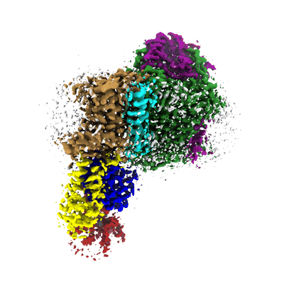 |
Single particle cryo-EM of KdpFABC WT (KdpB-Ser162-P) under turnover condition [multiple data sets in TIFF format] | Silberberg JM, Stock C, Hielkema L, Corey RA, Rheinberger J, Wunnicke D, Dubach VRA, Stansfeld PJ, Hänelt I, Paulino C [Pubmed: 36255052] [DOI: 10.7554/eLife.80988] |
2.9 TB | 3.1 - 3.5 Å |
| 2022-11-18 | 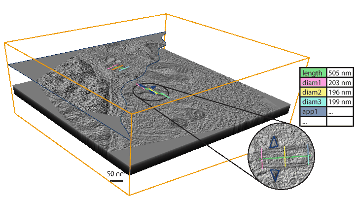 |
A high-throughput electron tomography workflow reveals over-elongated centrioles in relapsed-refractory multiple myeloma [multiple data sets in MRC format] | Köhrer S, Dittrich T, Schorb M, Weinhold N, Haberbosch I, Börmel M, Pajor G [Pubmed: 36452870] [DOI: 10.1016/j.crmeth.2022.100322] |
2.0 TB | — |
| 2022-11-18 | 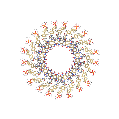 |
Micrographs of Rigid-Rod Aromatics pBP-NBD [8261 multi-frame micrographs composed of 40 frames each in TIFF format] | Yi M, Wang F, Tan W, Hsieh JT, Egelman EH, Xu B [Pubmed: 35849554] [DOI: 10.1021/jacs.2c05491] |
1.7 TB | 2.4 Å |
| 2022-11-17 | 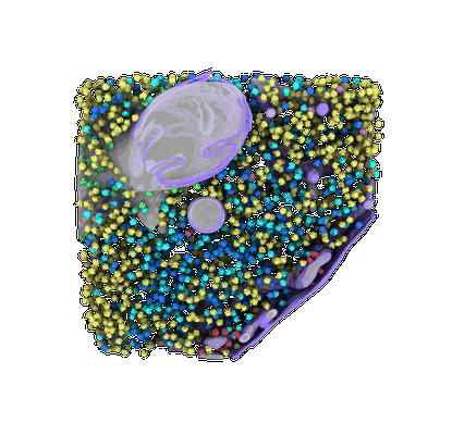 |
Defocus and Volta potential phase plate cryo-electron tomography of S. pombe cryo-FIB lamellae with comprehensive annotations of structures and macromolecules [multiple data sets in TIFF and MRC formats] | Goetz SK, Mahamid J [Pubmed: 36690741] [DOI: 10.1038/s41592-022-01746-2] |
305.5 GB | 9.3 - 34.0 Å |
| 2022-11-16 | 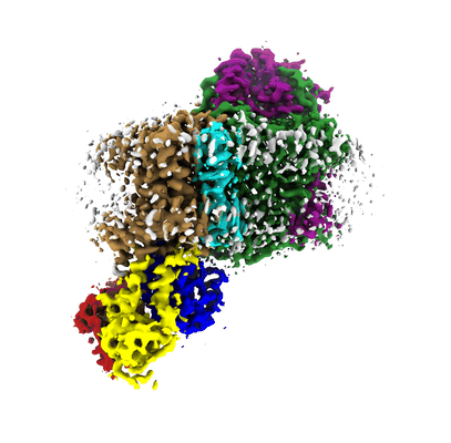 |
Single particle cryo-EM of KdpFAB(Ser162-P, D307N)C in apo condition [multiple data sets in TIFF format] | Silberberg JM, Stock C, Hielkema L, Corey RA, Rheinberger J, Wunnicke D, Dubach VRA, Stansfeld PJ, Hänelt I, Paulino C [Pubmed: 36255052] [DOI: 10.7554/eLife.80988] |
2.5 TB | 3.4 - 3.7 Å |
| 2022-11-16 | 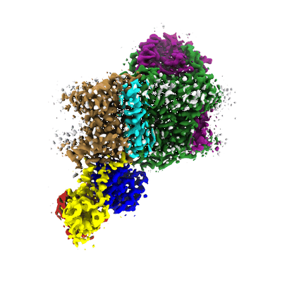 |
Single particle cryo-EM of KdpFABC WT (KdpB-Ser162-P) in presence of orthovanadate [multiple data sets in TIFF format] | Silberberg JM, Stock C, Hielkema L, Corey RA, Rheinberger J, Wunnicke D, Dubach VRA, Stansfeld PJ, Hänelt I, Paulino C [Pubmed: 36255052] [DOI: 10.7554/eLife.80988] |
399.3 GB | 3.3 - 7.4 Å |
| 2022-11-16 |  |
Single particle cryo-EM of KdpFAB(S162A)C under turnover condition [multiple data sets in TIFF format] | Silberberg JM, Stock C, Hielkema L, Corey RA, Rheinberger J, Wunnicke D, Dubach VRA, Stansfeld PJ, Hänelt I, Paulino C [Pubmed: 36255052] [DOI: 10.7554/eLife.80988] |
1.8 TB | 3.7 - 4.0 Å |
| 2022-11-16 |  |
Cryo-electron microscopy of Dicer-1 and Its Partner Protein Loqs-PB complex with model pre-miRNA in presence of Mg2+ ions [8128 multi-frame micrographs composed of 38 frames each in TIFF format] | Jouravleva K, Golovenko D, Demo G, Dutcher RC, Hall TMT, Zamore PD, Korostelev AA [Pubmed: 36182693] [DOI: 10.1016/j.molcel.2022.09.002] |
2.9 TB | 3.26 - 3.73 Å |
| 2022-11-16 |  |
Cryo-electron microscopy of Dicer-1 and Its Partner Protein Loqs-PB complex [2849 multi-frame micrographs composed of 30 frames each in TIFF format] | Jouravleva K, Golovenko D, Demo G, Dutcher RC, Hall TMT, Zamore PD, Korostelev AA [Pubmed: 36182693] [DOI: 10.1016/j.molcel.2022.09.002] |
1.1 TB | 3.94 - 4.02 Å |
| 2022-11-15 |  |
2.1 Å resolution structure of β-galactosidase obtained from Glacios equipped with Falcon 3 [multiple data sets in TIFF format] | Merk A, Darling JE, Grisshammer R, Ognjenović J | 4.8 TB | 2.1 Å |
| 2022-11-15 |  |
cryoEM movies from 30S-RbfA complex [3369 multi-frame micrographs composed of 20 frames each in MRC format] | Schedlbauer A, Fucini P, Connell SR [Pubmed: 34088665] [DOI: 10.1126/sciadv.abf7547] |
3.5 TB | 2.75 - 2.96 Å |
| 2022-11-15 | 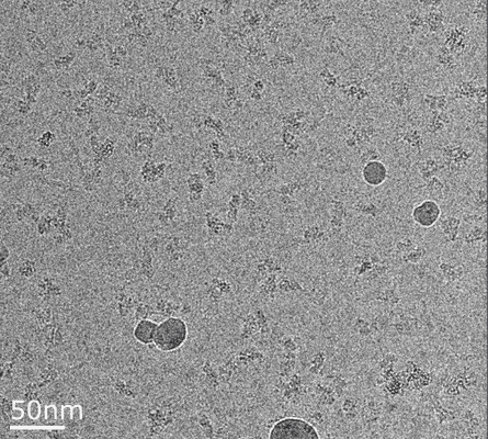 |
Movies from 30S-RbfA-RimP-RsmA dataset [4395 multi-frame micrographs composed of 27 frames each in MRCS format] | Schedlbauer A, Fucini P, Connell SR [Pubmed: 34088665] [DOI: 10.1126/sciadv.abf7547] |
3.6 TB | 3.86 - 3.93 Å |
| 2022-11-15 |  |
Amyloid fibril from the antimicrobial peptide uperin 3.5 [multiple data sets in TIFF, MRC and MRCS formats] | Bücker R, Seuring C, Cazey C, Veith K, Garcia-Alai M, Grünewald K, Landau M [Pubmed: 35896552] [DOI: 10.1038/s41467-022-32039-z] |
7.4 TB | 3.0 Å |
| 2022-11-15 | 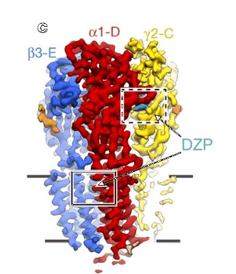 |
Cryo-EM movies of the human α1β3γ2 GABAA receptor in a lipid bilayer bound to diazepam and GABA [1472 multi-frame micrographs composed of 40 frames each in TIFF format] | Masiulis S, Desai R, Uchanski T, Serna Martin I, Laverty D, Karia D, Malinauskas T, Zivanov J, Pardon E, Kotecha A, Steyaert J, Miller KW, Aricescu AR [Pubmed: 30602790] [DOI: 10.1038/s41586-018-0832-5] |
1.8 TB | 3.58 Å |
| 2022-11-15 | 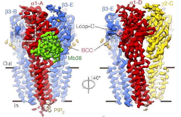 |
Cryo-EM movies of the human α1β3γ2 GABAA receptor in a lipid bilayer bound to bicuculline [988 multi-frame micrographs composed of 75 frames each in TIFF format] | Masiulis S, Desai R, Uchanski T, Serna Martin I, Laverty D, Karia D, Malinauskas T, Zivanov J, Pardon E, Kotecha A, Steyaert J, Miller KW, Aricescu AR [Pubmed: 30602790] [DOI: 10.1038/s41586-018-0832-5] |
1.2 TB | 3.69 Å |
