Electron Microscopy Public Image Archive
Electron Microscopy Public Image Archive
The EMPIAR-PDBj team at Osaka University assists Asian EM researchers with the transfer of big EM image data to EMPIAR. Instead of sending the data directly to the EBI (UK) via the internet, hard drives can also be sent to Osaka University by postal mail or via a courier service. As an alternative, internet transfer to our server in Osaka is also available. If you would like to take advantage of our submission services, please contact us first by e-mail before sending the data to us.
| Release date | Imageset | Title | Authors and references | Size | Resolution |
|---|---|---|---|---|---|
| 2022-12-13 | 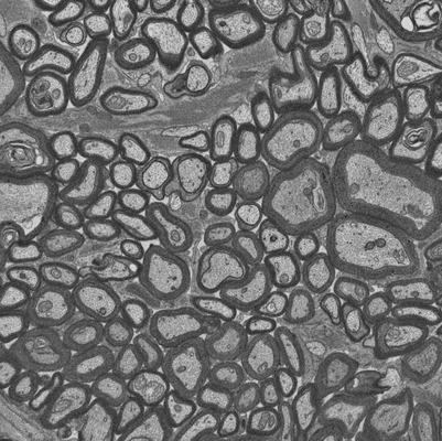 |
Focused ion beam-scanning electron microscopy links pathological myelin outfoldings to axonal changes in mice lacking Plp1 or Mag [910 micrographs in TIFF format] | Steyer AM, Möbius W [Pubmed: 36354016] [DOI: 10.1002/glia.24290] |
13.7 GB | — |
| 2022-12-13 |  |
Focused ion beam-scanning electron microscopy links pathological myelin outfoldings to axonal changes in mice lacking Plp1 or Mag [1186 micrographs in TIFF format] | Steyer AM, Möbius W [Pubmed: 36354016] [DOI: 10.1002/glia.24290] |
28.2 GB | — |
| 2022-12-16 | 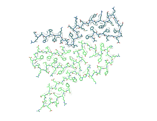 |
Amyloid fibril structure from the vascular variant of systemic AA amyloidosis [multiple data sets in TIFF format] | Banerjee S, Baur J, Daniel C, Pfeiffer PB, Hitzenberger M, Kuhn L, Wiese S, Bijzet J, Haupt C, Amann KU, Zacharias M, Hazenberg BPC, Westermark GT, Schmidt M, Fändrich M [Pubmed: 36433936] [DOI: 10.1038/s41467-022-34636-4] |
1009.8 GB | 2.56 Å |
| 2022-12-19 | 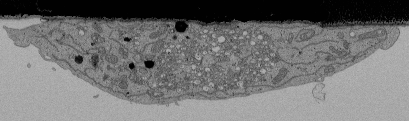 |
CLEMSite, a software for automated phenotypic screens using light microscopy and FIB-SEM. [multiple data sets in TIFF format] | Lleti JMSL, Steyer AMS, Schwab YS | 19.7 GB | — |
| 2023-01-03 | 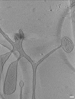 |
Cryo electron tomography of Ca. L. ossiferum [multiple data sets in MRC format] | Wollweber F, Xu J [Pubmed: 36544020] [DOI: 10.1038/s41586-022-05550-y] |
11.6 GB | 11.7 - 24.5 Å |
| 2023-01-03 | 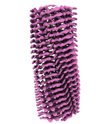 |
Cryo-EM structure of hnRNPDL amyloid fibrils [multiple data sets in TIFF format] | Chaves-Sanjuan A, Garcia-Pardo J, Bartolome-Nafria A, Gil-Garcia M, Visentin C, Bolognesi M, Ricagno S, Ventura S [Pubmed: 36646699] [DOI: 10.1038/s41467-023-35854-0] |
880.1 GB | 2.5 Å |
| 2023-01-03 | 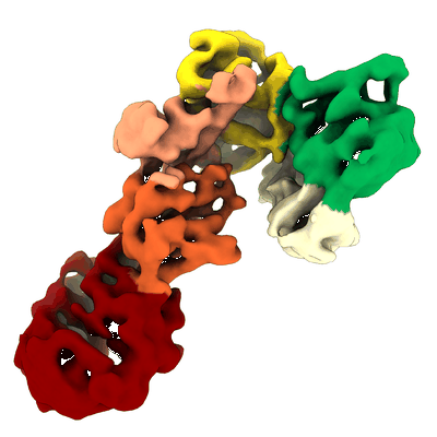 |
C-terminal half of Leucine Rich Repeat Kinase 1 (LRRK1) [3629 multi-frame micrographs composed of 50 frames each in TIFF format] | Matyszewski M, Leschziner AE [Pubmed: 36510024] [DOI: 10.1038/s41594-022-00863-y] |
1.2 TB | 5.8 Å |
| 2023-01-03 | 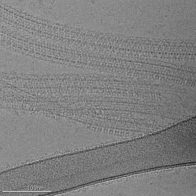 |
Apo C-terminal half of LRRK2 (I2020T) bound to microtubule [2379 multi-frame micrographs composed of 50 frames each in TIFF format] | Matyszewski M, Leschziner AE [Pubmed: 36510024] [DOI: 10.1038/s41594-022-00863-y] |
643.9 GB | 7.0 Å |
| 2023-01-03 | 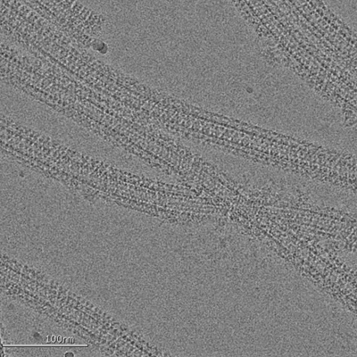 |
C-terminal half of LRRK2 (I2020T) bound to microtubule in presence of MLi-2 kinase inhibitor [2354 multi-frame micrographs composed of 55 frames each in TIFF format] | Matyszewski M, Leschziner AE [Pubmed: 36510024] [DOI: 10.1038/s41594-022-00863-y] |
684.8 GB | 4.5 - 18.0 Å |
| 2023-01-03 | 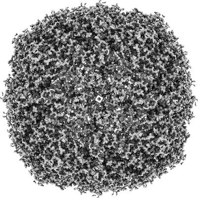 |
Movies of apoferritin collected at different dose rates on the Direct Electron Apollo direct detector - 15 eps [972 multi-frame micrographs composed of 76 frames each in TIFF format] | Peng R, Fu X, Mendez JH, Randolph PS, Bammes BE, Stagg SM [Pubmed: 36578473] [DOI: 10.1016/j.yjsbx.2022.100080] |
297.2 GB | — |
| 2023-01-10 | 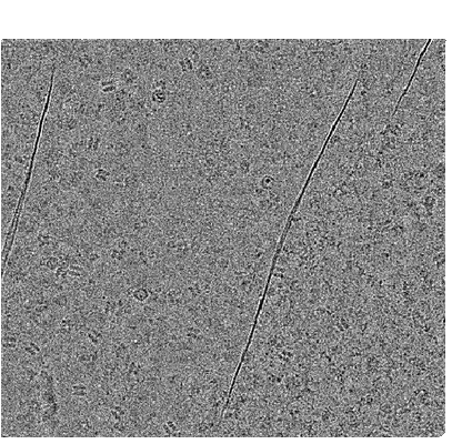 |
Cryo-EM raw image of Bovine retinal pigmented epithelium lysate [multiple data sets in TIFF format] | Zhang Z., Morgan C.E. [Pubmed: 36577381] [DOI: 10.1016/j.celrep.2022.111876] |
1.8 TB | 2.28 - 3.32 Å |
| 2023-01-16 | 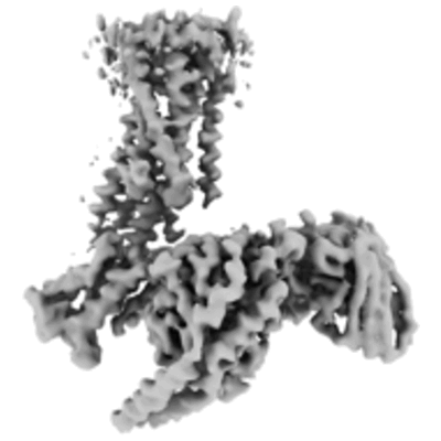 |
Structure of the active Gi-coupled human lysophosphatidic acid receptor 1 complexed with a potent agonist [6228 multi-frame micrographs composed of 48 frames each in TIFF format] | Akasaka H, Tanaka T, Sano FK, Matsuzaki Y, Shihoya W, Nureki O [Pubmed: 36109516] [DOI: 10.1038/s41467-022-33121-2] |
1.4 TB | 3.5 - 5.6 Å |
| 2023-01-16 | 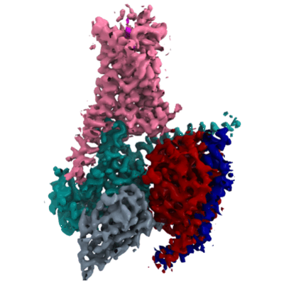 |
Cryo-EM structures of the β3 adrenergic receptor bound to solabegron and isoproterenol [multiple data sets in TIFF format] | Nagiri C, Kobayashi K, Tomita A, Kato M, Yamashita K, Nishizawa T, Inoue A, Shihoya W, Nureki O [Pubmed: 35489202] [DOI: 10.1016/j.bbrc.2022.04.065] |
2.5 TB | 3.3 - 3.9 Å |
| 2023-01-16 | 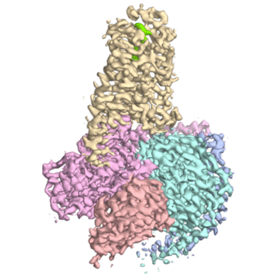 |
Dog beta3 adrenergic receptor bound to mirabegron in complex with a miniGs heterotrimer [2864 multi-frame micrographs composed of 48 frames each in TIFF format] | Nagiri C, Kobayashi K, Tomita A, Kato M, Yamashita K, Nishizawa T, Inoue A, Shihoya W, Nureki O [Pubmed: 34314699] [DOI: 10.1016/j.molcel.2021.06.024] |
671.1 GB | 3.16 Å |
| 2023-01-16 | 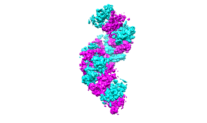 |
Structure of RecT protein from Listeria innoccua phage A118 in complex with 83-mer single stranded DNA [1619 multi-frame micrographs composed of 45 frames each in TIFF format] | Bell CE [Pubmed: 36543802] [DOI: 10.1038/s41467-022-35572-z] |
822.3 GB | 4.5 Å |
| 2023-01-16 | 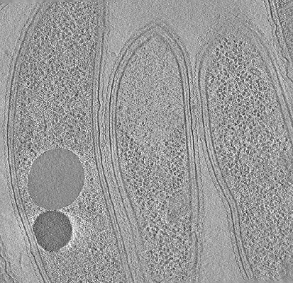 |
Cryo-electron tomography of FIB-milled Caulobacter crescentus expressing PopZ with IDR-156 and pentavalent OD [multiple data sets in MRC and TIFF formats] | Lasker K, Lam V, Villa E [Pubmed: 36163138] [DOI: 10.1038/s41467-022-33221-z] |
5.9 GB | — |
| 2023-01-16 | 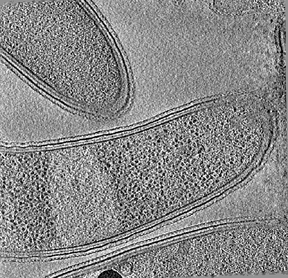 |
Cryo-electron tomography of FIB-milled Caulobacter crescentus expressing PopZ with IDR-156 [multiple data sets in MRC and TIFF formats] | Lasker K, Lam V, Villa E [Pubmed: 36163138] [DOI: 10.1038/s41467-022-33221-z] |
5.5 GB | — |
| 2023-01-17 | 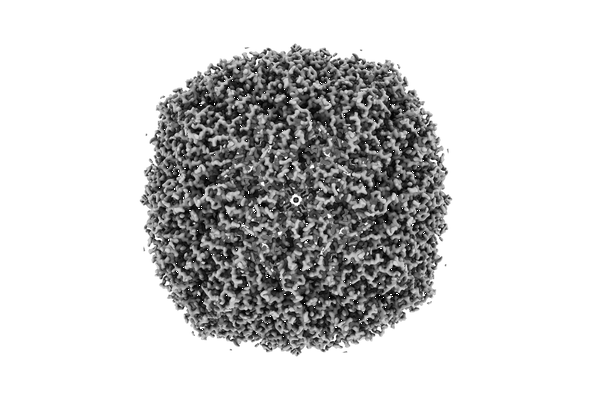 |
Movies of apoferritin collected at different dose rates on the Direct Electron Apollo direct detector - 60 eps [869 multi-frame micrographs composed of 20 frames each in TIFF format] | Peng R, Fu X, Mendez JH, Randolph PS, Bammes BE, Stagg SM [Pubmed: 36578473] [DOI: 10.1016/j.yjsbx.2022.100080] |
154.5 GB | 1.68 Å |
| 2023-01-18 | 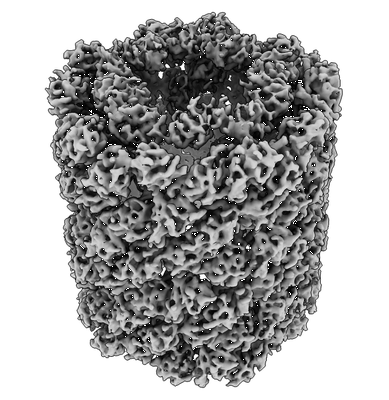 |
Cryo iDPC-STEM single particle analysis of keyhole limpet hemocyanin [multiple data sets in TIFF format] | Mann DM, Lazic I, Wirix M, de Haas F [Pubmed: 36064775] [DOI: 10.1038/s41592-022-01586-0] |
42.9 GB | 6.51 Å |
| 2023-01-18 | 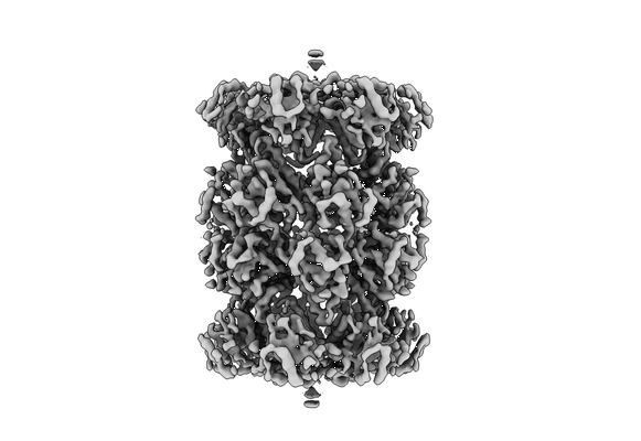 |
Multishot Tomography for High-Resolution In Situ Subtomogram Averaging: RiboProt singleshot [39 tilt series in MRC format] | Khavnekar S, Erdmann PSE, Plitzko J [Pubmed: 36343843] [DOI: 10.1016/j.jsb.2022.107911] |
122.2 GB | 4.7 - 7.8 Å |
| 2023-01-18 | 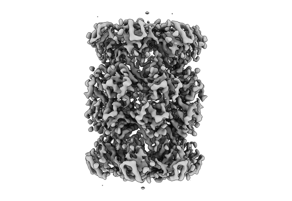 |
Multishot Tomography for High-Resolution In Situ Subtomogram Averaging: RiboProt multishot (2 shots) [26 tilt series in MRC format] | Khavnekar S, Erdmann PSE, Plitzko J [Pubmed: 36343843] [DOI: 10.1016/j.jsb.2022.107911] |
78.0 GB | 4.7 - 8.3 Å |
| 2023-01-18 | 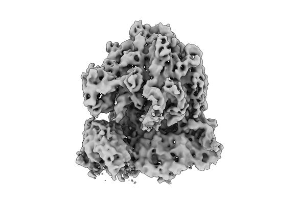 |
Multishot Tomography for High-Resolution In Situ Subtomogram Averaging: E.coli cryo-FIB lamellae multishot [30 tilt series in MRC format] | Khavnekar S, Erdmann PS, Plitzko JM [Pubmed: 36343843] [DOI: 10.1016/j.jsb.2022.107911] |
90.7 GB | 8.8 Å |
| 2023-01-18 | 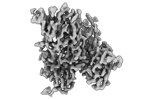 |
Organizing Structural Principles of the Interleukin-17 Ligand-Receptor Axis [multiple data sets in TIFF format] | Caveney NA, Wilson SC, Garcia KC [Pubmed: 35863378] [DOI: 10.1038/s41586-022-05116-y] |
17.7 TB | 2.5 - 4.4 Å |
| 2023-01-18 | 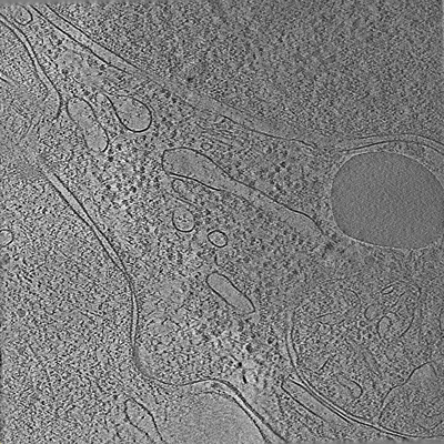 |
Tilt series of cell-cell contact of two PTK-1 cells [35 tilt series in MRC format] | Lemos M, Bezault A, Sauvanet C, Hanein D, Volkmann N [Pubmed: 36539423] [DOI: 10.1038/s41467-022-35409-9] |
1.1 GB | — |
| 2023-01-18 |  |
Movies of apoferritin collected at different dose rates on the Direct Electron Apollo direct detector - 30 eps [1478 multi-frame micrographs composed of 40 frames each in TIFF format] | Peng R, Fu X, Mendez JH, Randolph PS, Bammes BE, Stagg SM [Pubmed: 36578473] [DOI: 10.1016/j.yjsbx.2022.100080] |
348.5 GB | — |
