Electron Microscopy Public Image Archive
Electron Microscopy Public Image Archive
The EMPIAR-PDBj team at Osaka University assists Asian EM researchers with the transfer of big EM image data to EMPIAR. Instead of sending the data directly to the EBI (UK) via the internet, hard drives can also be sent to Osaka University by postal mail or via a courier service. As an alternative, internet transfer to our server in Osaka is also available. If you would like to take advantage of our submission services, please contact us first by e-mail before sending the data to us.
| Release date | Imageset | Title | Authors and references | Size | Resolution |
|---|---|---|---|---|---|
| 2022-06-01 | 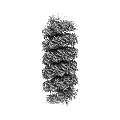 |
Micrographs of viruses ATV and AFV6 [791 multi-frame micrographs composed of 40 frames each in TIFF format] | Wang F, Cvirkaite-Krupovic V, Krupovic M, Egelman EH [Pubmed: 35325592] [DOI: 10.1016/j.cell.2022.02.019] |
218.0 GB | 3.9 Å |
| 2024-02-06 | 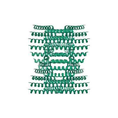 |
Raw micrographs of Form1-N2 peptide nanotube [7917 multi-frame micrographs composed of 40 frames each in TIFF format] | Wang F, Gnewou O, Conticello VP, Egelman EH [Pubmed: 35133794] [DOI: 10.1021/acs.chemrev.1c00753] |
1.8 TB | 3.4 Å |
| 2024-04-09 | 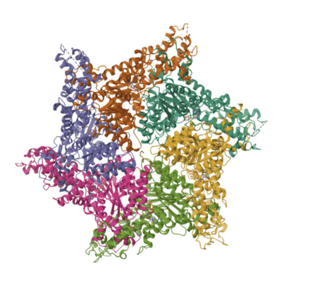 |
cryo-EM 3D maps of the S. cerevisiae Yta7 bound to ATPgS and histone H3 tail [12138 multi-frame micrographs composed of 75 frames each in TIFF format] | Wang FW [Pubmed: 36592926] [DOI: 10.1016/j.jbc.2022.102852] |
6.9 TB | 3.0 - 3.1 Å |
| 2024-03-26 | 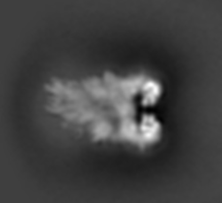 |
cryo-EM 3D maps of the S. cerevisiae Yta7 bound to the reconstituted nucleosome [16532 multi-frame micrographs composed of 75 frames each in TIFF format] | Wang FW [Pubmed: 36592926] [DOI: 10.1016/j.jbc.2022.102852] |
8.4 TB | 10.0 Å |
| 2024-02-16 |  |
Structure of the human UBR5 HECT-type E3 ubiquitin ligase in a dimeric form [13094 multi-frame micrographs composed of 75 frames each in TIFF format] | Wang FW [Pubmed: 37040767] [DOI: 10.1016/j.str.2023.03.010] |
7.0 TB | 2.66 - 2.8 Å |
| 2024-03-26 |  |
Structure of the human UBR5 HECT-type E3 ubiquitin ligase in a tetrameric form [17529 multi-frame micrographs composed of 75 frames each in TIFF format] | Wang FW [Pubmed: 37040767] [DOI: 10.1016/j.str.2023.03.010] |
7.6 TB | 3.5 Å |
| 2018-09-10 |  |
Cryo electron microscopy micrographs of yeast Exocyst complex [6472 multi-frame micrographs composed of 32 frames each in MRCS format] | Wang HW, Guo W, Li Y, Mei KR [Pubmed: 29335562] [DOI: 10.1038/s41594-017-0016-2] |
10.0 TB | 4.4 Å |
| 2017-07-26 |  |
Cryo EM of Type VI Secretion System VipA-N2/VipB contracted sheath [186 multi-frame micrographs composed of 30 frames each in MRC format] | Wang J, Basler M [Pubmed: 28947741] [DOI: 10.1038/s41564-017-0020-7] |
74.0 GB | 4.0 Å |
| 2017-07-26 | 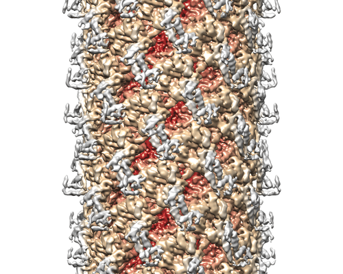 |
Cryo EM of Type VI Secretion System VipA-N3/VipB/Hcp complex [260 multi-frame micrographs composed of 30 frames each in TIFF format] | Wang J, Basler M [Pubmed: 28947741] [DOI: 10.1038/s41564-017-0020-7] |
26.2 GB | 3.7 Å |
| 2022-08-12 | 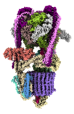 |
Cryo-EM structures of human V-ATPase [40113 multi-frame micrographs composed of 40 frames each in TIFF format] | Wang L, Wu H, Fu TM [Pubmed: 33065002] [DOI: 10.1016/j.molcel.2020.09.029] |
8.4 TB | 2.9 - 3.6 Å |
| 2022-12-06 |  |
Single particle cryo-EM structure of RIG-I in complex with p3dsRNA [3480 multi-frame micrographs composed of 34 frames each in TIFF format] | Wang W, Pyle AM [Pubmed: 36272408] [DOI: 10.1016/j.molcel.2022.09.029] |
1020.2 GB | 3.54 Å |
| 2022-12-06 |  |
Single particle cryo-EM structure of RIG-I bound to the end and internal sites of p3SLR30 (+ATP) [3417 multi-frame micrographs composed of 40 frames each in TIFF format] | Wang W, Pyle AM [Pubmed: 36272408] [DOI: 10.1016/j.molcel.2022.09.029] |
1.2 TB | 3.2 - 3.66 Å |
| 2022-12-06 | 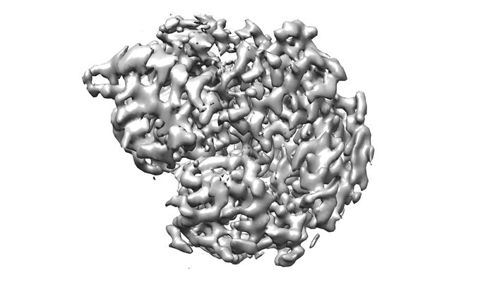 |
Single particle cryo-EM structure of RIG-I in complex with p1dsRNA [2460 multi-frame micrographs composed of 38 frames each in TIFF format] | Wang W, Pyle AM [Pubmed: 36272408] [DOI: 10.1016/j.molcel.2022.09.029] |
1.3 TB | 3.54 Å |
| 2022-12-06 | 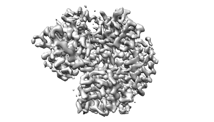 |
Single particle cryo-EM structure of RIG-I in complex with OHdsRNA [2838 multi-frame micrographs composed of 38 frames each in TIFF format] | Wang W, Pyle AM [Pubmed: 36272408] [DOI: 10.1016/j.molcel.2022.09.029] |
1.5 TB | 3.5 Å |
| 2022-12-06 | 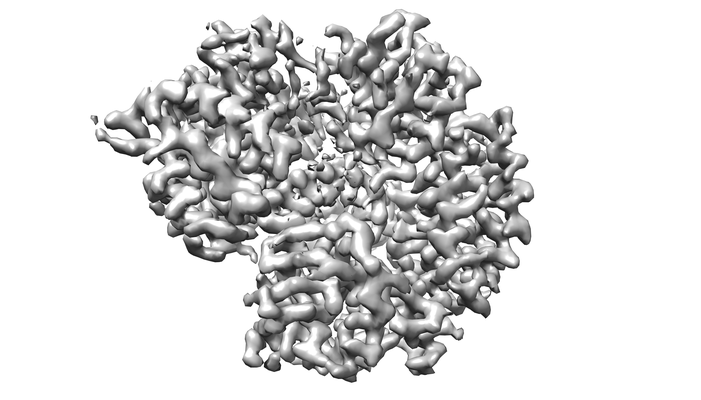 |
Single particle cryo-EM structure of RIG-I in complex with p2dsRNA [2586 multi-frame micrographs composed of 38 frames each in TIFF format] | Wang W, Pyle AM [Pubmed: 36272408] [DOI: 10.1016/j.molcel.2022.09.029] |
1.4 TB | 3.2 Å |
| 2022-12-06 | 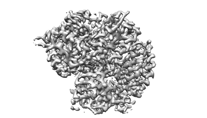 |
Single particle cryo-EM structure of RIG-I bound to the end and internal sites of OH3SLR30 (+ATP) [3663 multi-frame micrographs composed of 53 frames each in TIFF format] | Wang W, Pyle AM [Pubmed: 36272408] [DOI: 10.1016/j.molcel.2022.09.029] |
1.6 TB | 2.9 - 3.0 Å |
| 2022-12-06 | 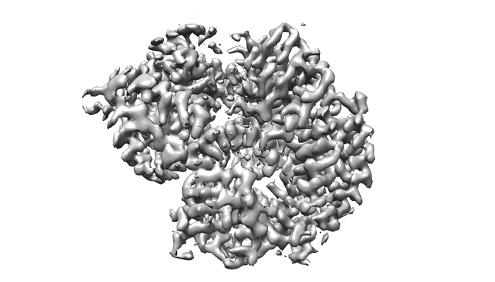 |
Single particle cryo-EM structure of RIG-I bound to the end of p3SLR30 (+AMPPNP) [2744 multi-frame micrographs composed of 38 frames each in TIFF format] | Wang W, Pyle AM [Pubmed: 36272408] [DOI: 10.1016/j.molcel.2022.09.029] |
1.5 TB | 3.3 Å |
| 2023-11-06 | 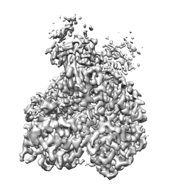 |
Single particle cryo-EM structure of RIG-I:RNA:Riplet ternary complex [3420 multi-frame micrographs composed of 40 frames each in TIFF format] | Wang W, Pyle AM [DOI: 10.1038/s41467-023-42982-0] |
1.5 TB | — |
| 2024-03-18 | 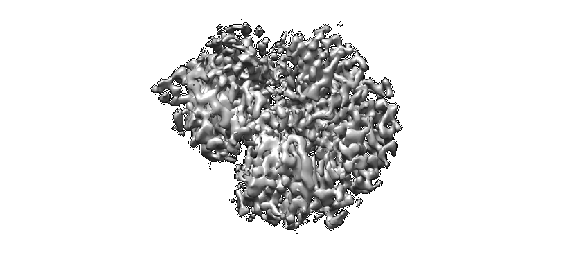 |
Single particle cryo-EM structure of 14-aa-GS-RIG-I in complex with p3SLR30 [5375 multi-frame micrographs composed of 38 frames each in TIFF format] | Wang W, Pyle AM | 2.7 TB | 3.4 Å |
| 2024-03-20 | 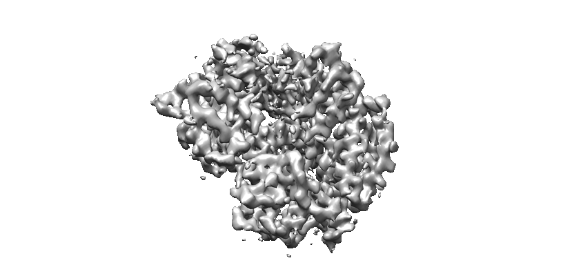 |
Single particle cryo-EM structure of RIG-I in complex with p3SLR14 [3531 multi-frame micrographs composed of 40 frames each in TIFF format] | Wang W, Pyle AM | 1.1 TB | 3.8 Å |
| 2014-09-07 |  |
Full virus map of Brome Mosaic Virus (micrographs and particle coordinates) [multiple data sets in TIFF and MRC formats] | Wang Z, Hryc C, Bammes B, Afonine P, Jakana J, Chen DH, Liu XA, Baker M, Kao C, Ludtke S, Schmid M, Adams P, Chiu W [Pubmed: 25185801] [DOI: 10.1038/ncomms5808] |
1.7 TB | 3.8 Å |
| 2014-09-09 | 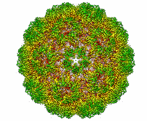 |
Full virus map of Brome Mosaic Virus (picked particles) [stack of 35142 particles in IMAGIC format] | Wang Z, Hryc C, Bammes B, Afonine P, Jakana J, Chen DH, Liu XA, Baker M, Kao C, Ludtke S, Schmid M, Adams P, Chiu W [Pubmed: 25185801] [DOI: 10.1038/ncomms5808] |
23.1 GB | 3.8 Å |
| 2021-11-08 | 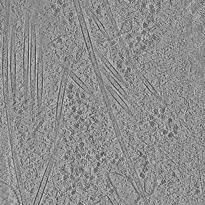 |
Frame-aligned tilt series of cryo-FIB-milled HEK293 cells treated with taxol [40 tilt series in MRC format] | Watanabe R, Buschauer R, Böhning J, Audagnotto M, Lasker K, Lu TW, Boassa D, Taylor S, Villa E [Pubmed: 32783917] [DOI: 10.1016/j.cell.2020.08.004] |
2.1 GB | — |
| 2021-11-08 | 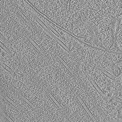 |
Frame aligned tilt series of cryo-FIB milled HEK293 cells expressing pathogenic LRRK2(I2020T)mutant [39 tilt series in MRC format] | Watanabe R, Buschauer R, Böhning J, Audagnotto M, Lasker K, Lu TW, Boassa D, Taylor S, Villa E [Pubmed: 32783917] [DOI: 10.1016/j.cell.2020.08.004] |
2.1 GB | — |
| 2020-09-30 | 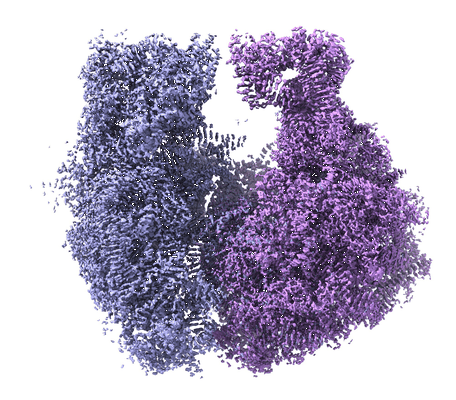 |
Structure of the Bacterial Ribosome at 2 Å Resolution [multiple data sets in TIFF format] | Watson ZL, Ward FR, Méheust R, Ad O, Schepartz A, Banfield JF, Cate JH [Pubmed: 32924932] [DOI: 10.7554/eLife.60482] |
2.1 TB | 1.98 Å |
