Electron Microscopy Public Image Archive
Electron Microscopy Public Image Archive
The EMPIAR-PDBj team at Osaka University assists Asian EM researchers with the transfer of big EM image data to EMPIAR. Instead of sending the data directly to the EBI (UK) via the internet, hard drives can also be sent to Osaka University by postal mail or via a courier service. As an alternative, internet transfer to our server in Osaka is also available. If you would like to take advantage of our submission services, please contact us first by e-mail before sending the data to us.
| Release date | Imageset | Title | Authors and references | Size | Resolution |
|---|---|---|---|---|---|
| 2022-03-21 | 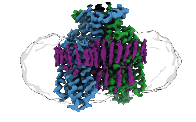 |
Structure of the ligand-free GPCR dimer Ste2 [9369 multi-frame micrographs composed of 53 frames each in EER format] | Velazhahan V, Tate CG [Pubmed: 35296853] [DOI: 10.1038/s41586-022-04498-3] |
8.2 TB | 3.1 Å |
| 2024-03-26 |  |
Structure of the human UBR5 HECT-type E3 ubiquitin ligase in a tetrameric form [17529 multi-frame micrographs composed of 75 frames each in TIFF format] | Wang FW [Pubmed: 37040767] [DOI: 10.1016/j.str.2023.03.010] |
7.6 TB | 3.5 Å |
| 2024-02-16 |  |
Structure of the human UBR5 HECT-type E3 ubiquitin ligase in a dimeric form [13094 multi-frame micrographs composed of 75 frames each in TIFF format] | Wang FW [Pubmed: 37040767] [DOI: 10.1016/j.str.2023.03.010] |
7.0 TB | 2.66 - 2.8 Å |
| 2021-03-05 |  |
Structure of the human U4/U6.U5 tri-snRNP [4812 micrographs in MRC format] | Stark H [Pubmed: 26912367] [DOI: 10.1126/science.aad2085] |
300.9 GB | 7.0 Å |
| 2022-07-22 |  |
Structure of the human RAD17-RFC clamp loader and 9-1-1 checkpoint clamp bound to a dsDNA-ssDNA junction [8271 multi-frame micrographs composed of 882 frames each in EER format] | Day M, Oliver AW, Pearl LH [Pubmed: 35819203] [DOI: 10.1093/nar/gkac588] |
4.4 TB | 3.59 Å |
| 2019-05-02 | 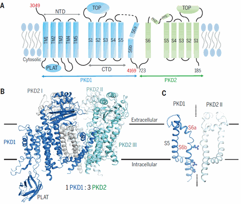 |
Structure of the human PKD1-PKD2 complex [stack of 3774 particles in MRCS format] | Su Q [Pubmed: 30093605] [DOI: 10.1126/science.aat9819] |
209.8 GB | 3.6 Å |
| 2017-02-09 | 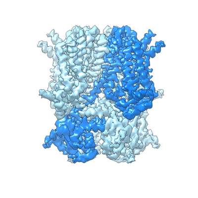 |
Structure of the human HCN1 hyperpolarization-activated cyclic nucleotide-gated ion channel [multiple data sets in MRC format] | Lee CH, MacKinnon R [Pubmed: 28086084] [DOI: 10.1016/j.cell.2016.12.023] |
66.5 GB | 3.5 Å |
| 2018-07-06 | 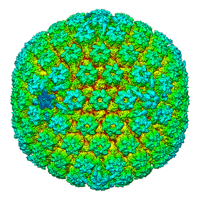 |
Structure of the herpes-simplex virus portal-vertex [3818 micrographs in MRC format] | McElwee M, Vijayakrishnan S, Rixon FJ, Bhella D [Pubmed: 29924793] [DOI: 10.1371/journal.pbio.2006191] |
238.6 GB | 7.7 Å |
| 2019-11-11 |  |
Structure of the green algal photosystem I supercomplex with a decameric light-harvesting complex I. [10989 multi-frame micrographs composed of 33 frames each in TIFF format] | Suga M, Ozawa S, Yoshida-Motomura K, Akita F, Miyazaki N, Takahashi Y [Pubmed: 31182847] [DOI: 10.1038/s41477-019-0438-4] |
8.0 TB | 2.8 - 2.9 Å |
| 2020-12-04 | 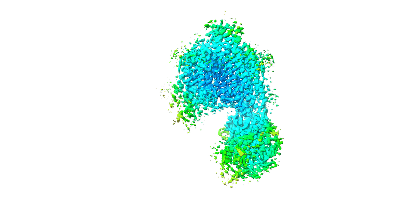 |
Structure of the class D GPCR Ste2 dimer coupled to two G proteins [multiple data sets in MRC and TIFF formats] | Velazhahan V, Ma N, Pándy-Szekeres G, Kooistra AJ, Lee Y, Gloriam DE, Vaidehi N, Tate CG [Pubmed: 33268889] [DOI: 10.1038/s41586-020-2994-1] |
1.3 TB | 3.3 Å |
| 2022-03-21 | 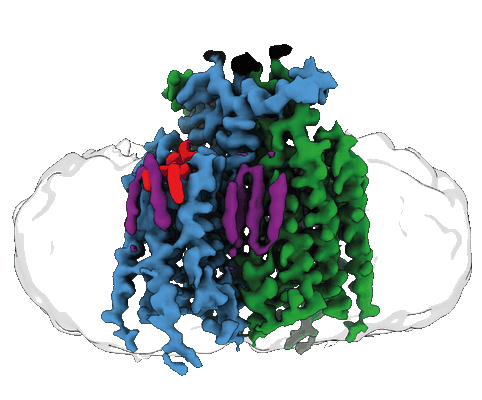 |
Structure of the agonist-bound GPCR dimer Ste2 [6944 multi-frame micrographs composed of 50 frames each in MRC format] | Velazhahan V, Tate CG [Pubmed: 35296853] [DOI: 10.1038/s41586-022-04498-3] |
1.3 TB | 3.46 - 3.53 Å |
| 2023-01-16 | 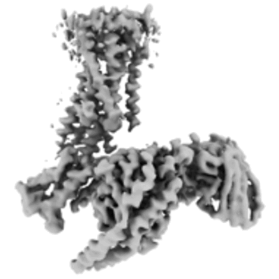 |
Structure of the active Gi-coupled human lysophosphatidic acid receptor 1 complexed with a potent agonist [6228 multi-frame micrographs composed of 48 frames each in TIFF format] | Akasaka H, Tanaka T, Sano FK, Matsuzaki Y, Shihoya W, Nureki O [Pubmed: 36109516] [DOI: 10.1038/s41467-022-33121-2] |
1.4 TB | 3.5 - 5.6 Å |
| 2018-01-03 |  |
Structure of the Z-disk isolated from the indirect flight muscle of the honey bee [96 class averages in MRC format] | Rusu M, Hu Z, Taylor KA, Trinick J [Pubmed: 28733815] [DOI: 10.1007/s10974-017-9477-5] |
3.0 GB | 60.0 Å |
| 2024-04-09 |  |
Structure of the Plasmodium falciparum 20S proteasome complexed with inhibitor TDI-8304 [29421 multi-frame micrographs composed of 75 frames each in TIFF format] | Hsu HC, Li H [Pubmed: 38097652] [DOI: 10.1038/s41467-023-44077-2] |
16.7 TB | 2.18 Å |
| 2024-04-03 |  |
Structure of the Plasmodium falciparum 20S proteasome beta-6 A117D mutant complexed with inhibitor WLW-vs [24086 multi-frame micrographs composed of 65 frames each in TIFF format] | Hsu HC, Li H [Pubmed: 38097652] [DOI: 10.1038/s41467-023-44077-2] |
11.3 TB | 2.58 Å |
| 2023-07-14 |  |
Structure of the Nucleosome Core Particle from Trypanosoma brucei [4913 multi-frame micrographs composed of 40 frames each in TIFF format] | Sandoval G, Deak G, Wapenaar H, Tuijtel MW, Wilson MD [Pubmed: 37427792] [DOI: 10.1093/nar/gkad577] |
1.4 TB | 3.3 Å |
| 2018-10-25 | 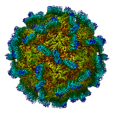 |
Structure of the Macrobrachium rosenbergii Nodavirus: A new genus within the Nodaviridae? [2459 micrographs in MRC format] | Ho KL, Gabrielsen M, Beh PL, Kueh CL, Thong QX, Streetley J, Tan WS, Bhella D [Pubmed: 30346944] [DOI: 10.1371/journal.pbio.3000038] |
130.4 GB | 3.28 Å |
| 2022-03-21 | 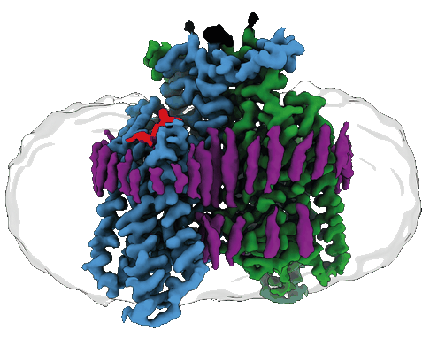 |
Structure of the GPCR dimer Ste2 bound to an antagonist [15751 multi-frame micrographs composed of 59 frames each in TIFF format] | Velazhahan V, Tate CG [Pubmed: 35296853] [DOI: 10.1038/s41586-022-04498-3] |
4.1 TB | 2.7 Å |
| 2022-07-18 | 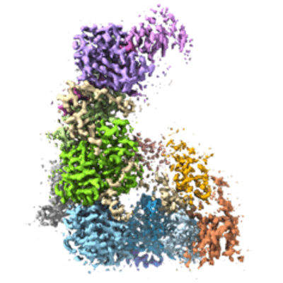 |
Structure of the Dicer-2-R2D2 heterodimer bound to a small RNA duplex [multiple data sets in TIFF format] | Yamaguchi S, Naganuma M, Nishizawa T, Kusakizako T, Tomari Y, Nishimasu H, Nureki O [Pubmed: 35768503] [DOI: 10.1038/s41586-022-04790-2] |
1.4 TB | 3.3 Å |
| 2020-09-30 | 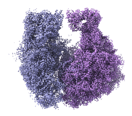 |
Structure of the Bacterial Ribosome at 2 Å Resolution [multiple data sets in TIFF format] | Watson ZL, Ward FR, Méheust R, Ad O, Schepartz A, Banfield JF, Cate JH [Pubmed: 32924932] [DOI: 10.7554/eLife.60482] |
2.1 TB | 1.98 Å |
| 2021-01-29 | 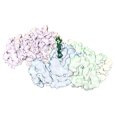 |
Structure of spastin bound to a glutamate-rich peptide implies a hand-over-hand mechanism of substrate translocation. [1200 multi-frame micrographs composed of 49 frames each in MRC format] | Han H, Schubert HL, Purdy MD, Yeager M, Sundquist WI, Hill CP [Pubmed: 31767681] [DOI: 10.1074/jbc.AC119.009890] |
1.8 TB | 4.2 Å |
| 2020-05-13 | 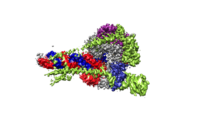 |
Structure of replicating SARS-CoV-2 polymerase [multiple data sets in TIFF and MRCS formats] | Hillen HS, Kokic G, Farnung L, Dienemann C, Tegunov D, Cramer P [Pubmed: 32438371] [DOI: 10.1038/s41586-020-2368-8] |
3.0 TB | 2.9 Å |
| 2022-09-23 |  |
Structure of pre-60S particle bound to DRG1(AFG2) [multiple data sets in TIFF format] | Prattes M, Grishkovskaya I, Hodirnau VV, Bergler H, Haselbach D [Pubmed: 36097293] [DOI: 10.1038/s41594-022-00832-5] |
3.8 TB | 3.2 - 3.8 Å |
| 2022-01-24 |  |
Structure of pathological TDP-43 filaments from ALS with FTLD (Individual 2, frontal cortex) [15991 multi-frame micrographs composed of 41 frames each in TIFF format] | Arseni D, Hasegawa M, Murzin AG, Kametani F, Arai M, Yoshida M, Ryskeldi-Falcon B [Pubmed: 34880495] [DOI: 10.1038/s41586-021-04199-3] |
2.9 TB | 2.94 Å |
| 2022-01-21 |  |
Structure of pathological TDP-43 filaments from ALS with FTLD (Individual 1, motor cortex) [12245 multi-frame micrographs composed of 40 frames each in TIFF format] | Arseni D, Hasegawa M, Murzin AG, Kametani F, Arai M, Yoshida M, Ryskeldi-Falcon B [Pubmed: 34880495] [DOI: 10.1038/s41586-021-04199-3] |
2.0 TB | 2.94 Å |
