Electron Microscopy Public Image Archive
Electron Microscopy Public Image Archive
The EMPIAR-PDBj team at Osaka University assists Asian EM researchers with the transfer of big EM image data to EMPIAR. Instead of sending the data directly to the EBI (UK) via the internet, hard drives can also be sent to Osaka University by postal mail or via a courier service. As an alternative, internet transfer to our server in Osaka is also available. If you would like to take advantage of our submission services, please contact us first by e-mail before sending the data to us.
| Release date | Imageset | Title | Authors and references | Size | Resolution |
|---|---|---|---|---|---|
| 2023-06-02 |  |
Cryo-ET tilt series from mouse islets lift-out sample [multiple data sets in TIFF format] | Wu Y, Qin C, Du W, Guo Z, Chen L, Guo Q [Pubmed: 37201639] [DOI: 10.1016/j.jsb.2023.107971] |
16.0 GB | — |
| 2018-10-17 |  |
Cryo-ET reveals the macromolecular reorganization of S. pombe mitotic chromosomes in vivo [25 tilt series in MRC format] | Cai S, Chen C, Tan ZY, Huang Y, Shi J [Pubmed: 30297429] [DOI: 10.1073/pnas.1720476115] |
35.1 GB | — |
| 2018-05-30 |  |
Cryo-ET of natural chromatin from Ostreococcus tauri and Saccharyomyces cerevisiae [25 class averages in MRC format] | Cai S, Song Y, Chen C, Shi J [Pubmed: 29742050] [DOI: 10.1091/mbc.E17-07-0449] |
40.1 GB | — |
| 2024-09-02 |  |
Cryo-ET of in vitro vaccinia core [38 tilt series in MRC format] | Liu J, Turoňová B [Pubmed: 38316878] [DOI: 10.1038/s41594-024-01218-5] |
42.9 GB | 7.7 - 13.4 Å |
| 2025-06-10 | 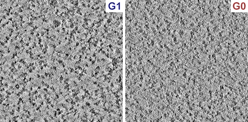 |
Cryo-ET of cryosections of S. pombe cells in G1 arrest and in G0 quiescence [20 tilt series in MRC format] | Tan ZY, Cai S, Paithankar SA, Liu T, Nie X, Shi J, Gan L [Pubmed: 40013339] [DOI: 10.1242/jcs.263654] |
439.8 GB | — |
| 2020-11-20 |  |
Cryo-ET of actin bundles induced by full length vinculin [2 tilt series in MRC format] | Ohad Medalia OM [Pubmed: 33185186] [DOI: 10.7554/eLife.53990] |
4.0 GB | 14.2 Å |
| 2025-04-03 |  |
Cryo-ET of RPE-1 G1 and metaphase cells and HeLa oligonucleosomes [multiple data sets in TIFF and MRC formats] | Chen JK, Liu T, Cai S, Ruan W, Ng CT, Shi J, Surana U, Gan L [Pubmed: 40097852] [DOI: 10.1038/s44318-025-00407-2] |
938.8 GB | 33.7 Å |
| 2025-01-24 |  |
Cryo-ET of Dictyostelium discoideum in distilled H2O (hypoosmotic stress) [59 tilt series in MRC format] | Hoffmann PC, Beck M [Pubmed: 39729993] [DOI: 10.1016/j.molcel.2024.11.038] |
241.1 GB | 37.0 Å |
| 2025-02-24 | 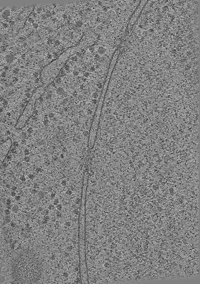 |
Cryo-ET of Dictyostelium discoideum in HL5 medium (control condition) [59 tilt series in MRC format] | Hoffmann PC, Beck M [Pubmed: 39729993] [DOI: 10.1016/j.molcel.2024.11.038] |
386.5 GB | 30.0 Å |
| 2025-02-24 | 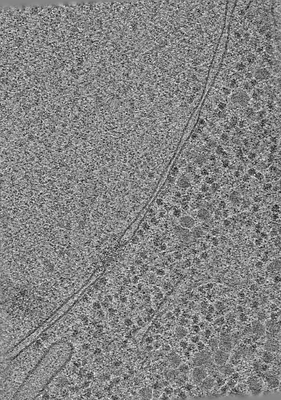 |
Cryo-ET of Dictyostelium discoideum in 0.4 M sorbitol (hyperosmotic stress) [59 tilt series in MRC format] | Hoffmann PC, Beck M [Pubmed: 39729993] [DOI: 10.1016/j.molcel.2024.11.038] |
332.1 GB | 38.0 Å |
| 2024-01-11 | 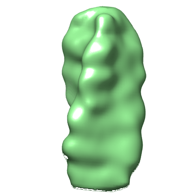 |
Cryo-ET detects bundled triple helices but not ladders in meiotic budding yeast [multiple data sets in MRC format] | Ma OX, Chong WG, Lee JKE, Cai S, Siebert CA, Howe A, Zhang P, Shi J, Surana U, Gan L [Pubmed: 35421110] [DOI: 10.1371/journal.pone.0266035] |
291.6 GB | 33.0 Å |
| 2025-05-22 | 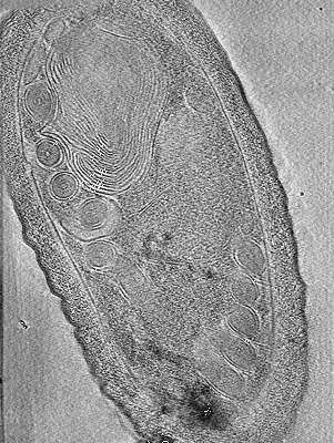 |
Cryo-ET datasets (1 and 2) of dormant microporidian spores from Encephalitozoon intestinalis [multiple data sets in TIFF and MRC formats] | Usmani M, Coudray N, Raghu R, Ramchandani H, Bobe D, Kopylov M, Zhong ED, Ekiert DC, Bhabha G [Pubmed: 39026755] [DOI: 10.1101/2024.07.13.603322] |
1.3 TB | 44.0 - 70.0 Å |
| 2024-05-16 |  |
Cryo-ET dataset on lamellae of Dictyostelium discoideum cells [213 tilt-series in MRC format] [213 tilt series in MRC format] | Tuijtel MW, Kreysing JP, Welsch S, Hummer G, Beck M, Turoňová B, Cruz-León S [Pubmed: 38669330] [DOI: 10.1126/sciadv.adk6285] |
382.6 GB | 3.9 Å |
| 2024-07-19 |  |
Cryo-ET dataset of purified SARS-CoV-2 double membrane vesicles formed by nsp3-4 [4635 tilt series in MRC format] | Huang YX, Ni T [DOI: 10.1016/s41586-024-07817-y] |
10.3 TB | — |
| 2025-05-22 |  |
Cryo-ET dataset of dormant microsporidian spores from Encephalitozoon hellem [multiple data sets in MRCS and MRC formats] | Kelley K, Bhabha G, Potter CS, Carragher B, Noble AJ [Pubmed: 40067903] [DOI: 10.1073/pnas.2415233122] |
252.1 GB | — |
| 2025-01-20 |  |
Cryo-ET dataset of FIB-milled mock infected (control) human monocyte-derived macrophages [multiple data sets in MRC format] | Kreysing JP, Welsch S, Turonova B, Beck M [Pubmed: 39826544] [DOI: 10.1016/j.cell.2024.12.008] |
324.5 GB | 27.9 - 36.7 Å |
| 2025-04-03 |  |
Cryo-ET dataset of FIB-milled HIV-1 infected human monocyte-derived macrophages [multiple data sets in MRC format] | Kreysing JP, Welsch S, Turoňová B, Beck M [Pubmed: 39826544] [DOI: 10.1016/j.cell.2024.12.008] |
560.6 GB | 24.5 - 33.2 Å |
| 2025-05-22 |  |
Cryo-ET dataset (3) of dormant microporidian spores from Encephalitozoon intestinalis [multiple data sets in TIFF and MRC formats] | Usmani M, Coudray N, Raghu R, Ramchandani H, Bobe D, Kopylov M, Zhong ED, Ekiert DC, Bhabha G [Pubmed: 39026755] [DOI: 10.1101/2024.07.13.603322] |
1.1 TB | 20.0 - 36.0 Å |
| 2023-06-20 |  |
Cryo-EPty SPA at CSA of 4.83 mrad [22 micrographs in MRC format] | Pei X, Zhou L, Huang C, Boyce M, Kim JS, Liberti E, Hu Y, Sasaki T, Nellist PD, Zhang P, Stuart DI, Kirkland AI, Wang P [Pubmed: 37230988] [DOI: 10.1038/s41467-023-38268-0] |
3.9 GB | 18.6 Å |
| 2023-06-20 |  |
Cryo-EPty SPA at CSA of 3.26 mrad [22 micrographs in MRC format] | Pei X, Zhou L, Huang C, Boyce M, Kim JS, Liberti E, Hu Y, Sasaki T, Nellist PD, Zhang P, Stuart DI, Kirkland AI, Wang P [Pubmed: 37230988] [DOI: 10.1038/s41467-023-38268-0] |
2.1 GB | 32.9 Å |
| 2023-06-20 |  |
Cryo-EPty SPA at CSA of 1.03 mrad [29 micrographs in MRC format] | Pei X, Zhou L, Huang C, Boyce M, Kim JS, Liberti E, Hu Y, Sasaki T, Nellist PD, Zhang P, Stuart DI, Kirkland AI, Wang P [Pubmed: 37230988] [DOI: 10.1038/s41467-023-38268-0] |
262.0 MB | 37.2 Å |
| 2023-08-18 |  |
Cryo-EM study on a single, highly heterogeneous cellular fraction with megadalton complexes derived from Chaetomium thermophilum [522 multi-frame micrographs composed of 30 frames each in MRC format] | Semchonok DA, Kyrilis FL, Hamdi F, Kastritis PL [DOI: 10.2139/ssrn.4211492] |
489.4 GB | 3.46 - 3.74 Å |
| 2023-01-16 |  |
Cryo-EM structures of the β3 adrenergic receptor bound to solabegron and isoproterenol [multiple data sets in TIFF format] | Nagiri C, Kobayashi K, Tomita A, Kato M, Yamashita K, Nishizawa T, Inoue A, Shihoya W, Nureki O [Pubmed: 35489202] [DOI: 10.1016/j.bbrc.2022.04.065] |
2.5 TB | 3.3 - 3.9 Å |
| 2022-11-14 |  |
Cryo-EM structures of the translocational binary toxin complex CDTa-bound CDTb-pore [11284 multi-frame micrographs composed of 84 frames each in TIFF format] | Kawamoto A, Yamada T, Yoshida T, Sato Y, Kato T, Tsuge H [Pubmed: 36253419] [DOI: 10.1038/s41467-022-33888-4] |
3.3 TB | 2.56 - 2.64 Å |
| 2020-09-11 | 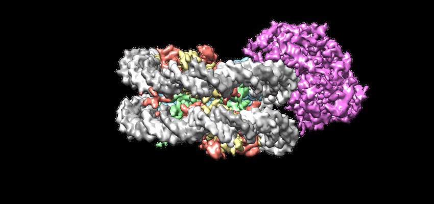 |
Cryo-EM structures of remodeler-nucleosome intermediates suggest allosteric control through the nucleosome [719 multi-frame micrographs composed of 30 frames each in MRCS format] | Armache J-P, Gamarra N, Johnson SL, Leonard JD, Wu S, Narlikar G, Cheng Y [Pubmed: 31210637] [DOI: 10.7554/eLife.46057] |
1.4 TB | 3.39 Å |
