Electron Microscopy Public Image Archive
Electron Microscopy Public Image Archive
The EMPIAR-PDBj team at Osaka University assists Asian EM researchers with the transfer of big EM image data to EMPIAR. Instead of sending the data directly to the EBI (UK) via the internet, hard drives can also be sent to Osaka University by postal mail or via a courier service. As an alternative, internet transfer to our server in Osaka is also available. If you would like to take advantage of our submission services, please contact us first by e-mail before sending the data to us.
| Release date | Imageset | Title | Authors and references | Size | Resolution |
|---|---|---|---|---|---|
| 2020-12-02 | 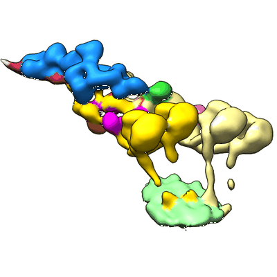 |
Cryo-electron tomography reveals that dynactin recruits a team of dyneins for processive motility. [127 tilt series in MRC format] | Grotjahn D.A, Lander G.C, Chowdhury S [Pubmed: 29416113] [DOI: 10.1038/s41594-018-0027-7] |
200.1 GB | 38.0 Å |
| 2020-10-09 |  |
Single particle cryo electron microscopy of aldolase (rabbit, muscle) using beam-tilt on Talos Arctica [1625 micrographs in MRC format] | Kearns S, Cianfrocco MA [Pubmed: 33209328] [DOI: 10.1107/S2052252520013482] |
87.6 GB | 2.8 - 4.9 Å |
| 2020-11-11 | 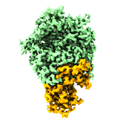 |
Cryo-EM structure of immunodominant protein P1 from human pathogen Mycoplasma pneumoniae on graphene oxide grid [multiple data sets in TIFF format] | Vizarraga D, Kawamoto A, Matsumoto U, Illanes R, Pérez-Luque R, Martín J, Mazzolini R, Bierge P, Pich OQ, Espasa M, Sanfeliu I, Esperalba J, Fernández-Huerta M, Scheffer MP, Pinyol J, Frangakis AS, Lluch-Senar M, Mori S, Shibayama K, Kenri T, Kato T, Namba K, Fita I, Miyata M, Aparicio D [Pubmed: 33057023] [DOI: 10.1038/s41467-020-18777-y] |
8.0 TB | 2.9 Å |
| 2020-11-27 | 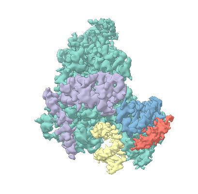 |
Movies of Nsp7-Nsp8-Nsp12 SARS-CoV2 RNA-dependent RNA polymerase in complex with template:primer dsRNA and favipiravir-RTP [63977 multi-frame micrographs composed of 24 frames each in MRCS format] | Naydenova K, Muir KW, Wu LF, Zhang Z, Coscia F, Peet MJ, Castro-Hartmann P, Qian P, Sader K, Dent K, Kimanius D, Sutherland JD, Löwe J, Barford D, Russo CJ [Pubmed: 33526596] [DOI: 10.1073/pnas.2021946118] |
23.9 TB | 2.5 Å |
| 2020-10-12 | 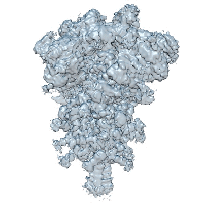 |
Cryo electron microscopy of SARS-CoV-2 spike in prefusion state [3207 multi-frame micrographs composed of 30 frames each in MRC format] | Carazo JM [Pubmed: 33063791] [DOI: 10.1107/S2052252520012725] |
2.1 TB | 3.0 - 3.3 Å |
| 2020-11-06 | 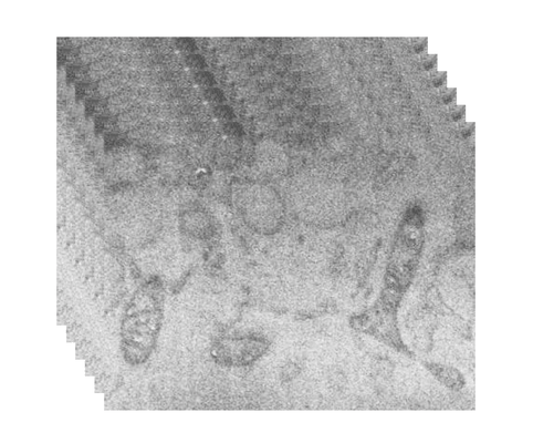 |
Serial cryoFIB/SEM reveals cytoarchitectural disruptions in Leigh syndrome patient cells [multiple data sets in TIFF format] | Zhu Y, Sun D, Schertel A, Fu X, Gwo P, Watson A, Zanetti-Domingues LC, Marisa L. Martin-Fernandez ML, Freyberg Z, Zhang P [Pubmed: 33096015] [DOI: 10.1016/j.str.2020.10.003] |
17.3 GB | — |
| 2020-09-28 | 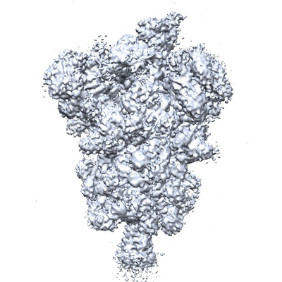 |
Cryo electron microscopy of SARS-CoV-2 stabilized spike in prefusion state [3511 multi-frame micrographs composed of 40 frames each in TIFF format] | Carazo JM [Pubmed: 33063791] [DOI: 10.1107/S2052252520012725] |
865.0 GB | 2.9 Å |
| 2020-10-09 | 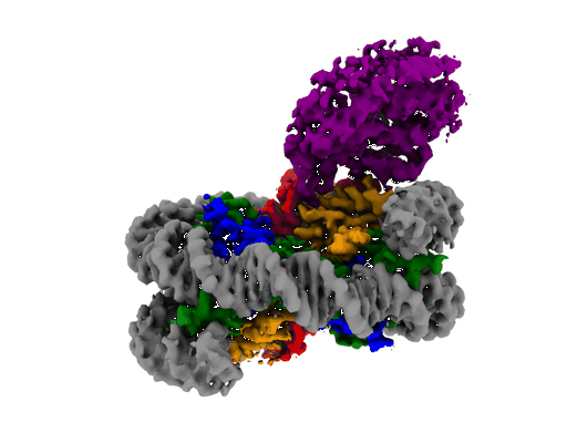 |
mouse cGAS with reconstituted nucleosome [5353 multi-frame micrographs composed of 40 frames each in MRC format] | Zhao B, Xu P [Pubmed: 32911481] [DOI: 10.1038/s41586-020-2749-z] |
767.6 GB | 2.98 Å |
| 2020-10-09 | 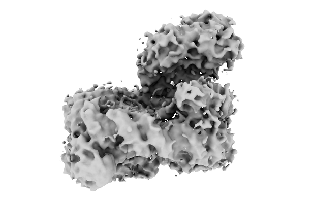 |
mouse cGAS with nucleosomes from 293T [2979 multi-frame micrographs composed of 40 frames each in MRC format] | Zhao B, Xu P, Rowlett CM, Jing T, Shinde O, Lei Y, West AP, Liu WR, Li P [Pubmed: 32911481] [DOI: 10.1038/s41586-020-2749-z] |
1.5 TB | 4.36 Å |
| 2021-03-24 | 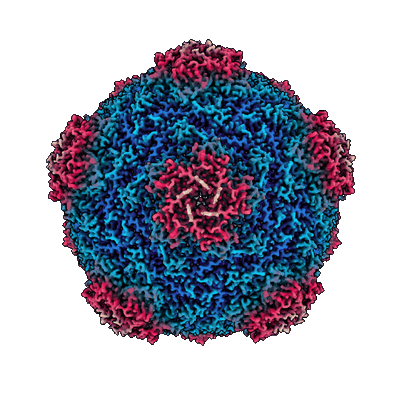 |
CryoEM SPA of Apo-SrpI Encapsulin Complex (Raw Frames) [2967 multi-frame micrographs composed of 39 frames each in TIFF format] | Nichols RJ, LaFrance BJ, Phillips NR, Oltrogge LM, Valentin-Alvarado LE, Bischoff AJ, Nogales E, Savage DF [Pubmed: 33821786] [DOI: 10.7554/eLife.59288] |
1.6 TB | 2.9 Å |
| 2020-09-30 | 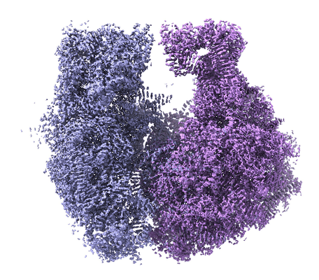 |
Structure of the Bacterial Ribosome at 2 Å Resolution [multiple data sets in TIFF format] | Watson ZL, Ward FR, Méheust R, Ad O, Schepartz A, Banfield JF, Cate JH [Pubmed: 32924932] [DOI: 10.7554/eLife.60482] |
2.1 TB | 1.98 Å |
| 2021-10-01 |  |
Cryo-EM structure of the SpCas9 delta4CE Ternary Complex [2570 multi-frame micrographs composed of 60 frames each in TIFF format] | Laughlin TG, Shams A, Savage DF [Pubmed: 34580310] [DOI: 10.1038/s41467-021-25992-8] |
1.5 TB | 6.2 Å |
| 2020-10-16 | 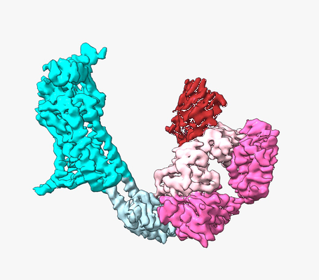 |
Structure of human Frizzled5 by fiducial-assisted cryo-EM [10922 multi-frame micrographs composed of 50 frames each in TIFF format] | Tsutsumi N, Gati C, Garcia KC [Pubmed: 32762848] [DOI: 10.7554/eLife.58464] |
6.0 TB | 3.7 Å |
| 2020-11-27 |  |
CryoEM SPA of Holo-SrpI Encapsulin Complex (Raw Frames) [1023 multi-frame micrographs composed of 33 frames each in TIFF format] | Nichols RJ, LaFrance BJ, Phillips NR, Oltrogge LM, Valentin-Alvarado LE, Bischoff AJ, Nogales E, Savage DF [Pubmed: 33821786] [DOI: 10.7554/eLife.59288] |
390.3 GB | 2.2 Å |
| 2021-11-01 | 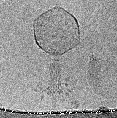 |
The contracted tail of myophage vb_EcoM_CBA120 (CBA120) [535 multi-frame micrographs composed of 20 frames each in MRCS format] | Nazarov S, Leiman P | 674.2 GB | 4.9 Å |
| 2020-11-20 |  |
CryoEM structure of Rubisco in the apo state [1834 multi-frame micrographs composed of 50 frames each in TIFF format] | He S, Chou HT, Matthies D, Wunder T, Meyer MT, Atkinson N, Martinez-Sanchez A, Jeffrey PD, Port SA, Patena W, He G, Chen VK, Hughson FM, McCormick AJ, Mueller-Cajar O, Engel BD, Yu Z, Jonikas MC [Pubmed: 33230314] [DOI: 10.1038/s41477-020-00811-y] |
895.3 GB | 2.68 Å |
| 2020-11-23 |  |
CryoEM structure of EPYC1(49-72) peptide-bound Rubisco [2500 multi-frame micrographs composed of 50 frames each in TIFF format] | He S, Chou HT, Matthies D, Wunder T, Meyer MT, Atkinson N, Martinez-Sanchez A, Jeffrey PD, Port SA, Patena W, He G, Chen VK, Hughson FM, McCormick AJ, Mueller-Cajar O, Engel BD, Yu Z, Jonikas MC [Pubmed: 33230314] [DOI: 10.1038/s41477-020-00811-y] |
1.2 TB | 2.13 Å |
| 2020-11-23 | 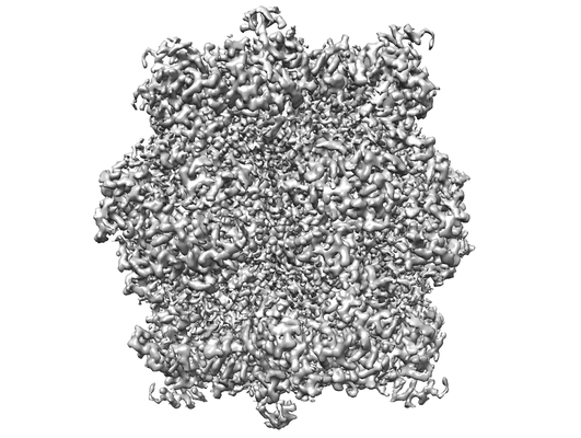 |
CryoEM structure of EPYC1(106-135) peptide-bound Rubisco [13727 multi-frame micrographs composed of 60 frames each in TIFF format] | He S, Chou HT, Matthies D, Wunder T, Meyer MT, Atkinson N, Martinez-Sanchez A, Jeffrey PD, Port SA, Patena W, He G, Chen VK, Hughson FM, McCormick AJ, Mueller-Cajar O, Engel BD, Yu Z, Jonikas MC [Pubmed: 33230314] [DOI: 10.1038/s41477-020-00811-y] |
5.8 TB | 2.06 Å |
| 2020-11-13 | 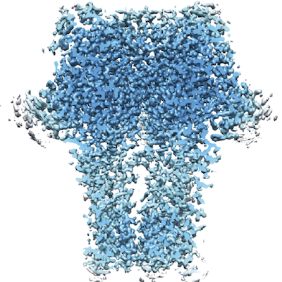 |
High resolution structure of GABAAR beta3 homopentamer [multiple data sets in EER and TIFF formats] | Nakane T, Kotecha A, Sente A, McMullan G, Masiulis S, Brown PMGE, Grigoras IT, Malinauskaite L, Malinauskas T, Miehling J, Uchański T, Yu L, Karia D, Pechnikova EV, de Jong E, Keizer J, Bischoff M, McCormack J, Tiemeijer P, Hardwick SW, Chirgadze DY, Murshudov G, Aricescu AR, Scheres SHW [Pubmed: 33087931] [DOI: 10.1038/s41586-020-2829-0] |
13.0 TB | 1.7 Å |
| 2020-12-04 | 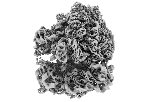 |
Tilt series of native M. pneumoniae cells treated with chloramphenicol [2666 tilt series in TIFF format] | Tegunov D, Xue L, Dienemann C, Cramer P, Mahamid J [Pubmed: 33542511] [DOI: 10.1038/s41592-020-01054-7] |
83.8 GB | 3.4 - 3.7 Å |
| 2021-11-15 | 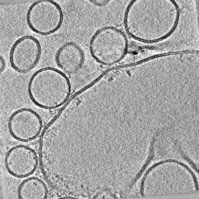 |
Arrangements of proteins at reconstituted synaptic vesicle fusion sites depend on membrane separation. [multiple data sets in MRC format] | Ginger L, Malsam J, Sonnen A.F.P., Morado D, Scheutzow A, Söllner T.H., Briggs J.A.G. [Pubmed: 32860428] [DOI: 10.1002/1873-3468.13916] |
70.0 GB | — |
| 2020-08-28 | 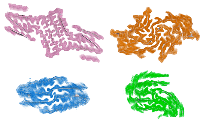 |
Cryo-EM structures of four polymorphic TDP-43 amyloid cores [multiple data sets in MRC format] | Cao Q, Boyer DR, Sawaya MR, Ge P, Eisenberg DS [Pubmed: 31235914] [DOI: 10.1038/s41594-019-0248-4] |
4.5 TB | 3.3 - 3.8 Å |
| 2020-12-22 | 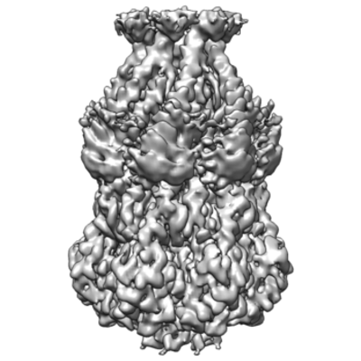 |
MscS Nanodisc with N-terminal His-Tag [2376 multi-frame micrographs composed of 50 frames each in MRCS format] | Reddy B, Perozo E [Pubmed: 31880537] [DOI: 10.7554/eLife.50486] |
762.3 GB | 3.1 Å |
| 2020-11-18 | 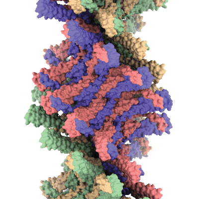 |
Structure and assembly of ESCRT-III helical Vps24 filaments [1320 micrographs in MRC format] | Huber ST, Mostafavi S, Mortensen SA, Sachse C [Pubmed: 32875105] [DOI: 10.1126/sciadv.aba4897] |
70.0 GB | 3.2 Å |
| 2020-10-28 | 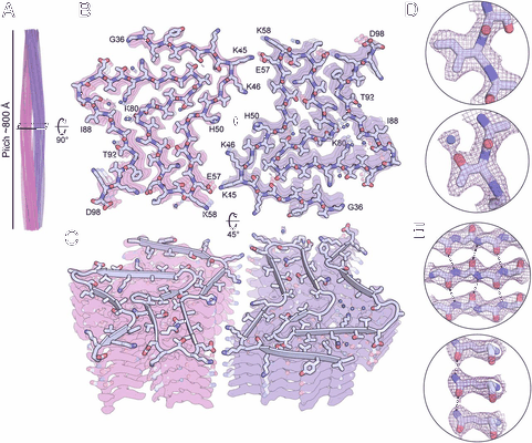 |
The α-synuclein hereditary mutation E46K unlocks a more stable, pathogenic fibril structure [4919 multi-frame micrographs composed of 30 frames each in TIFF format] | Boyer DR, Li B, Sun C, Fan W, Zhou K, Hughes MP, Sawaya MR, Jiang L, Eisenberg DS [Pubmed: 32015135] [DOI: 10.1073/pnas.1917914117] |
897.9 GB | 2.5 Å |
