Electron Microscopy Public Image Archive
Electron Microscopy Public Image Archive
The EMPIAR-PDBj team at Osaka University assists Asian EM researchers with the transfer of big EM image data to EMPIAR. Instead of sending the data directly to the EBI (UK) via the internet, hard drives can also be sent to Osaka University by postal mail or via a courier service. As an alternative, internet transfer to our server in Osaka is also available. If you would like to take advantage of our submission services, please contact us first by e-mail before sending the data to us.
| Release date | Imageset | Title | Authors and references | Size | Resolution |
|---|---|---|---|---|---|
| 2018-07-06 | 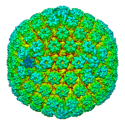 |
Structure of the herpes-simplex virus portal-vertex [3818 micrographs in MRC format] | McElwee M, Vijayakrishnan S, Rixon FJ, Bhella D [Pubmed: 29924793] [DOI: 10.1371/journal.pbio.2006191] |
238.6 GB | 7.7 Å |
| 2018-02-07 | 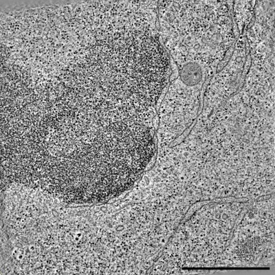 |
Raw 2d tomographic tilt series of a dividing cell [65 tilt series in ST format] | Otsuka S [Pubmed: 29323269] [DOI: 10.1038/s41594-017-0001-9] |
237.8 GB | — |
| 2017-02-20 | 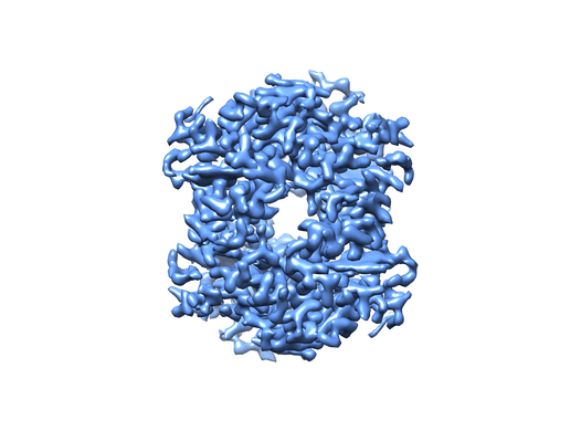 |
Cryo-EM structure of haemoglobin at 3.2 Å determined with the Volta phase plate [2261 multi-frame micrographs composed of 40 frames each in TIFF format] | Khoshouei M, Radjainia M, Baumeister W, Danev R [Pubmed: 28665412] [DOI: 10.1038/ncomms16099] |
237.1 GB | 3.2 Å |
| 2023-03-22 | 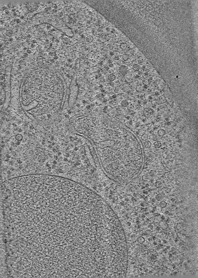 |
Electron cryo-tomography data on the ER-mitochondria encounter structure in cryo-FIB milled yeast cells [multiple data sets in MRC and TIFF formats] | Wozny MR, Di Luca A, Morado DR, Picco A, Khaddaj R, Campomanes P, Ivanovic L, Hoffmann PC, Miller EA, Vanni S, Kukulski W [Pubmed: 37165187] [DOI: 10.1038/s41586-023-06050-3] |
236.6 GB | — |
| 2017-12-18 |  |
CryoET of T20S proteasome single particle [multiple data sets in MRC format] | Noble AJ, Dandey VP, Wei H, Brasch J, Chase J, Acharya P, Tan YZ, Zhang Z, Kim LY, Scapin G, Rapp M, Eng ET, Rice WJ, Cheng A, Negro CJ, Shapiro L, Kwong PD, Jeruzalmi D, des Georges A, Potter CS, Carragher B [Pubmed: 29809143] [DOI: 10.7554/eLife.34257] |
235.3 GB | — |
| 2023-02-01 |  |
Cryo-EM data of alpha-synuclein A53T fibril [2663 micrographs in MRC format] | Wu KP, Huang JYC | 233.8 GB | 3.4 Å |
| 2023-07-10 |  |
Cryo electron tomography of Cytochalasin D-induced protrusions of Drosophila S2 alpha-tubulin acetyltransferase knock-out (dTAT KO) cells - Dataset 8 [multiple data sets in TIFF and MRC formats] | Ventura Santos C, Carter AP, Rogers SL [Pubmed: 37034688] [DOI: 10.1101/2023.03.31.535077] |
231.5 GB | — |
| 2021-03-19 |  |
GluK2/K5 apo [970 multi-frame micrographs composed of 40 frames each in TIFF format] | Khanra N, Meyerson J [Pubmed: 33724189] [DOI: 10.7554/eLife.66097] |
230.4 GB | 7.5 Å |
| 2020-10-09 | 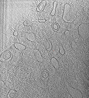 |
Human delta protocadherin 1 full ectodomains on membranes, tomogram 2 [multiple data sets in TIFF, JPEG and MRC formats] | Harrison OJ, Brasch J, Katsamba PS, Ahlsen G, Noble AJ, Dan H, Sampogna R, Potter CS, Carragher B, Honig B, Shapiro L [Pubmed: 32101743] [DOI: 10.1016/j.celrep.2020.02.003] |
230.3 GB | — |
| 2023-06-23 | 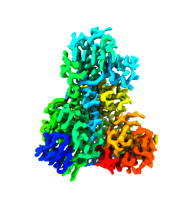 |
Unaligned and aligned cryo-EM micrographs of 82-kDa malate synthase G [multiple data sets in TIFF format] | Wu K.-P. [Pubmed: 36997036] [DOI: 10.1016/j.jsb.2023.107958] |
227.3 GB | 2.89 - 4.14 Å |
| 2022-09-20 | 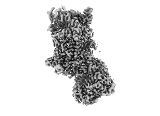 |
Cryo-EM dataset of Candida albicans CIII, inhibitor free [3634 micrographs in MRC format] | Di Trani J, Rubinstein JL [Pubmed: 34525326] [DOI: 10.1016/j.str.2021.08.006] |
227.1 GB | 3.0 Å |
| 2018-01-22 | 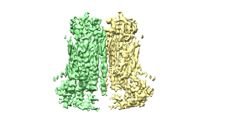 |
Cryo-EM structure of the TMEM16A in Nanodisc [stack of 3149 particles in MRCS format] | Dang S, Cheng Y [Pubmed: 29236684] [DOI: 10.1038/nature25024] |
226.7 GB | 3.8 Å |
| 2021-08-13 | 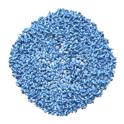 |
Human apo ferritin frozen on TEM grid with amorphous carbon supporting film [743 multi-frame micrographs composed of 32 frames each in TIFF format] | Huang X, Zhang L, Wen Z, Chen H, Li S, Ji G, Yin CC, Sun F [Pubmed: 32758492] [DOI: 10.1016/j.pbiomolbio.2020.07.009] |
226.5 GB | 2.6 Å |
| 2023-10-13 | 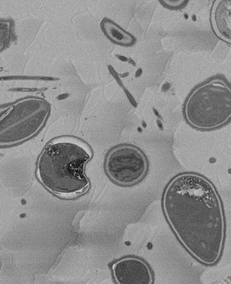 |
SBF-SEM micrographs of A. algerae microsporidia spores, 5 min germination [1215 micrographs in TIFF format] | Davydov A, Jaroenlak P, Ekiert D, Bhabha G [DOI: 10.7554/eLife.86638.1] |
226.3 GB | — |
| 2021-03-05 | 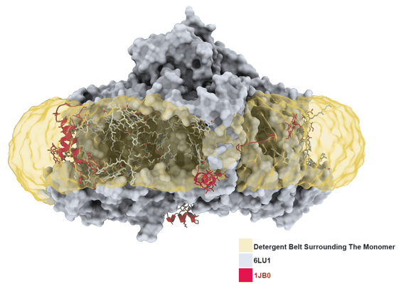 |
3.2 Å resolution structure of a functional monomeric Photosystem I from Thermosynechococcus elongatus BP-1 by single particle cryo-EM with a 200 kV CRYO ARM electron microscope [904 multi-frame micrographs composed of 60 frames each in TIFF format] | Coruh O, Frank A, Tanaka H, Kawamoto A, El-Mohsnawy E, Kato T, Namba K, Gerle C, Nowaczyk MM, Kurisu G [Pubmed: 33686186] [DOI: 10.1038/s42003-021-01808-9] |
226.1 GB | 3.2 Å |
| 2019-05-08 | 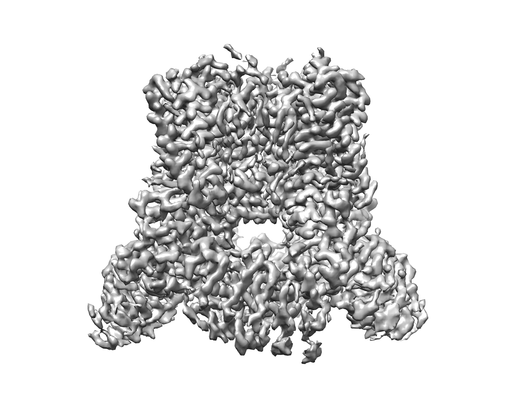 |
Cryo-EM structure of TRPV5 1-660 in nanodisc [stack of 2968 particles in MRCS format] | Dang S, van Goor MK, Asarnow D, Wang Y, Julius D, Cheng Y, van der Wijst J [Pubmed: 30975749] [DOI: 10.1073/pnas.1820323116] |
224.3 GB | 2.9 Å |
| 2022-05-20 | 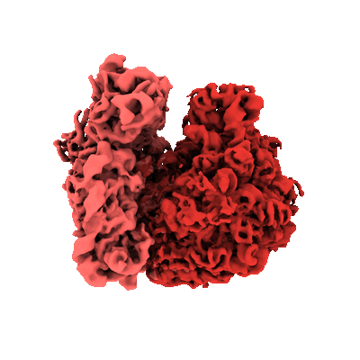 |
Parallel cryo electron tomography (PACE-tomo) of 70S ribosomes (200 kV, side-entry holder) [multiple data sets in MRC format] | Eisenstein F, Danev R [Pubmed: 36456783] [DOI: 10.1038/s41592-022-01690-1] |
222.8 GB | 5.8 - 6.5 Å |
| 2022-01-12 | 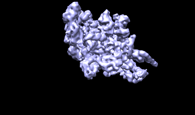 |
CryoEM of Mycobacterium tuberculosis WT RNAP holoenzyme/RbpA/Fdx [401 multi-frame micrographs composed of 50 frames each in TIFF format] | Boyaci H, Chen J, Darst SA, Campbell EA [Pubmed: 29480804] [DOI: 10.7554/eLife.34823] |
220.5 GB | 3.38 Å |
| 2022-10-31 | 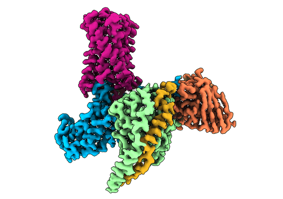 |
cryoEM structure of Gq-coupled MRGPRX1 with peptide agonist BAM8-22 & ML382 [2495 micrographs in MRC format] | Liu Y, Cao C, Huang XP, Gumpper RH, Rachman MM, Shih SL, Krumm BE, Zhang S, Shoichet BK, Fay JF, Roth BL [Pubmed: 36302898] [DOI: 10.1038/s41589-022-01173-6] |
219.1 GB | 2.87 Å |
| 2022-06-01 | 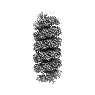 |
Micrographs of viruses ATV and AFV6 [791 multi-frame micrographs composed of 40 frames each in TIFF format] | Wang F, Cvirkaite-Krupovic V, Krupovic M, Egelman EH [Pubmed: 35325592] [DOI: 10.1016/j.cell.2022.02.019] |
218.0 GB | 3.9 Å |
| 2023-02-28 |  |
GroEL on EG-grid stored for 3 months after graphene oxidation [1209 multi-frame micrographs composed of 40 frames each in TIFF format] | Fujita J, Makino F, Asahara H, Moriguchi M, Kumano S, Anzai I, Kishikawa J, Matsuura Y, Kato T, Namba K, Inoue T [Pubmed: 36755111] [DOI: 10.1038/s41598-023-29396-0] |
217.8 GB | 2.06 Å |
| 2020-08-11 |  |
Human CALHM2 gap junction in nanodisc, 1 mM CaCl2 and 1 mM ATP [891 multi-frame micrographs composed of 50 frames each in TIFF format] | Syrjanen JL, Michalski K, Chou TH, Grant T, Rao S, Simorowski N, Tucker SJ, Grigorieff N, Furukawa H [Pubmed: 31988524] [DOI: 10.1038/s41594-019-0369-9] |
216.5 GB | 3.68 Å |
| 2022-10-31 |  |
Cryo-EM reconstruction of P.falciparum kinesin-8B motor domain in no nucleotide state bound to tubulin dimer [4075 micrographs in MRC format] | Liu T, Shilliday F, Cook AD, Moores CA [Pubmed: 36384964] [DOI: 10.1038/s41467-022-34710-x] |
216.2 GB | 3.3 - 4.3 Å |
| 2021-07-02 |  |
Affinity-purified RqcH-DR-ribosome-associated quality control complexes from Bacillus subtilis [2510 multi-frame micrographs composed of 30 frames each in TIFF format] | Crowe-McAuliffe C, Takada H, Murina V, Atkinson GC, Wilson DN, Hauryliuk V [Pubmed: 34255840] [DOI: 10.1093/nar/gkab589] |
213.5 GB | 3.2 Å |
| 2019-05-02 | 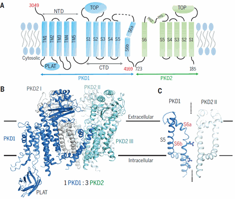 |
Structure of the human PKD1-PKD2 complex [stack of 3774 particles in MRCS format] | Su Q [Pubmed: 30093605] [DOI: 10.1126/science.aat9819] |
209.8 GB | 3.6 Å |
