Electron Microscopy Public Image Archive
Electron Microscopy Public Image Archive
The EMPIAR-PDBj team at Osaka University assists Asian EM researchers with the transfer of big EM image data to EMPIAR. Instead of sending the data directly to the EBI (UK) via the internet, hard drives can also be sent to Osaka University by postal mail or via a courier service. As an alternative, internet transfer to our server in Osaka is also available. If you would like to take advantage of our submission services, please contact us first by e-mail before sending the data to us.
| Release date | Imageset | Title | Authors and references | Size | Resolution |
|---|---|---|---|---|---|
| 2021-03-05 | 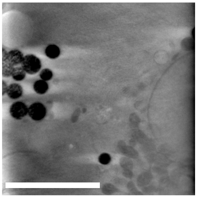 |
Fluorescent Au as Fiducials for SXT and SIM Correlation [700 tilt series in MRC format] | Okolo CA, Kounatidis I, Groen J, Nahas KL, Balint S, Fish T, Koronfel MA, López-Cortajarena A, Dobbie I, Pereiro E, Harkiolaki M | 3.6 GB | — |
| 2025-05-22 | 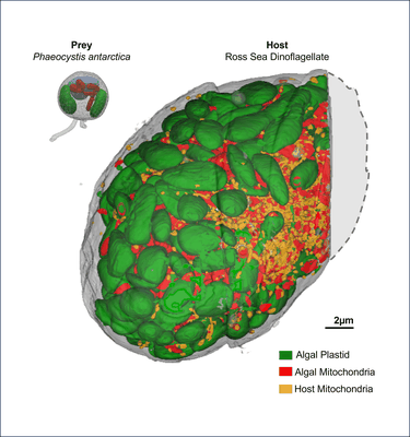 |
Fib-SEM stacks for Prey (Phaeocystis antarctica) and Host (Ross Sea Dinoflagellate; RSD) [multiple data sets in TIFF format] | Rao AK, Gallet B, Jouneau PH, Decelle J [Pubmed: 40250433] [DOI: 10.1101/2024.10.20.619283] |
272.6 GB | — |
| 2024-02-28 | 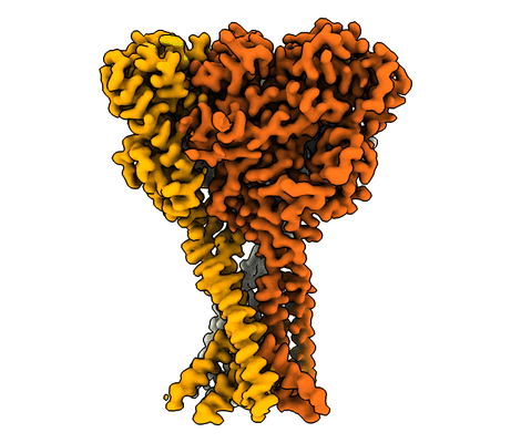 |
FMRFa-bound Malacoceros FaNaC1 in lipid nanodiscs in presence of diminazene [multiple data sets in TIFF format] | Kalienkova V, Dandamudi M, Paulino C, Lynagh T [Pubmed: 38337033] [DOI: 10.1038/s41594-023-01198-y] |
1.3 TB | 3.0 Å |
| 2024-02-28 | 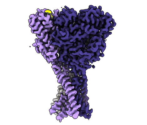 |
FMRFa-bound Malacoceros FaNaC1 in lipid nanodiscs [multiple data sets in TIFF format] | Kalienkova V, Dandamudi M, Paulino C, Lynagh T [Pubmed: 38337033] [DOI: 10.1038/s41594-023-01198-y] |
866.2 GB | 2.5 Å |
| 2023-03-17 | 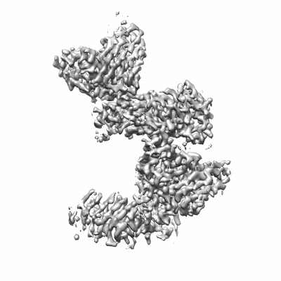 |
FMC63 scFv in complex with soluble CD19 [multiple data sets in TIFF format] | Meyerson JR, He C [Pubmed: 36867678] [DOI: 10.1126/sciimmunol.adf1426] |
1.4 TB | 3.05 Å |
| 2020-08-06 | 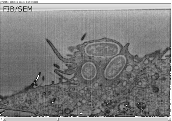 |
FIB/SEM sample dataset MCB-CLEM IV Weiner [1 multi-frame micrographs composed of 844 frames each in TIFF format] | Weiner A | 2.8 GB | — |
| 2020-02-07 | 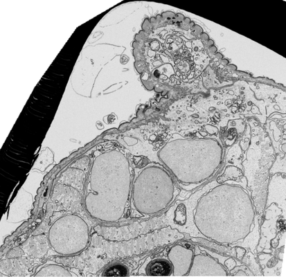 |
FIB-SEM of parapodia from Platynereis dumerilii [multiple data sets in TIFF format] | Hennies J, Lleti JMS, Schieber NL, Templin RM, Steyer AM, Schwab Y [Pubmed: 32029771] [DOI: 10.1038/s41598-020-58736-7] |
84.9 GB | — |
| 2022-01-12 | 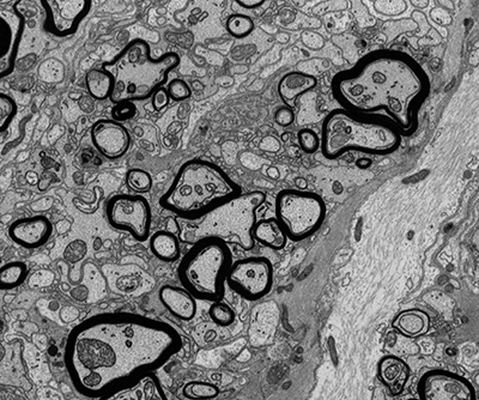 |
FIB-SEM of mouse optic nerve of an inducible conditional Mbp knock-out 26 weeks after induction [747 micrographs in TIFF format] | Meschkat M, Steyer AM, Ruhwedel T, Möbius W [DOI: 10.1101/2020.09.02.279612] |
21.5 GB | — |
| 2022-01-12 |  |
FIB-SEM of mouse optic nerve of an inducible conditional Mbp knock-out 16 weeks after induction [696 micrographs in TIFF format] | Meschkat M, Steyer AM, Ruhwedel T, Möbius W [Pubmed: 35246535] [DOI: 10.1038/s41467-022-28720-y] |
15.9 GB | — |
| 2022-01-12 |  |
FIB-SEM of an Mbp-deficient shiverer mouse optic nerve [857 micrographs in TIFF format] | Meschkat M, Steyer AM, Ruhwedel T, Möbius W [Pubmed: 35246535] [DOI: 10.1038/s41467-022-28720-y] |
18.8 GB | — |
| 2017-11-30 |  |
FIB-SEM of a dividing cell at 6.3 min after anaphase onset [3206 multi-frame micrographs composed of 1 frames each in TIFF format] | Otsuka S, Steyer AM, Schorb M, Hériché JK, Hossain MJ, Sethi S, Kueblbeck M, Schwab Y, Beck M, Ellenberg J [Pubmed: 29323269] [DOI: 10.1038/s41594-017-0001-9] |
83.2 GB | — |
| 2017-11-28 |  |
FIB-SEM of a dividing cell at 5.7 min after anaphase onset [2620 multi-frame micrographs composed of 1 frames each in TIFF format] | Otsuka S, Steyer AM, Schorb M, Hériché JK, Hossain MJ, Sethi S, Kueblbeck M, Schwab Y, Beck M, Ellenberg J [Pubmed: 29323269] [DOI: 10.1038/s41594-017-0001-9] |
63.0 GB | — |
| 2017-11-30 |  |
FIB-SEM of a dividing cell at 5.3 min after anaphase onset [1998 multi-frame micrographs composed of 1 frames each in TIFF format] | Otsuka S, Steyer AM, Schorb M, Hériché JK, Hossain MJ, Sethi S, Kueblbeck M, Schwab Y, Beck M, Ellenberg J [Pubmed: 29323269] [DOI: 10.1038/s41594-017-0001-9] |
31.2 GB | — |
| 2017-11-30 |  |
FIB-SEM of a dividing cell at 4.3 min after anaphase onset [1358 multi-frame micrographs composed of 1 frames each in TIFF format] | Otsuka S, Steyer AM, Schorb M, Hériché JK, Hossain MJ, Sethi S, Kueblbeck M, Schwab Y, Beck M, Ellenberg J [Pubmed: 29323269] [DOI: 10.1038/s41594-017-0001-9] |
14.0 GB | — |
| 2017-11-28 |  |
FIB-SEM of a dividing cell at 3.9 min after anaphase onset [2293 multi-frame micrographs composed of 1 frames each in TIFF format] | Otsuka S, Steyer AM, Schorb M, Hériché JK, Hossain MJ, Sethi S, Kueblbeck M, Schwab Y, Beck M, Ellenberg J [Pubmed: 29323269] [DOI: 10.1038/s41594-017-0001-9] |
26.8 GB | — |
| 2017-11-28 |  |
FIB-SEM of a dividing cell at 3.1 min after anaphase onset [1652 multi-frame micrographs composed of 1 frames each in TIFF format] | Otsuka S, Steyer AM, Schorb M, Hériché JK, Hossain MJ, Sethi S, Kueblbeck M, Schwab Y, Beck M, Ellenberg J [Pubmed: 29323269] [DOI: 10.1038/s41594-017-0001-9] |
27.0 GB | — |
| 2017-11-30 |  |
FIB-SEM of a dividing cell at 11.2 min after anaphase onset [777 multi-frame micrographs composed of 1 frames each in TIFF format] | Otsuka S, Steyer AM, Schorb M, Heriche JK, Hossain MJ, Sethi S, Kueblbeck M, Schwab Y, Beck M, Ellenberg J [DOI: 10.1038/s41594-017-0001-9] |
11.0 GB | — |
| 2020-02-07 |  |
FIB-SEM of a HeLa cell [multiple data sets in TIFF format] | Hennies J, Lleti JMS, Schieber NL, Templin RM, Steyer AM, Schwab Y [Pubmed: 32029771] [DOI: 10.1038/s41598-020-58736-7] |
94.0 GB | — |
| 2022-05-31 |  |
FIB-SEM of U2OS cell at leading edge of a wound assay, non-LPA stimulated, non-polarized [998 multi-frame micrographs composed of 1 frames each in TIFF format] | Costa J, Pinto A, Machado P | 5.7 GB | — |
| 2022-05-31 |  |
FIB-SEM of U2OS cell at leading edge of a wound assay, LPA stimulated, polarized [1798 multi-frame micrographs composed of 1 frames each in TIFF format] | Costa J, Pinto A, Machado P | 4.1 GB | — |
| 2021-04-30 |  |
FIB-SEM of Tuwongella immobilis - a species of the Planctomycetes phylum [1 multi-frame micrographs composed of 464 frames each in TIFF format] | Andersson SGE, Odelgard A, Dyrhage K, Mahajan M, Seeger C [DOI: 10.3389/fmicb.2021.643045] |
6.7 GB | — |
| 2021-04-30 |  |
FIB-SEM of Gemmata obscuriglobus - a species of the Planctomycetes phylum [1 stitched maps in TIFF format] | Seeger C, Dyrhage K, Mahajan M, Odelgard A, Bergström Lind S, Andersson SGE [DOI: 10.3389/fmicb.2021.643045] |
1.6 GB | — |
| 2023-03-13 |  |
FIB-SEM images about Control/OPA1 KD NIH-3T3 cells [1984 multi-frame micrographs composed of 1 frames each in TIFF format] | Suga S, Nakamura K, Kawai H, Hirabayashi Y [DOI: 10.1101/2021.06.11.448083] |
46.8 GB | — |
| 2024-12-02 |  |
FIB-SEM datasets of hippocampal neurons from WT and FAM92A1 knockout mice [multiple data sets in TIFF format] | Vihinen H, Jokitalo E, Wang L [Pubmed: 39043703] [DOI: 10.1038/s41467-024-50565-w] |
73.6 GB | — |
| 2024-02-15 |  |
FIB-SEM dataset showing localization of a Golgi matrix protein GM130 in human hepatocellular carcinoma cell (Huh-7) [564 reconstructed volumes in TIFF format] | Belevich I, Jokitalo E | 1.1 GB | — |
