Electron Microscopy Public Image Archive
Electron Microscopy Public Image Archive
大阪大學的EMPIAR-PDBj團隊為亞洲EM研究人員向EMPIAR傳送大型EM圖像數據提供服務。 除了通過互聯網將數據直接傳送到EBI(UK),研究人員還可以通過郵政或快遞服務將數據硬盤發送到大阪大學,或者通過互聯網傳送到設置於大阪大學的服務器,然後由我們代為傳送至數據登錄網站。 如果您想使用此項服務,請先通過 電子郵件 與我們聯繫。
| Release date | Imageset | Title | Authors and references | Size | Resolution |
|---|---|---|---|---|---|
| 2017-03-15 | 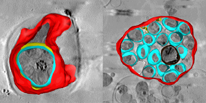 |
Soft X-ray tomography of Plasmodium falciparum infected human erythrocytes stalled in egress by the inhibitors Compound 2 and E64 [1 Soft X-ray tomograms in MRC format] | Hale VL, Saibil HR, Duke E, Fleck RA, Blackman MJ [Pubmed: 28292906] [DOI: 10.1073/pnas.1619441114] |
280.6 MB | — |
| 2023-10-13 | 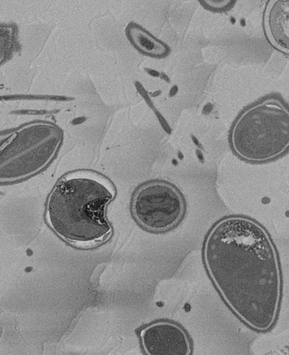 |
SBF-SEM micrographs of A. algerae microsporidia spores, 5 min germination [1215 micrographs in TIFF format] | Davydov A, Jaroenlak P, Ekiert D, Bhabha G [DOI: 10.7554/eLife.86638.1] |
226.3 GB | — |
| 2023-10-13 | 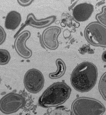 |
SBF-SEM micrographs of A. algerae microsporidia spores, 45 min germination [300 micrographs in TIFF format] | Davydov A, Jaroenlak P, Ekiert D, Bhabha G [DOI: 10.7554/eLife.86638.1] |
55.9 GB | — |
| 2021-01-08 | 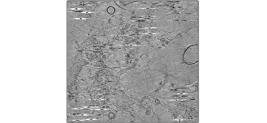 |
A lamin A/C variant causing striated muscle disease provides insights into filament organization [2 tilt series in MRC format] | Tatli M [Pubmed: 33536248] [DOI: 10.1242/jcs.256156] |
2.7 GB | — |
| 2023-10-10 | 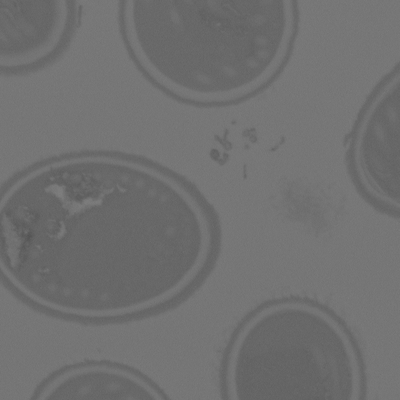 |
SBF-SEM micrographs of A. algerae spores, Ungerminated [250 micrographs in TIFF format] | Jaroenlak P, Cammer M, Davydov A, Sall J, Usmani M, Liang F, Ekiert D, Bhabha G [Pubmed: 32946515] [DOI: 10.1371/journal.ppat.1008738] |
46.6 GB | — |
| 2023-01-18 | 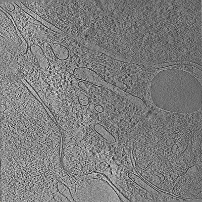 |
Tilt series of cell-cell contact of two PTK-1 cells [35 tilt series in MRC format] | Lemos M, Bezault A, Sauvanet C, Hanein D, Volkmann N [Pubmed: 36539423] [DOI: 10.1038/s41467-022-35409-9] |
1.1 GB | — |
| 2023-02-10 |  |
A surface morphometrics toolkit to quantify organellar membrane ultrastructure using cryo-electron tomography [multiple data sets in MRC format] | Barad BA, Medina M, Fuentes D, Wiseman RL, Grotjahn DA [DOI: 10.1101/2022.01.23.477440] |
126.9 GB | — |
| 2018-01-17 | 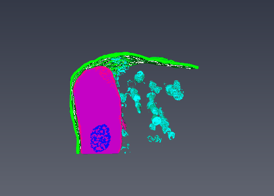 |
Serial Block Face Scanning Electron Micrscopy dataset of fetal day 64 guinea pig psoas muscle in transverse [93 micrographs in TIFF format] | Cocks ET [DOI: 10.1111/jmi.12676] |
264.4 MB | — |
| 2019-05-21 | 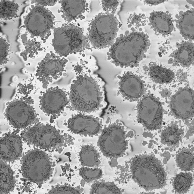 |
Serial Block Face SEM of HeLa cell pellet with 10 nm pixels and 50 nm slices (benchmark dataset) [518 micrographs in DM4 format] | Peddie CP, Jones ML, Collinson LM | 129.8 GB | — |
| 2021-02-12 |  |
Ultra-high voltage electron microscope tomography tilt series of neurite section [1 tilt series in MRC format] | Nishida T, Yoshimura R, Nishi R, Imoto Y, Endo Y [Pubmed: 32133640] [DOI: 10.1111/jmi.12885] |
120.3 MB | — |
| 2018-05-30 |  |
Cryo-ET of natural chromatin from Ostreococcus tauri and Saccharyomyces cerevisiae [25 class averages in MRC format] | Cai S, Song Y, Chen C, Shi J [Pubmed: 29742050] [DOI: 10.1091/mbc.E17-07-0449] |
40.1 GB | — |
| 2021-02-12 |  |
Ultra-high voltage electron microscope tomography tilt series of 0.7-μm-thick neurite section acquired at 6,000× magnification at an accelerating voltage of 1 MV [1 tilt series in MRC format] | Nishida T, Yoshimura R, Nishi R, Imoto Y, Endo Y [Pubmed: 32133640] [DOI: 10.1111/jmi.12885] |
120.3 MB | — |
| 2021-02-12 |  |
Ultra-high voltage electron microscope tomography tilt series of 0.7-μm-thick neurite section acquired at 15,000× magnification at an accelerating voltage of 1 MV [1 tilt series in MRC format] | Nishida T, Yoshimura R, Nishi R, Imoto Y, Endo Y [Pubmed: 32133640] [DOI: 10.1111/jmi.12885] |
120.3 MB | — |
| 2017-11-28 | 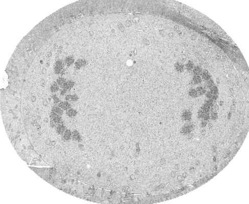 |
FIB-SEM of a dividing cell at 3.1 min after anaphase onset [1652 multi-frame micrographs composed of 1 frames each in TIFF format] | Otsuka S, Steyer AM, Schorb M, Hériché JK, Hossain MJ, Sethi S, Kueblbeck M, Schwab Y, Beck M, Ellenberg J [Pubmed: 29323269] [DOI: 10.1038/s41594-017-0001-9] |
27.0 GB | — |
| 2020-05-27 | 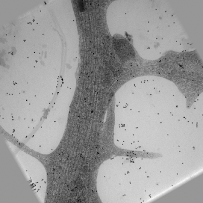 |
Ultra-high voltage electron microscope tomography tilt series of 0.7-μm-thick neurite section acquired at 20,000× magnification at an accelerating voltage of 1 MV [1 tilt series in MRC format] | Nishida T, Yoshimura R, Nishi R, Imoto Y, Endo Y [Pubmed: 32133640] [DOI: 10.1111/jmi.12885] |
120.3 MB | — |
| 2023-02-22 | 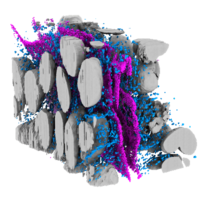 |
Light and electron microscopy continuum-resolution imaging of 3D cell cultures [19 multi-frame micrographs composed of 1 frames each in MRC format] | DImprima EDI, Garcia Montero MGM, Gawrzak, SG, Ronchi PR, Zagoriy IZ, Schwab YS, Jechlinger JS, Mahamid JM | 72.9 GB | — |
| 2017-11-30 | 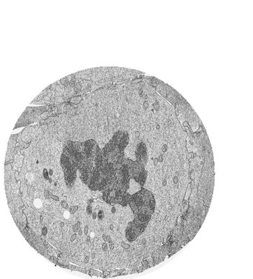 |
FIB-SEM of a dividing cell at 6.3 min after anaphase onset [3206 multi-frame micrographs composed of 1 frames each in TIFF format] | Otsuka S, Steyer AM, Schorb M, Hériché JK, Hossain MJ, Sethi S, Kueblbeck M, Schwab Y, Beck M, Ellenberg J [Pubmed: 29323269] [DOI: 10.1038/s41594-017-0001-9] |
83.2 GB | — |
| 2017-11-28 | 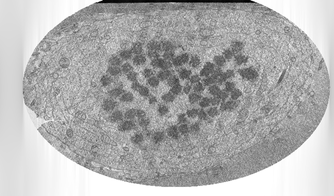 |
FIB-SEM of a dividing cell at 3.9 min after anaphase onset [2293 multi-frame micrographs composed of 1 frames each in TIFF format] | Otsuka S, Steyer AM, Schorb M, Hériché JK, Hossain MJ, Sethi S, Kueblbeck M, Schwab Y, Beck M, Ellenberg J [Pubmed: 29323269] [DOI: 10.1038/s41594-017-0001-9] |
26.8 GB | — |
| 2022-01-11 | 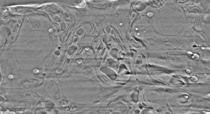 |
Cryo-FIB-SEM data on Chlamydomonas reinhardtii cells [37 micrographs in TIFF format] | Klumpe S [Pubmed: 34951584] [DOI: 10.7554/elife.70506] |
888.2 MB | — |
| 2017-11-30 | 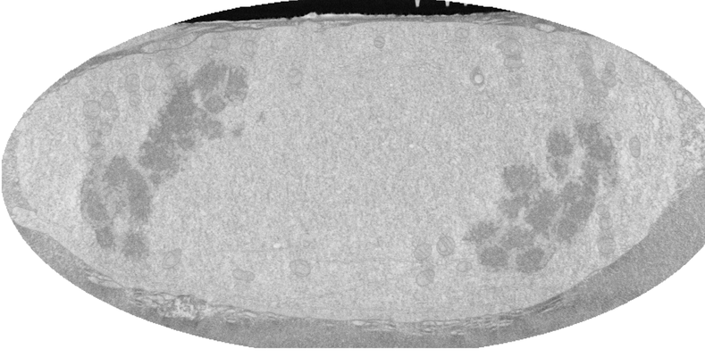 |
FIB-SEM of a dividing cell at 4.3 min after anaphase onset [1358 multi-frame micrographs composed of 1 frames each in TIFF format] | Otsuka S, Steyer AM, Schorb M, Hériché JK, Hossain MJ, Sethi S, Kueblbeck M, Schwab Y, Beck M, Ellenberg J [Pubmed: 29323269] [DOI: 10.1038/s41594-017-0001-9] |
14.0 GB | — |
| 2021-11-01 | 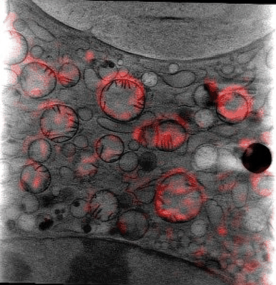 |
Cryo Soft X-ray data for tetraspeck correlation [multiple data sets in MRC format] | Groen J, Pereiro E [Pubmed: 33990802] [DOI: 10.1038/s41596-021-00522-4] |
15.1 GB | — |
| 2024-04-02 |  |
EMinsight: a tool to capture cryoEM microscope configuration and experimental outcomes for analysis and deposition [2605 multi-frame micrographs composed of 50 frames each in TIFF format] | Morris KL, Harrison PJ, Hatton D, Riggs S, Thiyagalingamc J | 1.2 TB | — |
| 2017-11-30 | 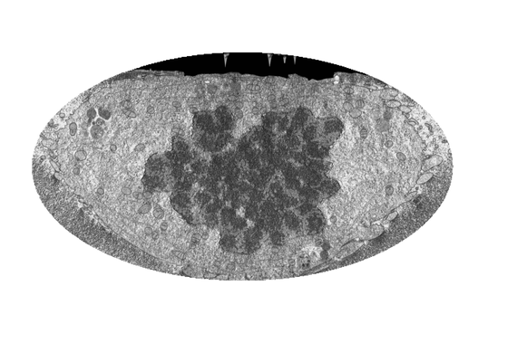 |
FIB-SEM of a dividing cell at 5.3 min after anaphase onset [1998 multi-frame micrographs composed of 1 frames each in TIFF format] | Otsuka S, Steyer AM, Schorb M, Hériché JK, Hossain MJ, Sethi S, Kueblbeck M, Schwab Y, Beck M, Ellenberg J [Pubmed: 29323269] [DOI: 10.1038/s41594-017-0001-9] |
31.2 GB | — |
| 2017-11-28 |  |
FIB-SEM of a dividing cell at 5.7 min after anaphase onset [2620 multi-frame micrographs composed of 1 frames each in TIFF format] | Otsuka S, Steyer AM, Schorb M, Hériché JK, Hossain MJ, Sethi S, Kueblbeck M, Schwab Y, Beck M, Ellenberg J [Pubmed: 29323269] [DOI: 10.1038/s41594-017-0001-9] |
63.0 GB | — |
| 2021-03-05 |  |
Dynabeads as Fiducials for SXT and SIM Correlation [700 tilt series in MRC format] | Okolo CA, Kounatidis I, Groen J, Nahas KL, Balint S, Fish T, Koronfel MA, López-Cortajarena A, Dobbie I, Pereiro E, Harkiolaki M | 1.2 GB | — |
